Transcription
Journal of Experimental & ClinicalCancer ResearchBioMed CentralOpen AccessResearchThe expression of PLK-1 in cervical carcinoma: a possible target forenhancing chemosensitivityYuan Zhang†1, Yu Liu†2, Yuan-Xian Yang1, Jia-Hong Xia3, Hong-Xiu Zhang4,Hua-Bin Li*3,4 and Chun-Zhao Yu*4Address: 1Department of Obstetrics and Gynecology, Union Hospital, Tongji Medical College, Huazhong University of Science and Technology,No. 1277 Jiefang Avenue, Wuhan, Hubei, PR China, 2Department of Medicine, Feinberg Medical School, Northwestern University, 745 NFairbanks, Chicago, IL, USA, 3Department of Surgery, Union Hospital, Tongji Medical College, Huazhong University of Science and Technology,No 1277 Jiefang Avenue, Wuhan, Hubei, PR China and 4Department of Surgery, The First Affiliated Hospital, Nanjing Medical University, No 300Guangzhou Road, Nanjing, Jiangsu, PR ChinaEmail: Yuan Zhang - yuanzhang75@hotmail.com; Yu Liu - hanliuyu@hotmail.com; Yuan-Xian Yang - zsuent@163.com; JiaHong Xia - xiajiahong@hotmail.com; Hong-Xiu Zhang - guangfa2000@sina.com; Hua-Bin Li* - allergyli@163.com; ChunZhao Yu* - chunzhaoyu@hotmail.com* Corresponding authors †Equal contributorsPublished: 23 September 2009Journal of Experimental & Clinical Cancer Research 2009, 28:130doi:10.1186/1756-9966-28-130Received: 13 May 2009Accepted: 23 September 2009This article is available from: http://www.jeccr.com/content/28/1/130 2009 Zhang et al; licensee BioMed Central Ltd.This is an Open Access article distributed under the terms of the Creative Commons Attribution License (http://creativecommons.org/licenses/by/2.0),which permits unrestricted use, distribution, and reproduction in any medium, provided the original work is properly cited.AbstractBackground: Polo-like kinase-1 (PLK-1) is reported to be upregulated in a variety of humantumors and is implicated in cell proliferation and survival. However, its importance in cervicalcarcinoma has not yet been fully elucidated.Methods: We examined PLK-1 expression in cervical carcinoma tissues usingimmunohistochemical staining. Furthermore, we blocked PLK-1 expression in HeLa cells usingspecific siRNA and detected the cell cycle, cell proliferation and chemosensitivity using westernblotting, MTT and flow cytometry.Results: We provide evidence that expression of PLK-1 exists in human cervical carcinoma tissuesand establish an association with tumor size. Furthermore, we show that PLK-1 knockdown bytransfection of siRNA induces accumulation of HeLa cells in the G2/M cell cycle phase and enhancescisplatin-induced apoptosis.Conclusion: Our results indicate that PLK-1 production in HeLa cells might be critical indetermining whether cells survive or undergo apoptosis. Therefore, targeting PLK-1 might be apromising strategy for enhancing sensitivity to chemotherapeutic reagents in cervical carcinoma.BackgroundCervical carcinoma is a common malignancy worldwideand its incidence has been increasing gradually. It poses asignificant health problem, especially in regions such asAsia and North America. Despite advances in diagnosticand treatment modalities, the proportion of failed treat-ments is still significant, with reported rates of 15.6% to58% [1]. To date, chemotherapy is the mainstay of treatment modalities for cervical carcinoma and cisplatin hasproven to be the most effective single cytotoxic agent forthe treatment of advanced or recurrent cervical cancer [2].However, the response rate is about 23%, due to chemorePage 1 of 10(page number not for citation purposes)
Journal of Experimental & Clinical Cancer Research 2009, 28:130sistance. Therefore, it is necessary to develop a novel strategy to overcome the chemoresistance of cervicalcarcinoma and improve clinical efficiency and prognosis.Although the molecular events responsible for the pathogenesis of cervical carcinoma remain to be elucidated, thefinal common pathway of carcinogenesis appears to be adisruption of the mechanisms involved in the regulationof cell cycle progression, leading to uncontrolled cell proliferation [3]. Critical cellular signaling underlying theregulation of cell cycle progression has been implicated ina number of cancers. With regard to tumorigenesis, it isworth noting that polo-like kinase 1 (PLK-1), a mitoticcyclin-independent serine-threonine kinase that isbelieved to be involved in the pathogenesis of numerouscarcinomas [4-6], has attracted much attention as a potential therapeutic target.PLK-1 is a member of the family of polo-like kinasesinvolved in a wide variety of cell cycle processes [7]. Inmammalian cells, PLK-1 is primarily localized in the centrosome, where it is responsible for centrosome separation and maturation. PLK-1-specific antibodiesintroduced into HeLa cells by microinjection prevent centrosome separation and reduce γ-tubulin accumulation,suggesting that PLK-1 functions in regulating centrosomefunction [8]. PLK-1 is also a target of the G2 DNA damagecheckpoint, where it undergoes ubiquitin-dependent proteolysis mediated by the checkpoint protein Chfr, implicating the loss of Plk-1 function as an important responseto DNA damage during the G2 phase of the cell cycle [9].Correspondingly, the elevation of PLK-1 expressionoccurs in a broad range of human tumors [10,11], and aclose correlation has been documented between mammalian PLK-1 expression and progression of endometrial andovarian cancers [12,13]. Therefore, PLK-1 is implicated asa critical candidate target for understanding the progression of cervical carcinoma and improving chemotherapy.However, little is known about the importance of PLK-1in the development and management of cervical carcinoma.To address this issue, we investigated the expression anddistribution of PLK-1 in cervical carcinoma tissues. Furthermore, in order to determine the importance of PLK-1in tumor progression, we investigated the effects of PLK-1knockdown on the biological characteristics of HeLa cellsby taking advantage of small interference RNA (siRNA)against PLK-1. Our results elucidate the pathogenesis ofcervical carcinoma and may help to develop a novel strategy to improve the efficiency of chemotherapy deliveredto patients with cervical erials and methodsImmunohistochemical stainingFor immunohistochemical staining, thirty-six surgicallyresected human cervical carcinoma tissue samples werecollected from the Department of Obstetrics and Gynecology, Wuhan Union Hospital. The study was approved bythe institutional review boards. Immunohistochemicalstaining was performed according to our previous protocol [14]. Briefly, human tumor tissues were embedded inparaffin and cut into 5-μm sections that were placed ontoglass slides. After antigen retrieval, sections were stainedfor the expression of PLK-1 (BD Biosciences, San Diego,CA) (1:100)detected by streptavidin-biotin-horseradishperoxidase complex formation. Tumor sections stainedfor IgG instead of primary antibodies were used as thenegative control. The immunoactivities of PLK-1 wereranked according to the percentage of positive tumor cells:score 3 ( 75%), score 2 (25-75%), score 1 ( 25%), andscore 0 (negative).Cell culture, transient transfection, RNA interference, andcisplatin treatmentHeLa cells were cultured in RPMI 1640 supplementedwith 10% fetal calf serum (FCS) (Invitrogen, Carlsbad,CA,). Plasmid construction and transfection were performed as previously described [4]. Briefly, PLK-1 cDNAwas cloned into the pcDNA3.2-DEST vector (Invitrogen),and the resulting expression plasmid (pcDNA3.2-DESTPlk1) was verified according to the reference sequence.PLK-1 (GenBank accession no. NM 005030) siRNAs, targeting regions of the Plk-1 transcript at positions 362-384,were also used in this study. HeLa cells were transfected at70% to 90% confluency using PLK-1 plasmid DNA (up to4 μg) mixed with Lipofectamine 2000 (Invitrogen) at aDNA (μg)/lipid (μL) ratio of 1:2.5. Similarly, PLK-1silencing was performed by transfecting HeLa cells withPLK-1 siRNA plasmids. At 4-6 h post-transfection, theplasmid- or siRNA-containing medium was replaced withnormal culture medium containing 10% FCS, and thecells were incubated in a 5% CO2 incubator at 37 C.Transfected cells were then cultured in fresh medium forup to 12-36 h and harvested for gene expression and otherassays. For cisplatin treatment, cisplatin (4 μg/ml) wasadded to HeLa cells, with DMSO as control. The timepoint chosen for the addition of cisplatin to the transfected cells was 24 h after transfection, and was based onpreliminary experiments (data not shown).Quantitative RT-PCR analysis for mRNA levelsReal-time RT-PCR was performed as detailed in our previous report [14]. Briefly, total RNA was extracted with TRIzol reagent (Invitrogen), following the manufacturer'sinstructions. Reverse transcription (RT) was performed,and the cDNA was synthesized from 2 μg of total RNA byusing an oligo (dT)18 primer and M-MLV reverse tran-Page 2 of 10(page number not for citation purposes)
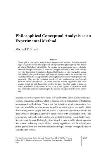
Journal of Experimental & Clinical Cancer Research 2009, 28:130scriptase (TAKARA, Syuzou, Shiga, Japan) for quantitativePCR. Expression of mRNA was determined using the ABIPRISM 7300 Detection System (Applied Biosystems, Foster City, CA) and SYBR Premix Taq (TAKARA). Thesequences of the primers were as follows: PLK1(NM 005030) forward: 5'-GGA CTA TTC GGA CAA GTACG-3'; PLK1 reverse: 5'-CGG AAA TAT TTA AGG AGGGTG A-3'; β-actin (NM 001101) forward: 5'-AAG ATGACC CAG ATC ATG TTT GAG ACC-3'; β-actin reverse: 5'AGC CAG GTC CAG ACG CAG GAT-3'. The mean value ofthe replicates for each sample was calculated andexpressed as cycle threshold (Ct). The amount of geneexpression was then calculated as the difference (ΔCt)between the Ct value of the target gene and the Ct value ofβ-actin.Assessment of cell viability by MTT AssayTreated or untreated cells were seeded into 96-well platesat 1 103 cells per well overnight and incubated with different concentrations of cisplatin (0 or 4 μg/ml) per treatment. After culture for 24 h, 20 μl MTT dye solution (5mg/ml) was added to each well and samples were incubated at 37 C for 4 h. The formazan product was dissolved by adding 200 μL of DMSO to each well. The plateswere read at 570 nm.http://www.jeccr.com/content/28/1/130Apoptosis Assay Kit (Invitrogen) according to the manufacturer's instructions.Hoechst 33258 staining and activity analysis of caspase-3The morphological alterations associated with apoptosiswere observed in transfected HeLa cells by microscopyusing the Hoechst 33258 staining approach. At 36 h posttransfection, cells were fixed (methanol/glacial acetic acidat 3:1) for 15 min at 4 C. Hoechst 33258 (Santa Cruz Biotechnology, Santa Cruz, CA) was added to the well at aconcentration of 10 μg/ml, and cells were then incubatedfor 20 min at 37 C. Before observation, cells were washedthree times with PBS. Caspase-3 activation was also testedwith the Caspase-3 Fluorescent Assay Kit (R&D, Minneapolis, MN). Transfected cells were harvested for the assay 36h after transfection, according to the manual.Statistical analysesImmunostaining of tissue sections was analyzed with theChi-square test. Differences between groups in terms ofmRNA analysis, cell proliferation, and apoptosis wereanalyzed using a two-tailed t-test or analysis of variance(ANOVA) using SPSS 13.0 software. The significance levelwas set at P 0.05.ResultsImmunoblotting analysisImmunoblotting was performed as previously described[14]. Briefly, treated and untreated HeLa cells were collected and the protein concentrations of lysates weredetermined by the Bradford method (Pierce, Rockford,IL). Samples containing 10 μg of protein were boiled andsubjected to sodium dodecyl sulfate polyacrylamide gelelectrophoresis (SDS-PAGE) on 10% Tris-glycine gels andtransferred electrophoretically to polyvinylidene fluoridemembranes. Primary antibodies (mouse anti-humanPLK-1 and β-actin monoclonal antibody, 1:2,000) (SantaCruz Biotechnology, Santa Cruz, CA) were used, followedby incubation with horseradish peroxidase-linked secondary antibody (goat anti-mouse IgG, 1:1,000). Blotswere visualized using an Enhanced Chemiluminescencekit (Cell Signaling, Danvers, MA). Therelative band density of PLK-1 to β-actin was quantified with Bio-RadQuantity One 1-D Analysis Software (Bio-Rad, Hercules,CA). The experiment was performed in triplicate.Cell cycle and apoptosis analysis by flow cytometryCell cycle and apoptosis status of HeLa cells after treatment were determined by flow cytometry. In brief, treatedcells were harvested and washed once with ice-cold 0.1 MPBS, fixed with 70% ethanol and stained with PI solution(50 μg/ml propidium iodide, 1 mg/ml RNase). Cells werethen analyzed for cell cycle status by flow cytometry (FACScan, Becton Dickinson, USA). To quantify apoptosis,cells were stained with annexin-V and PI using a VybrantExpression of PLK-1 in human cervical carcinoma tissuesTo investigate the presence of aberrant PLK-1 expressionin human cervical carcinoma tissues, we examined PLK-1expression by immunohistochemical staining. The clinical pathologic characteristics of specimens, includingtumor size, lymph node status, tumor grade, distantmetastasis and biomarker expression are listed in Table 1.Of the 36 tumor sections, 32 showed positive immunostaining for PLK-1, with a positive rate of 88.9%. Examplesof immunostained slides are shown in Fig. 1. Cytoplasmicand some brown nuclear staining in tumor cells served asan index of PLK-1 expression.To evaluate the possible importance of PLK-1 in tumorprogression, we then evaluated the relationship betweenPLK-1 intensity and tumor size. Using the Spearman rankcorrelation test, a statistically significant positive correlation between PLK-1 expression and primary tumor stage(r 0.605, P 0.002) but not metastasis was identified.Our results, therefore, provided clues that the expressionof PLK-1 is associated with the local expansion of cervicalcarcinoma.Levels of PLK-1 mRNA and protein in HeLa cells after PLK1 or siRNA transfectionTo evaluate the effects of PLK-1 siRNA on the biologicalcharacteristics of HeLa cells, we first transfected HeLa cellswith the PLK-1 plasmid and PLK-1 siRNA. We harvestedcells at different time points (0 h, 12 h, 24 h and 36 h) toPage 3 of 10(page number not for citation purposes)
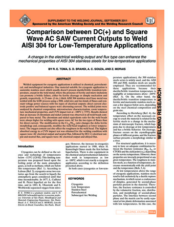
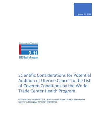
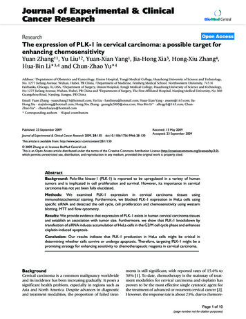
Journal of Experimental & Clinical Cancer Research 2009, 28:130Table 1: Description of the patient population and PLK-1expression.CharacteristicPatients (n 36)Age (Median SD)43.2 15.7HistologyUndifferentiatedDifferentiatedPrimary tumor stagesT(1)T(2)T(3)T(4)Nodular metastasisYesNoDistant metastasisYesNoPLK-1 expressionHigh (score 3)Middle (score 2)Low (score 1)Negative(score 0)21 (58.3%)15 (41.7%)6 (16.7%)7 (19.5%)9 (25%)14 (38.8%)6 (16.7%)30 (83.3%)3 (8.3%)33 (91.7%)17 (47.2%)8 (22.2%)7 (19.5%)4 (11.1%)measure PLK-1 gene and protein expression. As illustratedin Fig 2, levels of PLK-1 mRNA were significantly elevatedafter PLK-1 transfection compared to the control cellstransfected with empty plasmid, with an increase inexpression by 2.2-fold at 12 h, 3.5-fold at 24 h, and 4.7fold at 36 h (P 0.05). Similarly, an increase was alsoobserved in protein level at 24 h (2.1-fold) and 36 h (2.3fold). Conversely, siRNA was shown to inhibit PLK-1mRNA and protein expression. PLK-1 mRNA levels weresignificantly reduced after PLK-1 siRNA transfection compared to the control cells transfected with empty plasmid,with a decrease of 49% at 12 h, 62% at 24 h, 69% at 36 h(P 0.05). Similar decreases were also observed at theprotein level at 24 h (58%) and 48 h (76%). Our resultssuggest that PLK-1 siRNA transfection into HeLa cells isable to knock-down the expression of PLK-1.PLK-1 knock-down by siRNA transfection modulated HeLacell survivalWe next evaluated the functional consequences of PLK-1knock-down on the survival of HeLa cells by morphological examination. As illustrated in Fig 3, we observedenhanced apoptosis in HeLa cells after PLK-1 knock-downwith or without cisplatin treatment, as indicated by typical nuclear condensation and cellular shrinkage as determined by Hoechst staining. We then quantitated thenumber of condensed nuclei per field for several fields.The numbers of condensed nuclei in groups A (control),B (PLK-1), C (PLK-1 siRNA), D (PLK-1 plus cisplatin)were 2.5 (0-7), 6.2 (0-13), 22.7 (5-65), 35.5 (9-77) (con-http://www.jeccr.com/content/28/1/130densed nuclei/mm3), respectively; the results were significant (P 0.05).To determine whether PLK-1 influences HeLa cell survival,we examined cell cycle characteristics and apoptosis afterPLK-1 knockdown by flow cytometry. As shown in Fig. 4,we observed that PLK-1 siRNA significantly decreased G1/S arrest of HeLa cells from 64.5% to 32.5% (P 0.05).Conversely, G2/M arrest of HeLa cells increased significantly from 34.6% to 67.7% (P 0.05). These findingssuggested that PLK-1 knockdown contributed to cell cycleprogression. In contrast, PLK-1 transfection significantlyincreased G1/S arrest and decreased G2/M arrest in HeLacells.In addition, we also evaluated cell apoptosis after PLK-1knockdown by double-staining with PI/Annexin-V, followed by flow cytometric analysis. We observed a consistent pro-apoptotic effect of PLK-1 knockdown on HeLacells. The apoptotic rate of PLK-1 knockdown HeLa cellsincreased significantly from 4.2% to 12.5% (P 0.05),whereas PLK-1 transfection did not significantly affectHeLa cell apoptosis (Fig. 4). Interestingly, although cisplatin did not drive the cell cycle in combination with PLK1 siRNA, it acted synergistically with PLK-1 siRNA ininducing cell apoptosis (12.5% vs. 24.9%, P 0.05).PLK-1 knock-down inhibited cell proliferation andincreased caspase-3 activityTo further determine the effects of PLK-1 siRNA transfection on HeLa cells, we then examined cell proliferationand caspase-3 activity by MTT and fluorescent assay,respectively. As shown in Fig 5, PLK-1 knockdown significantly inhibited cell proliferation, as compared to thecontrol (P 0.05). However, PLK-1 transfection showedno significant effect. After treatment with cisplatin, weobserved a synergistic effect of PLK-1 siRNA and cisplatintreatment on HeLa cell proliferation (P 0.05). Furthermore, PLK-1 siRNA significantly increased caspase-3 activity in HeLa cells; caspase-3 activity was further enhancedby cisplatin compared to control and PLK-1 transfectedHeLa cells (P 0.05). These results were consistent withthose of the morphological examination, flow cytometricanalysis and proliferation assays, suggesting that PLK-1knock-down contributes to the induction of apoptosis inHeLa cells and to enhancing chemosensitivity.DiscussionIt is well-recognized that PLK-1 plays an important role incell cycle regulation by functioning in centrosome maturation, spindle formation, mitotic entry, and cytokinesis.When responding to DNA damage, PLK-1 triggers cellcycle arrest in the G2 and M phases, determining cell fate.The significance of PLK-1 has been demonstrated in a variety of tumors. For example, Takai et al. found that expres-Page 4 of 10(page number not for citation purposes)
Journal of Experimental & Clinical Cancer Research 2009, 28:130http://www.jeccr.com/content/28/1/130Figure 1Immunohistochemicalstaining of PLK-1 in human cervical carcinoma tissuesImmunohistochemical staining of PLK-1 in human cervical carcinoma tissues. Representative results of immunostaining are presented; cytoplasmic and some nuclear staining can be observed in tumor cells. A, Medium PLK-1 positive stainingin human cervical carcinoma tissues (original magnification, 200 ); B, low PLK-1 positive staining in human cervical carcinomatissues (original magnification, 200 ); C, PLK-1 negative control staining in human cervical carcinoma tissues (original magnification, 200 ); D, Association of PLK-1 expression and primary tumor stage (* P 0.05 compared to other group).sion of PLK-1 in ovarian cancer is associated withhistological grade and clinical stage [13]. Feng et al.reported that overexpression of PLK1 is associated withpoor survival due to the inhibition of apoptosis viaenhancement of survivin levels in esophageal squamouscell carcinoma [15]. PLK-1 has also been demonstrated tobe associated with cancer invasion and metastasis [16,17].However, the significance of PLK-1 in the pathogenesisand management of cervical carcinoma is not well-understood. In the present study, we demonstrated, for the firsttime, that PLK-1 is expressed in cervical carcinoma with apositive rate of 88.9%, and PLK-1 expression in tumorswas associated with primary tumor progression (T stage).Interestingly, we found four samples that were negativefor PLK-1 staining, which were later found to be the differ-entiated samples. These results suggest that PLK-1 expression might be associated with the inactivity of cell mitosis.Therefore, our results indicate that PLK-1 may be a potential target for tumor evaluation and management of cervical carcinoma.PLK is a well-conserved family that has four known members in humans: PLK1, PLK2, PLK3, and PLK4 [10]. PLK1expression is regulated during cell cycle progression. Levels are low in G0, G1, and S, but begin to increase in G2and peak in M phase. PLK-1 has attracted much attentionin the field of carcinogenesis and cancer therapy due to itsknown functions. Blocking PLK-1 through RNA interference has shown promise as a way to intervene in cancerprogression [18,19]. RNA interference is a newly discov-Page 5 of 10(page number not for citation purposes)
Journal of Experimental & Clinical Cancer Research 2009, 28:130http://www.jeccr.com/content/28/1/130Figure 2 of PLK-1 gene and protein expression in HeLa cells after PLK-1 or siRNA transfectionAlterationAlteration of PLK-1 gene and protein expression in HeLa cells after PLK-1 or siRNA transfection. PLK-1 production in HeLa cells increased after PLK-1 transfection, but was inhibited by siRNA transfection. A, PLK-1 mRNA and protein levels in HeLa cells increased after PLK-1 transfection, but decreased following siRNA transfection. The level of mRNA wasdetermined by real-time RT-PCR. A time-dependent induction was observed; B, Representative results of immunoblotting ofPLK-1 expression in HeLa cells were shown. C, PLK-1 protein in HeLa cells increased after PLK-1 transfection, but decreasedfollowing siRNA transfection. The level of protein was determined by immunoblotting. A time-dependent modulation wasobserved. Data were the means of three independent experiments. * P 0.05 compared to the control.ered cellular pathway for silencing genes in a sequencespecific manner at the mRNA level through the introduction of cognate double-stranded small interfering RNA(siRNA). This method is significantly more efficient thantraditional antisense approaches. In our previous study[4], we knocked down PLK-1 production in pancreaticcancer cells by utilizing siRNA transfection, and observedenhanced chemosensitivity to therapeutic agents. To further understand the importance of PLK-1 in the management of cervical carcinoma, we used siRNA transfection toknock down PLK-1 production in HeLa cells.It has been demonstrated that PLK-1 mRNA expression iselevated in proliferating cells, such as various cancer celllines and tumors of different origins. Here, we observedthe expression of PLK-1 mRNA in HeLa cells. We thentransfected PLK-1 plasmids and PLK-1 siRNA into HeLacells, to evaluate the effects of PLK-1 up- or down-regulation on the biological characteristics of HeLa cells. As weexpected, PLK-1 mRNA was significantly elevated afterPLK-1 transfection, compared to the control cells transfected with empty plasmid. In contrast, PLK-1 siRNA significantly inhibited PLK-1 production in HeLa cells. Theseresults showed that siRNA transfection of HeLa cells isable to knock down the expression of PLK-1. Based onthese findings, we then performed morphological examinations to evaluate the functional consequences of PLK-1knock-down on HeLa cell survival. We observed enhancedapoptosis in HeLa cells after PLK-1 knock-down with orwithout cisplatin treatment, as indicated by typicalnuclear condensation and cellular shrinkage visualized byHoechst staining.Eukaryotic cells have developed a network of checkpointsto ensure their survival and the propagation of accuratecopies of the genome to the next generation, even whensuffering from a variety of stresses [19]. PLK-1 is a criticalcomponent responsible for tumor progression. SilencingPage 6 of 10(page number not for citation purposes)
Journal of Experimental & Clinical Cancer Research 2009, nock-downPLK-13by siRNA transfection modulated apoptosis in HeLa cellsPLK-1 knock-down by siRNA transfection modulated apoptosis in HeLa cells. A, Control; B, Cells transfected withPLK-1; C, Cells transfected with PLK-1 siRNA; D, Cells transfected with PLK-1 siRNA and treated with cisplatin (4 μg/ml)(original magnification, 200 ); Enhanced apoptosis was demonstrated in B, C and D by typical nuclear condensation aftersiRNA transfection, as determined by Hoechst staining. Three independent experiments were performed. Representative fluorescent images are presented.PLK1 expression by RNA interference inhibits tumor cellproliferation and induces G2/M arrest. To determinewhether PLK-1 influences HeLa survival, we examined cellcycle characteristics and apoptosis after PLK-1 knockdown by using flow cytometry. Importantly, we observedthat PLK-1 siRNA significantly decreased the G1/S arrestof HeLa cells from 64.5% to 32.5%. Conversely, G2/Marrest of HeLa cells increased significantly from 34.6% to67.7%. These findings suggested that PLK-1 contributes toHeLa cell cycle progression.Currently, cervical carcinoma is the second most commoncancer worldwide among women and one of the leadingcauses of death in relatively young women. Chemotherapy represents a crucial strategy for the management ofboth primary and recurrent cervical carcinoma [20]. However, some types of cervical carcinoma exhibit limited sensitivity to cytotoxic agents and easily develop drugresistance during long-term chemotherapy [21]. For thisreason, enhancing chemosensitivity is essential forimproved prognosis. According to the literature, investigating the importance of PLK-1 in the prevention of othercancers, we believe PLK-1 can be considered an importantcandidate for the enhancement of chemosensitivity in cervical carcinoma.To examine this possibility, we investigated the apoptosisof HeLa cells after PLK-1 knockdown by RNA interference.Importantly, we observed a consistent pro-apoptoticeffect of PLK-1 knock-down in HeLa cells. The apoptoticrate in HeLa cells increased significantly from 4.2% to12.5% after PLK-1 knockdown, whereas transfection withPLK-1 did not affect HeLa cell apoptosis. Although cisplatin did not drive the cell cycle, when used in combinationwith PLK-1 siRNA, the compound demonstrated a synergistic effect with PLK-1 siRNA in inducing cell apoptosis(12.5% vs. 24.9%). Consistently, we observed that PLK-1knockdown significantly inhibited cell proliferation andPage 7 of 10(page number not for citation purposes)
Journal of Experimental & Clinical Cancer Research 2009, nock-downPLK-14modulated cell cycle characteristics and apoptosis in cisplatin-treated HeLa cellsPLK-1 knock-down modulated cell cycle characteristics and apoptosis in cisplatin-treated HeLa cells. A synergistic effect with cisplatin treatment (4 μg/ml) was demonstrated. A, PLK-1 siRNA significantly decreased G1/S arrest butenhanced G2/M arrest of HeLa cells; B, PLK-1 siRNA significantly enhanced the apoptosis of HeLa cells, demonstrating a synergistic effect with cisplatin treatment. Representative results of flow cytometric analysis are presented. Data were the means ofthree independent experiments. * P 0.05, compared to the control cells transfected with empty plasmid; ** P 0.05, compared to the cells transfected with PLK-1 siRNA alone.Page 8 of 10(page number not for citation purposes)
Journal of Experimental & Clinical Cancer Research 2009, nockdownPLK-15by siRNA transfection modulated proliferation and caspase-3 activity in HeLa cellsPLK-1 knockdown by siRNA transfection modulated proliferation and caspase-3 activity in HeLa cells. A, PLK-1knockdown significantly inhibited cell proliferation, as determined by MTT assay; B, Cell proliferation curve for four groups ofHeLa cells was presented, as determined by MTT assay; C, PLK-1 knockdown significantly increased caspase-3 activity in HeLacells, as determined by Fluorescent Assay. Data are the means of three independent experiments. * P 0.05 compared to thecontrol cells.induced apoptosis, displaying a synergistic effect with cisplatin treatment. Based on these results, PLK-1 knockdown shows promise as an adjuvant chemotherapy forcervical carcinoma. It will be of great interest to furtherinvestigate the possible mechanisms underlying PLK-1driven cell survival.designed the study and prepared the manuscript. Allauthors read and approved the final manuscript.In conclusion, we have provided evidence that there is acorrelation between overexpressed PLK-1 and the primarycancer stage in cervical carcinoma tissues. To further characterize the role of PLK-1 in the carcinogenesis of cervicalcarcinoma and the importance of PLK-1 knockdown inthe prevention of cervical carcinoma, we investigated theeffects of PLK-1 RNA interference on cell cycle characteristics and apoptosis in HeLa cells. We demonstrated thatPLK-1 knockdown contributed to G2/M arrest and to theinduction of apoptosis in HeLa cells, as well as to the inhibition of cell viability and to the enhancement of chemosensitivity. Therefore, PLK-1 can be thought of as apotential target for preventing cervical carcinoma.ReferencesAcknowledgementsThis study was supported by grants from the Natio
Fairbanks, Chicago, IL, USA, 3Department of Surgery, Union Hospital , . transfection of siRNA induces a ccumulation of HeLa cells in th e G2/M cell cycle phase and enhances cisplatin-induced apoptosis. . Plasmid construction and transfection were per-fo