Transcription
Saladin: Anatomy &Physiology: The Unity ofForm and Function, ThirdEditionAtlas B Surface Anatomy The McGraw HillCompanies, 2003TextATLASSurface AnatomyThe Importance of External Anatomy 392Head and Neck (fig. B.1) 393Trunk 394 Thorax and Abdomen (fig. B.2) 394 Back and Gluteal Region (fig. B.3) 395 Pelvic Region (fig. B.4) 396 Axillary Region (fig. B.5) 397Upper Limb 398 Lateral Aspect (fig. B.6) 398 Antebrachium (forearm) (fig. B.7) 398 Wrist and Hand (fig. B.8) 399Lower Limb 400 Thigh and Knee (fig. B.9) 400 Leg (figs. B.10–B.12) 401 Foot (figs. B.13–14) 404Muscle Test (fig. B.15) 406B
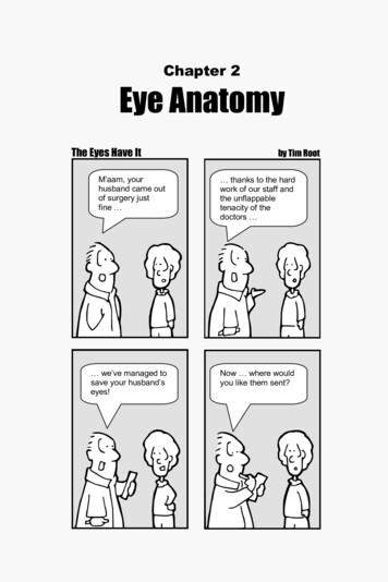
Saladin: Anatomy &Physiology: The Unity ofForm and Function, ThirdEditionAtlas B Surface AnatomyText The McGraw HillCompanies, 2003392 Part Two Support and MovementThe Importance of ExternalAnatomyAtlas BIn the study of human anatomy, it is easy to become so preoccupied with internal structure that we forget the importance of what we can see and feel externally. Yet externalanatomy and appearance are major concerns in giving aphysical examination and in many aspects of patient care.A knowledge of the body’s surface landmarks is essentialto one’s competence in physical therapy, cardiopulmonary resuscitation, surgery, making X rays and electrocardiograms, giving injections, drawing blood, listening toheart and respiratory sounds, measuring the pulse andblood pressure, and finding pressure points to stop arterialbleeding, among other procedures. A misguided attemptto perform some of these procedures while disregarding ormisunderstanding external anatomy can be very harmfuland even fatal to a patient.Having just studied skeletal and muscular anatomyin the preceding chapters, this is an opportune time foryou to study the body surface. Much of what we see therereflects the underlying structure of the superficial bonesand muscles. A broad photographic overview of surfaceanatomy is given in atlas A (see fig. A.5). In the followingpages, we examine the body literally from head (fig. B.1)to toe (fig. B.14), studying its regions in more detail. Tomake the most profitable use of this atlas, refer to theskeletal and muscular anatomy in chapters 8 to 10. Relatedrawings of the clavicles in chapter 8 to the photograph infigure B.1, for example. Study the shape of the scapula inchapter 8 and see how much of it you can trace on the photographs in figure B.3. See if you can relate the tendonsvisible on the hand (fig. B.8) to the muscles of the forearmillustrated in chapter 10, and the external markings of thepelvis (fig. B.4) to bone structure in chapter 8.For learning surface anatomy, there is a resourceavailable to you that is far more valuable than any laboratory model or textbook illustration—your own body.For the best understanding of human structure, comparethe art and photographs in this book with your body orwith structures visible on a study partner. In addition tobones and muscles, you can palpate a number of superficial arteries, veins, tendons, ligaments, and cartilages,among other structures. By palpating regions such as theshoulder, elbow, or ankle, you can develop a mentalimage of the subsurface structures better than you canobtain by looking at two-dimensional textbook images.And the more you can study with other people, the moreyou will appreciate the variations in human structureand be able to apply your knowledge to your futurepatients or clients, who will not look quite like any textbook diagram or photograph you have ever seen.Through comparisons of art, photography, and the livingbody, you will get a much deeper understanding of thebody than if you were to study this atlas in isolation fromthe earlier chapters.At the end of this atlas, you can test your knowledgeof externally visible muscle anatomy. The two photographs in figure B.15 have 30 numbered muscles and a listof 26 names, some of which are shown more than once inthe photographs and some of which are not shown at all.Identify the muscles to your best ability without lookingback at the previous illustrations, and then check youranswers in appendix B at the back of the book.
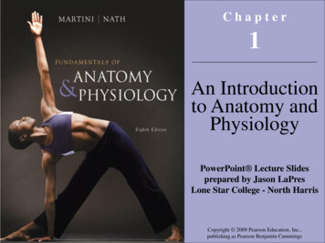
Saladin: Anatomy &Physiology: The Unity ofForm and Function, ThirdEditionAtlas B Surface Anatomy The McGraw HillCompanies, 2003TextAtlas B Surface Anatomy alBuccal (cheek)MentalNuchal (posterior cervical)CervicalAtlas B(a)Frons (forehead)Root of noseBridge of noseSuperciliaryridgeSuperior palpebralsulcusInferior palpebralsulcusAuricle (pinna)of earPhiltrumLabia (lips)Lateral commissureTrapezius avicleSuprasternal notchMedial commissureDorsum nasiApex of noseAla nasiMentolabial sulcusMentum (chin)Sternum(b)Figure B.1 The Head and Neck. (a) Anatomical regions of the head, lateral aspect. (b) Features of the facial region and upper thorax.
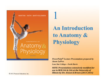
Saladin: Anatomy &Physiology: The Unity ofForm and Function, ThirdEditionAtlas B Surface Anatomy The McGraw HillCompanies, 2003Text394 Part Two Support and vicleTrapeziusThyroid cartilageSuprasternal notchDeltoidAcromionManubriumPectoralis sSerratus anteriorTendinousinsertion ofrectus abdominisLinea semilunarisLinea albaUmbilicusAtlas BAnterior superiorspine of iliumExternal abdominalobliqueIliac crestInguinal pezius m.Sternum:Suprasternal notchDeltoid m.ManubriumAngleBreast:Body (gladiolus)Axillary tailXiphoid processNippleAreolaCorpus (body)Linea albaCostal marginLinea semilunarisRectusabdominis m.UmbilicusExternal abdominaloblique m.Anterior superiorspine of ilium(b)Figure B.2 The Thorax and Abdomen, Ventral Aspect. (a) Male; (b) female. All of the features labeled are common to both sexes, thoughsome are labeled only on the photograph that shows them best.
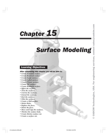
Saladin: Anatomy &Physiology: The Unity ofForm and Function, ThirdEditionAtlas B Surface AnatomyText The McGraw HillCompanies, 2003Atlas B Surface Anatomy 395Flexor carpi ulnarisBrachioradialisBiceps brachiiTriceps brachiiDeltoidAnterior partMiddle partPosterior partTeres majorInfraspinatusMedial borderof scapulaTrapeziusVertebral furrowErector spinaeLatissimus dorsiAtlas BIliac crest(a)AcromionInfraspinatusMedial borderof scapulaTrapeziusInferior angleof scapulaOlecranonLatissimusdorsiErector spinaeIliac crestGluteus mediusGluteus maximusSacrumCoccyxNatal cleftGreatertrochanterof femurHamstring musclesGluteal fold(b)Figure B.3 The Back and Gluteal Region. (a) Male; (b) female. All of the features labeled are common to both sexes, though some are labeledonly on the photograph that shows them best.
Saladin: Anatomy &Physiology: The Unity ofForm and Function, ThirdEditionAtlas B Surface AnatomyText The McGraw HillCompanies, 2003396 Part Two Support and MovementAtlas B(a)(b)Figure B.4 The Pelvic Region. (a) The anterior superior spines of the ilium are marked by anterolateral protuberances (arrows). (b) The posteriorsuperior spines are marked in some people by dimples in the sacral region (arrows).
Saladin: Anatomy &Physiology: The Unity ofForm and Function, ThirdEditionAtlas B Surface Anatomy The McGraw HillCompanies, 2003TextAtlas B Surface Anatomy 397OlecranonBiceps brachiiTriceps brachiiAnterior axillary fold (pectoralis major)DeltoidPosterior axillary fold (latissimus dorsi)Pectoralis majorLatissimus dorsiSerratus anteriorRectus abdominisExternal abdominal obliqueFigure B.5 The Axillary Region.Atlas BAxilla (armpit)
Saladin: Anatomy &Physiology: The Unity ofForm and Function, ThirdEditionAtlas B Surface Anatomy The McGraw HillCompanies, 2003Text398 Part Two Support and oidMetacarpophalangealjointsBiceps brachiiPectoralis majorTriceps brachiiLong headLateral headBrachioradialisExtensor carpiradialis longusLateral epicondyleof humerusOlecranonStyloid processof ulnaExtensor digitorumFigure B.6 The Upper Limb, Lateral Aspect.Atlas BCubital fossaTriceps brachiiBiceps brachiiMedial epicondyleof humerusCephalic veinOlecranonMedian cubital veinHead of radiusBrachioradialisBrachioradialisFlexor carpi radialisPalmaris longusFlexor carpi ulnarisFlexor carpi ulnarisExtensor carpi ulnarisExtensor digitorumStyloid process of radiusStyloid process ofulnaHypothenar eminenceThenar eminenceTendons of extensor digitorumFlexion creasesPalmar surface of handDorsum of handPollex (thumb)Volar surface of fingersFlexion lines(a)Figure B.7 The Antebrachium (forearm). (a) Ventral aspect; (b) dorsal aspect.(b)
Saladin: Anatomy &Physiology: The Unity ofForm and Function, ThirdEditionAtlas B Surface Anatomy The McGraw HillCompanies, 2003TextPalmaris longus tendonFlexor carpi radialis tendonFlexion creasesThenar eminenceHypothenar eminenceIPollex (thumb)Flexion tsAtlas BVIIIVIII(a)( )Styloid process of radiusStyloid process of ulnaExtensor pollicis brevis tendonAnatomical snuffboxExtensor pollicis longus tendonExtensor digiti minimi tendonExtensor digitorum tendonsAdductor pollicis(b)Figure B.8 The Wrist and Hand. (a) Ventral aspect; (b) dorsal aspect.399
Saladin: Anatomy &Physiology: The Unity ofForm and Function, ThirdEditionAtlas B Surface Anatomy The McGraw HillCompanies, 2003Text400 Part Two Support and MovementLateralLateralMedialMedialVastus lateralisTensor fasciae lataeBiceps femoris(long head)SemitendinosusRectus femorisSemimembranosusGracilisGracilisAtlas BVastus lateralisVastus medialisQuadricepsfemoris tendonPopliteal fossaIliotibial bandPatellaPatellar ligamentTibial tuberosity(a)Figure B.9 The Thigh and Knee. (a) Ventral aspect; (b) dorsal aspect.(b)
Saladin: Anatomy &Physiology: The Unity ofForm and Function, ThirdEditionAtlas B Surface Anatomy The McGraw HillCompanies, 2003TextAtlas B Surface Anatomy 401Vastus lateralisBiceps femorisIliotibial bandLateral epicondyle of femurHead of fibulaPatellar ligamentLateral head of gastrocnemiusFibularis longusTibialis anteriorFibularis longus and fibularisbrevis tendonsCalcaneal tendonLateral malleolusCalcaneusFigure B.10 The Leg and Foot, Lateral Aspect.Atlas BSoleus
Saladin: Anatomy &Physiology: The Unity ofForm and Function, ThirdEditionAtlas B Surface Anatomy402 Part Two Support and MovementSemimembranosusSemimembranosus tendonPatellaMedial epicondyle of femurSemitendinosus tendonMedial condyle of tibiaMedial head of gastrocnemiusTibiaAtlas BSoleusMedial malleolusExtensor hallucis longus tendonMedial longitudinal archHead of metatarsal IAbductor hallucisFigure B.11 The Leg and Foot, Medial Aspect.Text The McGraw HillCompanies, 2003
Saladin: Anatomy &Physiology: The Unity ofForm and Function, ThirdEditionAtlas B Surface Anatomy The McGraw HillCompanies, 2003TextAtlas B Surface Anatomy 403MedialLateralHamstring musclesBiceps femoris tendonSemitendinosus tendonPopliteal fossaAtlas BGastrocnemiusMedial headLateral headSoleusFibularis longusTibialis anteriorCalcaneal tendonLateral malleolusExtensor digitorum brevisCalcaneusFigure B.12 The Leg and Foot, Dorsal Aspect.
Saladin: Anatomy &Physiology: The Unity ofForm and Function, ThirdEditionAtlas B Surface AnatomyText The McGraw HillCompanies, 2003404 Part Two Support and MovementCalcaneal tendonLateral malleolusExtensor digitorum brevisLateral longitudinal archAtlas BExtensor digitorum longustendons(a)Medial malleolusCalcaneal tendonMedial longitudinal archCalcaneusHead of metatarsal I(b)Figure B.13 The Foot. (a) Lateral aspect; (b) medial aspect.
Saladin: Anatomy &Physiology: The Unity ofForm and Function, ThirdEditionAtlas B Surface Anatomy The McGraw HillCompanies, 2003TextSoleusTibiaTibialis anteriorMedial malleolusLateral malleolusSite for palpating dorsal pedal arteryExtensor hallucis longus tendonExtensor digitorum longus tendonsDigits (I–V)IIHallux (great toe)VIVIIIIIAtlas BHead of metatarsal IIIIIIIVHallux (great toe)VHead of metatarsal I(a)Transverse archHead of metatarsal VAbductor digiti minimiAbductor hallucisMedial longitudinal archLateral longitudinal archLateral malleolusCalcaneus(b)Figure B.14 The Right Foot. (a) Dorsal aspect, (b) plantar aspect.405
Saladin: Anatomy &Physiology: The Unity ofForm and Function, ThirdEditionAtlas B Surface Anatomy The McGraw HillCompanies, 2003Text406 Part Two Support and s B5628729301516(a)(b)Figure B.15 Muscle Test. To test your knowledge of muscle anatomy, match the 30 labeled muscles on these photographs to the alphabetical listof muscles below. Answer as many as possible without referring to the previous illustrations. Some of these names will be used more than once, since thesame muscle may be shown from different perspectives, and some of these names will not be used at all. The answers are in appendix B.a. biceps brachiij.b. brachioradialisk. latissimus dorsit. subscapularisc. deltoidl.u. teres majord. erector spinaem. pectoralis majorv. tibialis anteriore. external abdominal obliquen. rectus abdominisw. transversus abdominisf. flexor carpi ulnariso. rectus femorisx. trapeziusg. gastrocnemiusp. serratus anteriory. triceps brachiih. gracilisq. soleusz. vastus ius capitiss. sternocleidomastoid
Anatomy In the study of human anatomy, it is easy to become so pre-occupied with internal structure that we forget the impor-tance of what we can see and feel externally. Yet external . cardiograms, giving injections, drawing blood, listening to heart and respiratory sounds, measuring the pulse and blood pressure, and finding pressure points .