Transcription
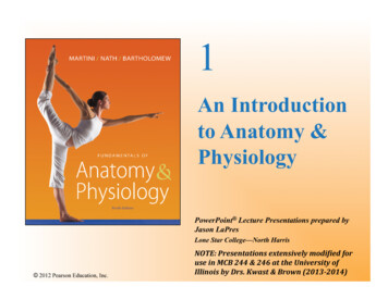
1An Introductionto Anatomy &PhysiologyPowerPoint Lecture Presentations prepared byJason LaPresLone Star College—North Harris 2012 Pearson Education, n(2013- ‐2014)
Chapter 1 Learning Objectives Describe the basic functions of organisms. Define anatomy & physiology and the various specialties ofeach. Identify and understand the major levels of organization ofour bodies. Identify and describe the 11 organ systems of the body. Understand and be able to explain the concept of“homeostasis” and describe the roles of negative andpositive feedback in regulating body functions. Identify the major body cavities using proper anatomicalterms. 2012 Pearson Education, Inc.
Anatomy & Physiology: The study of structurefunction relationships in biology Anatomy Describes the structures of the body including What they are made of Where they are located Associated structures Physiology Is the study of the function of biological systems including,of course, anatomical structures It includes both individual and cooperative functions Anatomy & Physiology: forms the foundation forunderstanding the body’s parts and functions in concert. 2012 Pearson Education, Inc.
Introduction – A Brief History of Anatomy Anatomy (anatome to cut up): study of “cutting up” of the structural parts Oldest medical science; cadaver dissection (dis apart; secare to cut)Egypt: Anatomical or Edwin Smith Surgical Papyrus (1600 BCE): Contains 48 case histories of medical trauma and their treatment; describes closingwounds with sutures, preventing and curing infection with honey, stopping bleedingwith raw meat as well as immobilizing the head and neck to prevent spinal cordinjuries during transport. Also contain the first known descriptions of the cranial sutures, meninges, externalsurface of the brain, cerebrospinal fluid, and intracranial pulsations. Also basicanatomy of major organs and blood vessels as well as use of plants for treatingmedical conditions.Greece: Hippocrates (5th & 4th century BCE) – Greek physician/medical scientist Aristotle (4th century BCE) – text based in animal dissections: arteries, veins,organs and organ systems Herophilos & Erasistratus (4th century BCE) – extensive cadaver dissections 2012 Pearson Education, Inc.
Introduction – A Brief History of Anatomy Cont Greece Cont.: Herophilos & Erasistratus even performed vivisections oncriminals! Claudius Galenus (a.k.a. Galen of Pergamon (Turkey)--2nd century BCE) –compiled previous knowledge and filled in gaps with animal (e.g., monkey & pig)dissections (“Ancient World’s Gray’s Anatomy” [1500 years]) – physician toRoman Emperors Renaissance (1500s)— Andreas Vesalius (“De humani corporis fabrica” – Onthe workings of the human body) – founder of modern human anatomy Gallows (Roman 14th – 16th BCE) & Graves (Michelangelo 17th – 18th century) Galileo charged admission for traveling cadaver dissections Anatomy Act of 1832 (UK) – finally provided for an adequate legal supply ofcadavers for medicine (lead to Gray’s Anatomy)[MurderAct of 1752 --stipulated that only the bodies of convicted murderers were allowedfor legal dissection] What about recent advances? Are there any or has it all been done? . 2012 Pearson Education, Inc.
Introduction – Modern Anatomical Projects 2012 Pearson Education, Inc.
Introduction – Human Anatomy as Art? Body Worlds , Bodies , etc. travelingexhibitions of preserved human bodiesprepared using a technique calledplastination (German anatomist Gunthervon Hagens - water and fat replaced byacetone then plastics [silicone rubber,polyester & epoxy resins] – up to 12month process); have done animals aslarge as horses. Controversial: Who are these people andwere they willing participants? Also debate over Texas inmate used inVisible Human Project Finally, there continue to be advances inpaleopathology, showing evolution of thehuman form (not only from distantrelatives but of modern humans—height) 2012 Pearson Education, Inc.
Introduction – Human Physiology Physiology comes from Ancient Greek: physis, "nature, origin";and -logia, "study of". Anatomy & Physiology together is the study of structure-functionrelationships in biological systems Human Physiology is the study of the mechanical, physical, andbiochemical functions of humans, their organs, and the cells ofwhich they are composed. Physiology includes: Biochemistry, Biophysics, Cell Biology &Chemistry, Endocrinology, Genetics, Genomics, Immunology,Kinesiology, Neurobiology, Pathology, etc. 2012 Pearson Education, Inc.
Introduction – Brief History of Human Physiology Human physiology dates back to the time of Hippocrates — father ofmodern medicine (5th century BCE) Claudius Galenus [a.k.a. Galen of Pergamon] (c. 126-199 A.D.) usedexperiments to probe body functions; the founder of experimentalphysiology. Middle Ages — the Muslim physician Avicenna (980-1037) introducedexperimentation and quantification in The Canon of Medicine. Ibn al-Nafis (1213–1288) — first physician to correctly describe theanatomy of the heart, the coronary circulation, the structure of thelungs, and the pulmonary circulation Renaissance (1500s)— Andreas Vesalius (De humani corporis fabrica)– founder of modern human anatomy Herman Boerhaave (Leiden 1708) — father of clinical physiology —textbook Institutiones medicae 2012 Pearson Education, Inc.
Introduction – Brief History of Human Physiology 19th century — Cell theory of Schleiden & Schwann, which “radically”stated that organisms are made up of units called cells. Claude Bernard's (1813–1878) concept of milieu interieur (internalenvironment), which would later be taken up and championed as"homeostasis" by American physiologist Walter Cannon (1871–1945) 20th century — comparative physiology and ecophysiology (KnutSchmidt-Nielsen and George Bartholomew). Most recently, evolutionaryphysiology has become a distinct subdiscipline. Recent advances are in the Systems Biology subdisciplines, such asphysiological genomics (functional genomics) In addition, advances in cell physiology/ biology can be expected fordecades (centuries?) Physiology IS at the center of systems biology and, indeed,personalized medicine. 2012 Pearson Education, Inc.
1-4 Relationships between Anatomy & Physiology Anatomy Gross anatomy, or macroscopic anatomy,examines large, visible structures Surface anatomy: exterior features Regional anatomy: body areas Systemic anatomy: organ systems Developmental anatomy: from conception to death Clinical anatomy: medical specialties 2012 Pearson Education, Inc.
1-4 Relationships between Anatomy & Physiology Anatomy Microscopic anatomy examines cells andmolecules Cytology: study of cells and their structures cyt- cell Histology: study hist- tissue 2012 Pearson Education, Inc.
1-4 Relationships between Anatomy & Physiology Physiology Subdisciplines Cell physiology: processes within and between cells Organ physiology: functions of specific organs Systemic physiology: functions of an organ system Pathological physiology: effects of diseases 2012 Pearson Education, Inc.
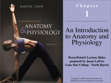
1-5 Levels of Organization Chemical (or Molecular) Chemical, Mechanical and Electrical events that occur within and between cells Cellular The fundamental compartments of all known living organisms and themolecules and organelles within working together Tissue Group of cells working together in a concerted manner Organ A group of tissues working together to perform specific functions Organ System An organ system is a group of organs working together Humans have 11 organ systems Organism A human is an organism 2012 Pearson Education, Inc.
Figure 1-1 Levels of OrganizationTissue LevelCardiac muscletissueOrgan systemlevelOrgan LevelOrganismlevelThe heartThecardiovascularsystemCellular LevelChemical and Molecular LevelsHeart musclecellComplex protein molecule 2012 PearsonEducation, Inc.Atomsin combinationProtein filaments
1-5 Levels of Organization – Organ Systems (11) Integumentary (Chpt 5) Skeletal (Chpts 6-9) Major Organs Major Organs Skin Bones ( 270) Hair Cartilages Associatedligaments Bone marrow Sweat glands Nails Functions Protects againstenvironmental hazards Helps regulate bodytemperature Provides sensoryinformation 2012 Pearson Education, Inc. Functions Provides support andprotection for other tissues Stores calcium and otherminerals Forms blood cells
1-5 Levels of Organization – Organ Systems (11) Muscular (Chpts 10-11) Nervous (Chpts 12-17) Major Organs Skeletal muscles( 650) and associatedtendons Functions Provides movement Provides protectionand support for othertissues Generates heat thatmaintains bodytemperature 2012 Pearson Education, Inc. Major Organs Brain Spinal cord Peripheralnerves Sense organs Functions Directs immediateresponses to stimuli Coordinates or moderatesactivities of other organsystems Provides and interpretssensory information aboutexternal conditions
1-5 Levels of Organization – Organ Systems (11) Endocrine (Chpt 18) Cardiovascular (Chpts 19-21) Major Organs Major Organs Pituitary gland Heart Pancreas Thyroid gland Blood Gonads Adrenal glands Blood vessels Endocrine tissues in othersystems Functions Directs long-term changes inthe activities of other organs Adjusts metabolic activityand energy use by the body Controls structural &functional changes duringdevelopment 2012 Pearson Education, Inc. Functions Distributes bloodcells, water anddissolved materialsincluding nutrients, wasteproducts, oxygen, andcarbon dioxide Distributes heat and assists incontrol of body temperature
1-5 Levels of Organization – Organ Systems (11) Lymphatic (Chpt 22) Respiratory (Chpt 23) Major Organs Major Organs Spleen Nasal cavities Thymus Sinuses Bronchi Lymphatic vessels Larynx Lymph nodes Trachea Alveoli Tonsils Functions Defends againstinfection and disease Returns tissue fluids tothe bloodstream 2012 Pearson Education, Inc. Lungs Functions Delivers air to alveoli Provides oxygen to bloodstream Removes carbon dioxide frombloodstream Produces sounds forcommunication
1-5 Levels of Organization – Organ Systems (11) Digestive (Chpts 24-25) Urinary (Chpts 26-27) Major Organs Major Organs Teeth Small intestine Kidneys Tongue Large intestine Ureters Pharynx Liver Urinary bladder Esophagus Gallbladder Stomach Pancreas Urethra Functions Functions Processes and digests food Absorbs and conserves water Absorbs nutrients Stores energy reserves 2012 Pearson Education, Inc. Excretes wastefrom the bloodproducts Controls water balance byregulating volume of urineproduced Stores urine prior tovoluntary elimination Regulates blood ionconcentrations and pH
1-5 Levels of Organization – Organ Systems (11) Male & Female Reproduction (Chpts 28-29) Major Organs Major Organs Testes Ovaries Epididymides Uterine tubes Labia Ductus deferentia Uterus Seminal vesicles Mammary glands Prostate gland Functions Produces male sex cells(sperm), suspendingfluids, and hormones 2012 Pearson Education, Inc. Clitoris Functions Penis and Scrotum Sexual intercourse Vagina Produces female sex cells(oocytes) and hormones Supports developing embryo fromconception to delivery Provides milk to nourish newborninfant Sexual intercourse
1-6 Homeostasis – Keeping our organ systems inbalance Homeostasis: the ability of an organism to harness mechanisms forthe preservation (maintenance) of an almost constant internal state inthe face of perturbations Homeostasis first put forth by Claude Bernard and later championed byWalter Cannon Systems respond to external and internal changes to function within anormal range (body temperature, fluid balance, etc.) Both passive and active mechanisms involved 2012 Pearson Education, Inc.
1-6 Homeostasis Mechanisms of Regulation Autoregulation (intrinsic) Automatic response in a cell, tissue, or organ to someenvironmental change (e.g., cells release chemicals in responseto decline in O2 during exercise that increase blood vesseldilation and thus blood flow to active tissues) Extrinsic regulation Simultaneous control of several systems by nervous orendocrine input (e.g., nervous system control of heart rate andcentral and peripheral blood flow to active tissues in low O2) 2012 Pearson Education, Inc.
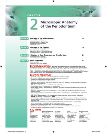
Figure 1-2 The Control ofRoom eter Required Parts for Control: Receptor – Receives stimulus Control center - processes signal &sends instructionsInformation Effector – Carries out instructionsaffectsSTIMULUS:Room temperaturerisesNormal roomtemperatureRESPONSE:Room temperaturedropsNormalconditionrestoredEFFECTORAir conditionerturns on20 30 40 SendscommandstoIn response to input from a receptor (a thermometer), a thermostat(the control center) triggers an effector response (either an air conditioner or a heater) that restores normal temperature. In this case,when room temperature rises above the set point, the thermostatturns on the air conditioner, and the temperature returns to normal. 2012 Pearson Education, Inc.Room temperature ( C)CONTROL CENTER(Thermostat)HOMEOSTASISAirAirconditioner conditionerturns onturns off22NormalrangeTimeWith this regulatory system, roomtemperature fluctuates around theset point.
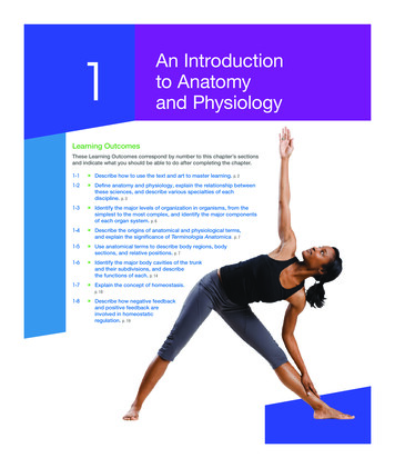
Figure 1-3 Negative Feedback in the Control of Body TemperatureRECEPTORSTemperaturesensors in mationaffectsCONTROLCENTERSTIMULUS:Body temperaturerisesHOMEOSTASISRESPONSE:Increased heat loss,body temperaturedropsNormaltemperaturerestoredEFFECTORS Sweat glandsin skin increasesecretion Blood vesselsin skin dilateSendscommandstoEvents in the regulation of body temperature, which arecomparable to those shown in Figure 1-2. A control centerin the brain (the hypothalamus) functions as a thermostatwith a set point of 37 C. If body temperature exceeds37.2 C, heat loss is increased through enhanced blood flowto the skin and increased sweating. 2012 Pearson Education, Inc.Body temperature ( C)Thermoregulatorycenter in brainNormal trict,sweating sweatingincreases decreasesNormalrangeTimeThe thermoregulatory center keepsbody temperature fluctuatingwithin an acceptable range, usuallybetween 36.7 and 37.2 C.
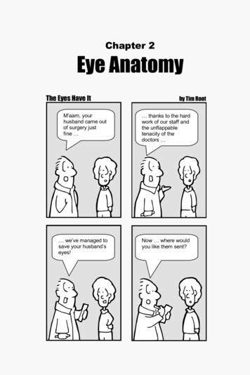
1-7 Negative and Positive Feedback The Role of Negative Feedback The response of the effector negates the stimulus or disturbance (i.e.,inverts the signal) Body is brought back into homeostasis Normal range is achieved The Role of Positive Feedback The response of the effector increases and amplifies the stimulus ordisturbance (i.e., in the same direction as the original signal) Body is moved away from current “set point” Normal range is lost Used to speed up certain processes (e.g., blood clotting, child birth) 2012 Pearson Education, Inc.
Figure 1-4 Positive Feedback: Blood micalsChemicalsDamage to cells in theblood vessel wall releaseschemicals that begin theprocess of blood clotting. 2012 Pearson Education, Inc.The chemicals start chainreactions in which cells,cell fragments, andsoluble proteins in theblood begin to form a clot.As clotting continues,each step releaseschemicals that furtheraccelerate the process.Blood clotThis escalating processis a positive feedbackloop that ends with theformation of a blood clot,which patches the vesselwall and stops the bleeding.
1-7 Negative and Positive Feedback Systems Integration Systems work together to maintain homeostasis Homeostasis is a state of equilibrium Opposing forces are in balance Dynamic equilibrium — continual adaptation Physiological systems work to restore balance Failure results in disease or death 2012 Pearson Education, Inc.
Table 1-1 The Roles of Organ Systems in Homeostatic Regulation 2012 Pearson Education, Inc.
1-8 Anatomical Terminology Although we will often examine the integration of variousorgan systems in the maintenance of whole-bodyhomeostasis, it is easier for introductory students to learnthe anatomy and physiology of each organ system oneat a time (Chapters 5 – 29). Thus, your text book begins with some basic anatomicalterminology in Chapter 1 that we will now examine as itwill be used throughout the two semestesr. 2012 Pearson Education, Inc.
1-8 Anatomical Terminology Anatomical position: hands and arms extended at sides, palmsforward, legs straight, feet togetherSupine: lying down, face upProne: lying down, face down Superficial Anatomy – structures on or near the body surface Anatomical Landmarks References to palpable (those that can be felt or touched) structures Anatomical Regions 4 Abdominopelvic quadrants – often used by clinicians 9 Abdominopelvic regions – often used by anatomists Anatomical Directions Reference terms based on subject 2012 Pearson Education, Inc.
Frontal orforeheadFigure 1-5a:Anatomical LandmarksCranialor skullCephalic or headOtic or earBuccal or cheekFacialor faceCervical or neckOral or mouthMental or chinThoracic orthorax, chestAxillary or armpitMammaryor breastBrachialor armAbdominal(abdomen)Umbilicalor navelAntecubitalor front ofelbowAnterior view 2012 Pearson Education, Inc.Nasal or noseOcular, orbitalor eyeTrunk
Figure 1-5a Anatomical LandmarksAntebrachialor forearmPelvic(pelvis)TrunkCarpal or wristPalmar or palmManualor handPollexDigitsor thumb (phalanges)or fingers (digitalor phalangeal)Patellaror kneecapInguinalor groinPubic(pubis)Femoralor thighCruralor legTarsal orankleDigits (phalanges)or toes (digital orphalangeal)Hallux orgreat toe 2012 Pearson Education, Inc.Pedalor footAnterior view
Figure 1-5b Anatomical LandmarksCephalicor headAcromial orshoulderDorsal orbackCervicalor neckOlecranalor backof elbowUpperlimbPosterior view 2012 Pearson Education, Inc.
Figure 1-5b Anatomical LandmarksUpperlimbLumbaror loinGlutealor buttockLowerlimbPopliteal orback of kneeSuralor calfCalcaneal orheel of footPlantar orsole of foot 2012 Pearson Education, Inc.Posterior view
Figure 1-6a Abdominopelvic Quadrants and RegionsRight UpperQuadrant(RUQ)Left UpperQuadrant(LUQ)Right LowerQuadrant(RLQ)Left LowerQuadrant(LLQ)Abdominopelvic quadrants. The fourabdominopelvic quadrants are formed by twoperpendicular lines that intersect at the navel. Theterms for these quadrants, or their abbreviations,are most often used in clinical discussions. 2012 Pearson Education, Inc.
Figure 1-6b Abdominopelvic Quadrants and RegionsRighthypochondriacregionRight iacregionLeft lumbarregionLeft inguinalregionAbdominopelvic regions. The nine abdominopelvicregions provide more precise regional descriptions. 2012 Pearson Education, Inc.
Figure 1-6c Abdominopelvic Quadrants and RegionsLiverGallbladderStomachSpleenLarge intestineSmall intestineAppendixUrinarybladderAnatomical relationships. The relationship betweenthe abdominopelvic quadrants and regions and thelocations of the internal organs are shown here. 2012 Pearson Education, Inc.
Figure 1-7 Directional or ventralPosterioror dorsalLateralCaudalMedialProximalDistalInferiorA lateral view. 2012 Pearson Education, Inc.DistalAn anterior view. Arrowsindicate important directionalterms used in this text;definitions and descriptionsare given in Table 1-2.
Table 1-2 Directional Terms 2012 Pearson Education, Inc.
Figure 1-8 Sectional PlanesFrontal planeSagittal planeTransverse plane Plane: a three-dimensional axis Section: a slice parallel to a plane Important in radiological techniques(e.g., MRI, PET, CT) 2012 Pearson Education, Inc.
Table 1-3 Terms That Indicate Sectional Planes 2012 Pearson Education, Inc.
1-9 Body Cavities Essential Functions of Body Cavities1. Protect organs from accidental shocks2. Permit changes in size and shape of internal organs Ventral body cavity (coelom) Divided by the diaphragm Thoracic cavity Abdominopelvic cavity 2012 Pearson Education, Inc.
Figure 1-9 Relationships among the Subdivisions of the Ventral Body CavityVentral Body Cavity Provides protection Allows organ movement Linings prevent frictionSubdivides during development intoAbdominopelvic CavityThoracic CavitySurrounded by chest wall anddiaphragmPeritoneal CavityRight Pleural CavityMediastinumLeft Pleural CavitySurrounds right lungContains thetrachea, esophagus,and major vesselsSurrounds left lungPericardial CavitySurrounds heart 2012 Pearson Education, Inc.Extendsthroughoutabdominal cavityand into superiorportion of pelviccavityAbdominal CavityPelvic CavityContains manydigestive glandsand organsContains urinarybladder,reproductiveorgans, lastportion ofdigestive tract
1-9 Body Cavities Serous Membranes Line body cavities and cover organs Consist of parietal layer and visceral layer Parietal layer — lines cavity Visceral layer — covers organs For example within the Abdominopelvic Cavity: Peritoneal cavity — chamber within abdominopelviccavity Parietal peritoneum lines the internal body wall 2012 Pearson Education, Inc. Visceral peritoneum covers the organs
Figure 1-10a The Ventral Body Cavity and Its Subdivisions 2012 Pearson Education, Inc.
1-9 Body Cavities – Abdominopelvic Cavity Abdominal cavity — superior portion Diaphragm to top of pelvic bones Contains digestive organs Retroperitoneal space Area posterior to peritoneum and anterior to muscularbody wall Contains pancreas, kidneys, ureters, and parts of thedigestive tract Pelvic cavity — inferior portion Within pelvic bones Contains reproductive organs, rectum and bladder 2012 Pearson Education, Inc.
Chapter 1 Objective Summary Review Be able to name the various specialties of anatomyand physiology. Be able to name the major levels of organization inorganisms, from molecular to organisms. Be familiar with the 11 organ systems of the bodyand their major components. (MURDERS LINC) Be able to explain the concept of homeostasis,including both positive and negative feedback. Be able to identify the major body cavities usingproper anatomical terms. 2012 Pearson Education, Inc.
1-4 Relationships between Anatomy & Physiology Anatomy Gross anatomy, or macroscopic anatomy, examines large, visible structures Surface anatomy: exterior features Regional anatomy: body areas Systemic anatomy: organ systems Developmental anatomy: from conception to death