Transcription
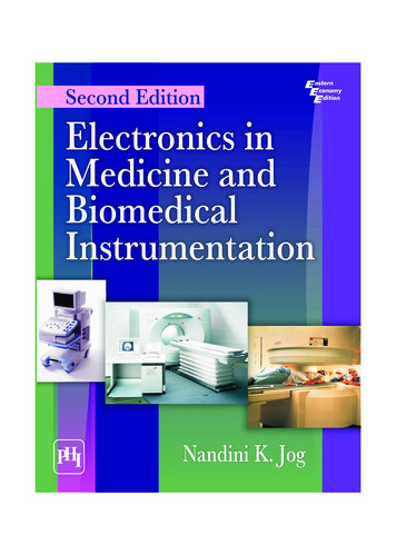
Second EditionElectronics inMedicine andBiomedicalInstrumentationNandini K. Jog
Electronics in Medicine andBiomedical InstrumentationSECOND EDITIONNANDINI K. JOGAdjunct ProfessorDepartment of Electronics EngineeringMukesh Patel School of Technology Management and EngineeringNMIMS UniversityMumbaiDelhi–1100922013
ELECTRONICS IN MEDICINE AND BIOMEDICAL INSTRUMENTATION, Second EditionNandini K. Jog 2013 by PHI Learning Private Limited, Delhi. All rights reserved. No part of this book maybe reproduced in any form, by mimeograph or any other means, without permission in writingfrom the publisher.ISBN-978-81-203-4724-3The export rights of this book are vested solely with the publisher.Fifth Printing (Second Edition). . . . . . . . .March, 2013Published by Asoke K. Ghosh, PHI Learning Private Limited, Rimjhim House, 111, PatparganjIndustrial Estate, Delhi-110092 and Printed by Mudrak 30-A, Patparganj, Delhi-110091.
To the fond memory of my parentsLate Professor L.V. Gurjar&Late Mrs. Sundara Gurjar
ContentsPreface . xiPreface to the First Edition . xiii1. INTRODUCTION TO BIOMEDICAL INSTRUMENTATION . 1–201.11.2Basics of Biomedical Instrumentation Systems . 2Anatomy and Physiology of the Human Body . 31.2.1 The Human Body . 31.3 Body Systems . 41.3.1 The Lymphvascular System . 41.3.2 The Endocrine System . 61.4 Homeostasis . 81.5 Membranes . 111.6 Cells and Chromosomes . 111.7 How Potentials in the Body are Generated? . 121.7.1 Body Potentials . 121.8 Bioelectrodes . 161.8.1 Properties of Bioelectrodes . 161.8.2 Different Types of Electrodes . 181.9 Conclusion . 20Exercises . 202. TRANSDUCERS, AMPLIFIERS, RECORDERS AND DISPLAYS . 21–592.12.22.3Sensors . 212.1.1 Diaphragms . 212.1.2 Force Sensors . 23Transducers . 242.2.1 Classification of Transducers . 262.2.2 Passive Transducers . 262.2.3 Active Transducers . 412.2.4 Production of an EMF . 43Photodiodes and Phototransistors . 452.3.1 Photoconductive Cells . 462.3.2 Photomultiplier Tubes . 46v
viContents2.3.3 Piezoelectric Transducers . 462.3.4 Crystal Microphone . 482.4 Ultrasonic Transducers . 482.4.1 Electrochemical Transducers . 492.4.2 Digital Transducers . 512.4.3 Transducers Used in Nuclear Radiation Measurement . 522.4.4 Transducers Based on Optoelectronic Principle . 532.4.5 Optical Fiber Transducers and Sensors . 532.4.6 Fiber Optic Microphone . 542.5 Amplifiers . 542.5.1 Chopper Amplifiers . 572.6 Recorders and Displays . 582.7 Conclusion . 59Exercises . 593. BLOOD .60–843.1Measurement of Blood Flow . 623.1.1 Radiographic Techniques . 633.1.2 Indicator Dye Dilution Method . 633.1.3 Thermal Convection . 643.1.4 Magnetic Blood Flow Rate . 643.1.5 Ultrasonic Blood Flow Meter . 663.2 Blood Pressure . 673.2.1 Measurement of Blood Pressure . 673.3 Blood Gas Analysers . 723.3.1 pH Measurement of Blood . 733.4 Oximetry . 733.4.1 Measurement of Partial Pressure of CO2 in Blood . 733.4.2 Measurement of Blood PaO2 . 743.4.3 In vitro Oximetry . 743.5 Analysis of Molecules in Medicine . 793.5.1 Photometry . 793.5.2 Flame Photometry . 823.6 Conclusion . 83Exercises . 834. CARDIOVASCULAR SYSTEM OR CIRCULATORY SYSTEM . 85–1174.14.24.34.4The Anatomy of the Heart . 86Function of the Heart . 884.2.1 Systemic Circulatory System . 884.2.2 Pulmonary Circulation . 88Action of the Heart . 89Cardiography . 924.4.1 Frontal Plane ECG . 934.4.2 The Transverse Plane ECG . 954.4.3 Sagittal Plane ECG Measurements . 97
Contentsvii4.4.4 Interpretation of the ECG . 974.4.5 Common Terms and Interpretation of the ECG . 984.5 ECG Amplifier . 994.6 Abnormalities—Heart Blocks . 1014.6.1 Phonocardiography . 1024.6.2 Echocardiography . 1024.6.3 Vector Cardiography . 1044.6.4 Stress Testing . 1054.7 Heart Rate Measurement . 1064.7.1 Cardiotachometers . 1064.7.2 Average Heart Rate Meter . 1084.8 Cardiac Pacemakers . 1084.8.1 Pacemakers . 1094.8.2 Types of Pacemakers . 1094.9 Defibrillators . 1134.9.1 Ventricular Fibrillation . 1134.9.2 Placement of Electrodes or Paddles of a Defibrillator . 1154.10 Bedside Monitor . 1164.11 Conclusion . 117Exercises . 1175. NERVOUS SYSTEM . 118–1465.15.25.35.45.55.65.75.85.9The Nervous System . 1185.1.1 The Peripheral Nervous System . 1195.1.2 The Central Nervous System . 119The Human Brain . 1245.2.1 Cerebrum . 1245.2.2 Cerebellum . 1255.2.3 Medulla Oblongata . 125Study of Brain Signals . 1275.3.1 Different Waves from Different Parts of The Brain . 1275.3.2 Types of Electrodes . 1285.3.3 Evoked Potentials . 1295.3.4 Connection of Electrodes to the Amplifier . 131EEG Amplifiers . 131Recording the EEG Signal . 1325.5.1 Characteristics of a Recording System . 132Artifacts . 1335.6.1 Processing Artifacts . 134Analysis of Diseases Using EEG . 134Sleep Analysis . 1385.8.1 Magneto Encephalography or MEG . 139Electromyography . 1415.9.1 How Muscles Work . 1415.9.2 Paralysis . 143
viiiContents5.9.3 Myograph . 1435.9.4 Nerve Conduction Velocity . 1445.10 Conclusion . 146Exercises . 1466. RESPIRATORY AND OTHER MEASUREMENTS . 147–1666.16.26.3Respiratory Measurements . 148Spirometers . 148Respiratory Gas Analysers . 1496.3.1 Infrared Gas Analysers . 1506.3.2 Oxygen Analyser . 1506.3.3 Nitrogen Analyser . 1516.4 Galvanic Skin Resistance . 1516.4.1 Lie Detector . 1526.5 Patient Safety . 1536.5.1 Macro Shock . 1536.5.2 Microcurrent Shock . 1536.6 Telemedicine and Telemetry . 1546.7 The Ear . 1556.7.1 Anatomy of the Ear and the Mechanism of Hearing . 1556.7.2 Frequency Encoding by the Auditory System . 1566.7.3 Deafness . 1576.8 Kidneys . 1596.8.1 Hemodialysis . 1606.8.2 Peritoneal Dialysis . 1626.9 Electroculogram . 1636.10 Electroretinogram . 1636.11 Use of Lasers in Medicine . 1636.11.1 Ruby Laser . 1636.11.2 Argon Laser . 1646.11.3 Carbon Dioxide Laser . 1646.12 Diathermy . 1646.12.1 Short Wave Diathermy . 1646.12.2 Microwave Diathermy . 1656.12.3 Ultrasound Therapy . 1656.13 Conclusion . 165Exercises . 1657. IMAGING TECHNIQUES . 167–1927.1X-Ray Imaging and CT Scan . 1677.1.1 Properties of X-rays . 1697.1.2 Production of X-rays . 1697.1.3 Applications of X-rays in Medicine . 1717.1.4 CAT Scan . 1717.1.5 X-ray Therapy . 172
Contentsix7.2Magnetic Resonance Imaging (MRI) . 1727.2.1 MRI Procedure . 1737.2.2 Patient Safety . 1757.2.3 Benefits, Risks and Limitations of MRI . 1767.2.4 Mediscope . 1767.3 Positron Emission Tomography (PET) . 1777.3.1 Scanning . 1797.3.2 Patient Preparation for the Test . 1807.3.3 PET Instrumentation System . 1807.3.4 Advantages of PET Scan . 1827.3.5 Risks . 1827.3.6 Single Photon Emission Computed Tomography (SPECT) . 1827.3.7 History of PET and Advantages of PET Scanning . 1827.4 Ultrasound in Medicine . 1837.4.1 Physics of Ultrasound . 1837.4.2 Doppler Effect . 1857.4.3 Ultrasound Imaging . 1857.4.4 A-Scan Display . 1877.4.5 B-Scan . 1897.4.6 Multiarray Scanning . 1907.4.7 M-Mode Scan . 1917.4.8 Advantages and Disadvantages of Ultrasound Scanning . 1917.5 Conclusion . 192Exercises . 192Bibliography . 193–194Index . 195–197
PrefaceAfter teaching the subject of medical electronics, I felt the need for updating this book toinclude certain topics like magnetoencephalography, features of mediscope software usedfor medical imaging and fiber optic microphones used in MRI scanning.The recent topics in vogue like phonocardiography, vector cardiography, nuclear stresstest, MRI stress test and details of pacemakers and defibrillators have also been added.Chapter six is updated with the processes of dialysis, hemodialysis and peritoneal dialysis.Other medical instruments introduced and explained in the new edition are colorimeter,spectrophotometer, flame photometry and auto analyzers for the study of toxic levels in thebody.NANDINI K. JOGxi
Preface to the First EditionThe leaps and bounds with which progress in the field of electronics has taken place arereflected in the number of electronic gadgets which have become a part of our daily lives.The radio, TV, telephone, mobile communications and computers have now become essentialcommodities which did not commonly exist in every household 50 years ago. A lot ofelectronic instruments are used by doctors and in hospitals. Electronic instruments measuringblood pressure, pulse rate and blood flow, and machines used for X-rays, CAT scans, MRIscans, PET scans, ultrasound scanning, bone density measurement, etc. are some of themodern electronic gadgets used. Maintenance of these biomedical instruments is generallydone by trained technicians from the company which manufactures the product andneither the doctor nor the ordinary engineer has detailed knowledge about the gadget orthe working of the internal circuitry apart from its application. Though tracing the circuitand repairing it may not be difficult for a skilled engineer, a little knowledge of the humanbody, its working and various diseases is essential for the engineer to understand thedesign of the instrument. Similarly, a doctor whose hobby may be electronics, could havea deeper interest in the instruments he uses, apart from their applications.In this book, an attempt is made to make the electronics engineer aware of the functioningof the human body and the development of biomedical instrumentation. It may be interestingto note that the subject of biomedical instrumentation developed when engineers encounteredproblems selling the excess amplifiers and other electronic equipment manufactured forwarfare after the 2nd World War ended. This marked the commencement of research in thefield of biomedical instrumentation. With extensive research in all aspects of biomedicalinstrumentation, especially image processing, we now have state-of-the-art equipments invarious hospitals around the world.A course on biomedical instrumentation is offered by various universities and in myopinion, every engineer who studies electronics should take up this course.I am grateful to Dr. Charu Apte for permitting me to take photographs of variousbiomedical instruments used in the hospital. I would like to thank Mr. Hiroo Jhangiani andMr. Anant Nimkar for helping me with the photographs. I would also like to thankVenkat Islur for typing the manuscript of this book. I am grateful to PHI Learning forundertaking the publication of this book. I would finally like to thank my daughter,Dr. Suneeti Jog, son, Ninad Jog, and husband, Krishna Jog, for taking interest in my book.NANDINI K. JOGxiii
1Introduction toBiomedicalInstrumentationThis chapter deals with:· Basics of biomedicalinstrumentationsystems.· Types of electrodesused for measuringbio-potentials.· Introduction to theanatomy andphysiology of thehuman body.In the last fifty years, there has been a tremendousprogress in the field of electronics. We see a large numberof elelctronic gadgets in the market. Electronics haspervaded all fields of engineering, and instrumentationis no exception. Manually operated instruments,mechanical instruments are all being replaced byelectronic instruments. In the field of medicine wenow find electronic blood pressure measuringinstruments, blood glucose monitors and a wide varietyof hospital equipment that requires knowledge ofelectronic systems. It is imperative for an electronicsengineer to learn the working and design of biomedicalinstrumentation systems. For a thorough understanding,it is also imperative to have knowledge of the humanbody, its anatomy and physiology.1
2Electronics in Medicine and Biomedical Instrumentation1.1BASICS OF BIOMEDICAL INSTRUMENTATION SYSTEMSBiomedical instrumentation deals with the measurement and analysis of current orvoltage signals from different parts of the body. The human body is like a powerstation which generates a variety of voltages. However, these voltages are extremelysmall.In most of the biomedical instrumentation systems, currents between two points on thesurface of the body due to potential differences between the two points are measured. Thesecurrents are very small, of the order of mamps, and are therefore amplified by instrumentationamplifiers.After amplification, the signal is processed, noise is filtered, bandwidth is restrictedand then it is displayed either on a CRT, a strip chart recorder or a camera, or a magnetictape. An example of a continuous signal is the ECG signal obtained due to the electricalactivity of the heart. In some cases, an external stimulus is given to the patient and theresponse to the stimulus, which is a voltage signal, is recorded. Sometimes transducers arerequired to obtain an electrical signal. Figure 1.1 depicts the generalized block diagram ofa biomedical instrumentation system. Many physiological processes in the body areaccompanied with electrical changes. Signals are produced in the body by muscles cesCRTX-YRecorderCameraFig. 1.1StripchartrecorderPrinterMagnetictapeBlock Diagram of a Biomedical Instrumentation System.Some of the biomedical signals and their characteristics are listed in Table 1.1.However, there are many more kinds of signals.The questions which come to the mind are: ‘From where do these signals originate?’‘How do they originate?’ These signals originate from the cell due to action potential. Itis therefore necessary to study the human cell. But before we come to action potential,knowledge of anatomy and physiology of the human body is essential.
3Introduction to Biomedical InstrumentationTable 1.1Sr.No.Various Body SignalsParameterSigna
In this book, an attempt is made to make the electronics engineer aware of the functioning of the human body and the development of biomedical instrumentation. It may be interesting to note that the subject of biomedical inst


![History of Electronics electricity [Read-Only]](/img/18/history-20of-20electronics-20-20electricity.jpg)


