Transcription
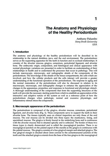
1The Anatomy and Physiologyof the Healthy PeriodontiumAnthony PalumboStony Brook UniversityUSA1. IntroductionThe anatomy and physiology of the healthy periodontium will be described in itsrelationship to the natural dentition, jaws, and the oral environment. The periodontiumserves as the supporting apparatus for the teeth in function and in occlusal relationships. Itconsists of the alveolar mucosa, gingiva, cementum, periodontal ligament, and alveolarbone. The embryonic origin, composition, and histological and clinical appearance withnormal physiologic variations are presented in order to facilitate an understanding of theirrelationships in health and to understand the processes that occur in pathology. This willinclude macroscopic, microscopic, and radiographic details of the components of theperiodontium. The knowledge of the details of the tissue compartments, the cells which areinvolved, and how the cellular products and the cells interact will provide a greaterunderstanding of the functional operation of the periodontium. The response to aging andthe normal maturation of the periodontium will also be discussed in relation to itsmacroscopic, microscopic, and radiographic changes to enhance the appreciation of thechanges in the appearance, properties, and responses to functional and physiologic stimuli.A thorough understanding of the components that form the supporting structures of theteeth will provide the necessary starting point from which to establish an appreciation of theinteractive and adaptive nature of the system as well as a reference point of how theperiodontium changes when pathologic, normal and excessive physiologic, andinflammatory stimuli stress the components.2. Macroscopic appearance of the periodontiumThe periodontium is composed of the gingiva, alveolar mucosa, cementum, periodontalligament, and alveolar bone (Fig. 1). These components serve to support the teeth in theiralveolar bone. The tissues typically seen on clinical inspection are only those of the oralmucosa. The oral mucosa can be divided into three types: the masticatory, lining, andspecialized mucosa. The gingiva is firmly bound to the underlying bone and is continuouswith the alveolar mucosa that is situated apically and is unbound. The border of these twotissue types is clearly demarcated and is called the mucogingival junction. There is nomucogingival junction on the palatal aspect of the maxilla as the gingiva is continuous withthe palatal mucosa. The gingiva consists of a free gingival margin and attached gingiva. Thefree gingival margin is situated about 2mm coronal to the cementoenamel junction of thetooth and the attached gingiva extends from the base of the free gingiva to the mucogingivalwww.intechopen.com
4Gingival Diseases – Their Aetiology, Prevention and Treatmentjunction (Ainamo and Loe 1966). The gingiva is typically coral pink in color, but may varydue to physiologic pigmentation among some races, whereas the alveolar mucosa is deepred in color (Fig2a/b).Fig. 1. Components of the periodontium (Garant 2003)a) Normal pigmentationb) Physiologic pigmentationFig. 2. Normal variation in the appearance of the gingival tissuesThe tissue that resides in the interproximal embrasure is called the interproximal papilla. Theshape of this tissue is influenced by the shape of the interproximal contact, the width of thewww.intechopen.com
The Anatomy and Physiology of the Healthy Periodontium5interproximal area, and the position of the cementoenamel junction of the involved teeth. Theshape of this papilla varies from triangular and knife-edge in the anterior regions due to pointsized contacts of the teeth to broader and more square shaped tissue in the posterior sextantsdue to the teeth having broad contact areas. Also present in the wider papillary areas is the col.This is a valley-like structure situated apical to the contact area (Fig 3).Fig. 3. Interdental tissue shapes (Garant 2003)The texture of the gingiva varies with age and is typically smooth in youth, stippled inadulthood, and again becomes smoother with advanced age. Stippled tissue has a texturesimilar to the rind of an orange and its presence does not necessarily mean health. (Fig 4).Another feature that does not appear in all of healthy periodontiums is the free gingivalgroove. The free gingival groove is a depression that appears in about 50% of population.The groove appears at the border of the free and attached gingiva and usually represents thebase of the gingival sulcus. The gingival sulcus is the invagination around a tooth boundedby the free gingival margin.Fig. 4. Stippling of gingival tissue.www.intechopen.com
6Gingival Diseases – Their Aetiology, Prevention and TreatmentFig. 5. Width of attached gingiva in specific areaswww.intechopen.com
The Anatomy and Physiology of the Healthy Periodontium7When a periodontal probe is placed into this space, a measure may be recorded which isvery useful for diagnosis. Studies have shown an average depth of 0.7mm but variationsmay range from 0 to 6mm (Gargiulo 1961)). The width of the attached gingiva varies withthe location in the oral cavity as well as with physiologic age. The facial gingiva is typicallywidest in the incisor region and narrowest in the premolar region for the maxillary arch andranged from 1-9mm. In the mandible, the facial attached gingiva is narrowest in thepremolar and canine regions (Bowers 1963). When the lingual attached gingiva wasexamined, it was found that the widest areas were on the mandibular molars and thenarrowest were on the incisor and canine regions, about 1.8mm (Voigt 1978) (Fig. 5). Thereis a general increase from the primary to permanent dentition as well as with increasing age(Ainamo and Talari 1976)(Fig. 6).Fig. 6. Changes in the amount of attached tissue with age.Using radiographs one can visualize several of the components of the periodontium aswell as their size and relation to the teeth (Fig. 7). Although the radiograph is a twodimensional depiction of a three-dimensional object, the location of the alveolar bone crestrelative to the cementoenamel junction is seen along with the space occupied by theperiodontal ligament. Since the periodontal ligament itself is not a mineralized tissue, theradiograph will show a radiolucent area that it occupies. The cortical bone that houses theteeth is known as the lamina dura. The alveolar bone also follows a path that parallels thepositions of the cementoenamel junctions of teeth (Ritchey and Orban 1953). In health theinterdental bone is 1.0mm from the cementoenamel junction and increases with age to2.8mm (Gargiulo 1961).www.intechopen.com
8Gingival Diseases – Their Aetiology, Prevention and TreatmentFig. 7. Appearance and location of periodontal components on a periapical radiograph3. Microscopic appearance of the periodontiumGreater detail of the periodontium is obtained histologically. The schematic cross-section ofthe periodontal attachment and components is seen in Fig. 1. The components are again thealveolar bone, gingiva, periodontal ligament, and cementum. The gingiva consists of asurface epithelium and underlying connective tissue termed the lamina propria. There arethree types of epithelium present, the oral, sulcular, and the junctional epithelium. The oralwww.intechopen.com
The Anatomy and Physiology of the Healthy Periodontium9epithelium is continuous with the epithelium of the oral cavity. The sulcular epithelium isadjacent to the tooth but not connected or attached to the tooth surface. The junctionalepithelium is at the base of the sulcus and is in direct contact with the tooth (Carranza2002)(Fig. 8).Fig. 8. Types of epithelium in the periodontium (Garant 2003)The border of the connective tissue and epithelium is undulating (Fig. 9). These epithelialextensions are known as epithelial ridges or rete pegs. The connective tissue layer is alsotermed the lamina propria or the dental papillae. In health, this is a characteristic finding inthe attached gingiva, but are absent in the sulcular and junctional epithelium.The gingival epithelium is quite similar to the epidermis in its structure. The gingivaconsists of keratinized, stratified, squamous epithelium. The major cell type is thekeratinocyte. There are four distinct layers; the stratum basale, stratum spinosum, stratumgranulosum, and the stratum corneum (Fig. 10). The stratum basale or basal layer consists ofone to two layers of cells cuboidal in shape. These are the most undifferentiated of the cellsand serve to replenish cells as they are shed during their maturation and exfoliation. Thebasal cells are immediately adjacent to the connective tissue from which it is separated by abasement membrane. The basement membrane consists of a two zones, the lamina lucidaand lamina densa. The lamina lucida contacts the cell surface and has manyhemidesmosomes. Hemidesmosomes are specialized structures that connect an epithelialwww.intechopen.com
10Gingival Diseases – Their Aetiology, Prevention and TreatmentFig. 9. Rete pegs (Garant 2003)Fig. 10. Cellular layers of epithelium (Garant 2003)www.intechopen.com
The Anatomy and Physiology of the Healthy Periodontium11cell to the basement membrane. In the lamina densa, anchoring fibrils formed from Type VIIcollagen bind to the Type I and III collagen of the extracellular matrix (Listgarten 1972;Schroeder 1997).The stratum spinosum consists of larger cells with cytoplasmic processes that resemblespines. There are typically 10-20 layers of cell in this stratum. The cells are bound to eachother by desmosomes, which are in essence pairs of hemidesmosomes. The cells containmany keratin filament bundles known as tonofibrils. Other cells found in this layer includemelanocytes, Langerhan's cells and Merkel cells. Melanocytes produce the pigment melaninwhich is contained in granules. Langerhan's cells are part of the immune system and serveas antigen presenting cells. The Merkel cells are responsible for the perception of sensation.In the stratum granulosum, keratohyalin bodies and tonofibrils are seen extensively. As thecells proceed from the basal layer and reach the stratum granulosum, a dramatic decrease inorganelles can be observed. The stratum corneum is seen abruptly after the stratumgranulosum. It consists of layers of flattened cells that may exhibit different patterns ofkeratinization depending on location and external stimuli (Carranza 2002).This keratinization process as the cells mature through the layers is considereddifferentiation. Orthokeratinized cells are flattened and have no discernible nuclei andcyptoplasmic organelles. Parakeratinized refers to cells that exhibit incompletekeratinization and cells that contain remnants of nuclei and cellular organelles. The locationmost keratinized is the palate, followed by the gingiva and tongue, and finally the buccalmucosa(Miller 1951). The degree of keratinization of the oral mucosa generally decreaseswith age and with the onset of menopause (Papic 1950). The sulcular epithelium is thin,non-keratinized epithelium.In health, the depth of the sulcular epithelium is less than 3mm and ends at the cornoalsurface of the junctional epithelium. Cadaver studies found the depth of the sulcus to be anaverage of 0.69mm (Gargiulo 1961). Rete pegs are not present in the sulcular epithelium. Thejunctional epithelium contains cells that are directly attached to the tooth surface. Aninternal basal lamina attaches the cells to the tooth surface through hemidesmosomes andan external basal lamina attaches the cells to the underlying connective tissue. Early in life ittypically consists of a few stratified squamous cell layers, but with age the number of layersincreases to between 10-20. The average width of the junctional epithelium is 1mm(Gargiulo 1961). The junctional epithelium also has wide intercellular spaces and functionsas a permeable barrier. This is an important property since it acts as a semi-permeablebarrier through which bacteria and their components and byproducts may pass into andinvade the tissue. It also facilitates the passage of leukocytes (e.g. neutrophils) and immunecomponents (e.g. complement), enzymes, and gingival crevicular fluid. Gingival crevicularfluid is a modified inflammatory exudate produced that resembles serum. The col areasshare similar characteristics to the junctional epithelium. These areas are also nonkeratinized and have a high level of turnover (Garant 2003). In summary, the junctionalepithelium differs from the oral epithelium in having cells of smaller size, largerintercellular spaces, and fewer desmosomes.Beneath the epithelial layer is a connective tissue layer also known as the lamina propria.This layer is composed of a papillary and a reticular layer. The papillary layer is adjacent tothe basal cells of the epithelium and their rete pegs. The reticular layer is adjacent to theunderlying alveolar bone. Collagen Type I is the predominant component of the laminapropria. Also residing in this layer are cells, nerves, blood vessels, and ground substance.The cells present are fibroblasts, mast cells, and immunologic cells. Mast cells containvesicles with vasoactive substances such as histamine and proteolytic enzymes. Oncewww.intechopen.com
12Gingival Diseases – Their Aetiology, Prevention and Treatmentactivated by stimuli, the cells can degranulate and induce changes in blood flow to the areaand increase tissue permeability. The immunologic cells present are macrophages,neutrophils, lymphocytes, and plasma cells. These cells are present to initiate and maintain aresponse to a foreign substance or cell present in the area. Ground substance is a gel-likesubstance composed of glycosaminoglycans and proteoglycans. These substances cause alarge amount of water retention which maintains the shape and structure of the area whenforce is applied. This substance also serves as a medium for the transportation ofelectrolytes, nutrients, and metabolites (Rose 2004).The fibroblasts are the predominant cells and function to synthesize collagen andextracellular matrix. These cells are elongated and elliptical in shape and their microscopicappearance is characteristic of a cell producing large amounts of cellular products, a welldeveloped rough endoplasmic reticulum and Golgi apparatus, and many mitochondria.Collagen is formed by both an intracellular and extracellular process. Intracellularlytropocollagen, the smallest unit of collagen, is produced. Tropocollagen consists of threepolypeptide chains of 1000 amino acids in an -helical formation and is 3000 Å long and hasa 15Å diameter. A significant percentage of the amino acid composition is glycine, proline,and hydroxyproline. The later is unique to collagen and when assayed can be used todetermine the amount of collagen in the sample. The tropocollagen is excreted into theextracellular environment, where the remainder of the formation takes place. Tropocollagenis arranged into protofibrils and then collagen fibrils. The fibrils are then bundled togetherFig. 11. Formation of collagen. (top to bottom) (tropocollagen, protofibril, collagen fibril,and collagen fiber)www.intechopen.com
The Anatomy and Physiology of the Healthy Periodontium13and collagen fibers are created with a typical cross-banding pattern of 700 A. (Fig 11). Ascollagen matures and ages it develops greater cross-linking making the collagen less solubleand resistant to breakdown (Rose 2004).Most collagen present in the gingiva and connective tissue is irregularly arranged but somedistinct arrangements of fibers can be observed. These include the dentogingival, circular,and transseptal group. The dentogingival group fibers may run from the root surface to theperiosteum of the bone, from the root surface to the gingiva, and from the alveolar bone tothe gingiva. Circular fibers run circumferentially around the tooth within the gingiva and donot touch the tooth itself. The transseptal group runs from the root surface of one tooth tothe root surface of another tooth transversing the alveolar bone (Garant 2003) (Fig. 12).Fig. 12. Gingival fiber groups. (Garant 2003)The periodontal ligament is the connective tissue that connects the tooth to the alveolarbone. The periodontal ligament serves to allow forces to be distributed to the alveolar boneduring mastication and occlusal function. The majority of the volume of the ligament isoccupied by dense connective tissue and the minority by loose connective tissue withneurovascular structures. Cells present in this tissue include osteoblasts, cementoblasts,osteoclasts, multipotent stem cells, epithelial remnants, and fibroblasts, which are the mostabundant (Carranza 2003) Since the periodontal ligament contains such a variety of cells, itplays an important role in healing and repair. This potential is also a focus for to periodontalregenerative procedures (Melcher 1976). The ligament is about 0.15mm to 0.25mm in widthand has an hourglass shape with the mid root level having the narrowest width. The widthof the ligament can adapt to forces by decreasing in lowered function and a widening of theligament with increased occlusal load or hyperfunction. With age there is a decrease invascularity, cell mitotic activity, fiber number and in fibroblasts there is a slight decrease inwidthn (Van der Velden 2004).With root development principle fibers, which are collagenous bundles, insert their terminalends into the root cementum and alveolar bone and are termed Sharpey’s fibers orperiodontal ligament fibers. These collagen fibers are produced by fibroblasts,chondroblasts, osteoblasts and other cells in a manner described previously. The fibers arewww.intechopen.com
14Gingival Diseases – Their Aetiology, Prevention and Treatmenttypically Type I collagen. The fibers can also be arranged by their position and orientation.The six groups are the transseptal, horizontal, alveolar, oblique, apical, and radicular groups(Fig. 13). The transseptal group extends from the cementum of one tooth over the interseptalbone to the cementum of an adjacent tooth. The horizontal group attaches the cementum tothe alveolar crest and run perpendicular to the root and alveolar surfaces. The alveolargroup attaches the cementum to the alveolar bone and originates apical of thecementoenamel junction. The oblique group constitutes the majority of the fibers and runobliquely from the root cementum to the alveolar bone. These fibers provide support fromintrusive forces from mastication. The apical fiber group emerges from the cementum nearthe apex of the root and connects to the alveolar bone. The radicular group is seen inmultirooted teeth near the furcation and connects the cementum of that area to theneighboring bone. Other fibers present include oxytalan fibers, which run parallel to theroot surface vertically, and elaunin fibers that are similar to immature elastic fibers (Rose2004; Garant 2003).Fig. 13. Periodontal fiber groups (Garant 2003)The cementum is a mineralized tissue covering the anatomic root of the tooth. Cementum isavascular and has no direct innervation. It is made of collagen fibers within a mineralizedmatrix. Fibers present in the cementum may be classified as extrinsic or intrinsic. Theextrinsic fibers are created by the fibroblasts in the periodontal ligament and the intrinsicfibers are produced by cementoblasts. The mineralized matrix is composed of mainlyhydroxyapatite [Ca10(P04)6(OH)2]. Cementum has some characteristics that are bothwww.intechopen.com
The Anatomy and Physiology of the Healthy Periodontium15biochemically and physically similar to bone due to its composition. Cementum iscontinuously deposited throughout life and the apical third of the root typically has thethickest deposition. In doing so, the deposited cementum compensates for the eruption ofteeth from attrition. The thickness of the cementum varies from 15 to 150 microns dependingon the location on the root and age of the patient. There is some permeability of cementumto organic substances, ions, and bacterial products. Typically the permeability of cementumdiminishes with age. The extent of cementum coronally exhibits different patterns (Fig. 14).In most instances the cementum overlaps the enamel ( 60%), and less frequently it has abutt-joint ( 30%), and least frequently it ends short of the enamel (5-10%). This anatomicalvariation among the position the enamel and cementum board is clinically relevant whengingival recession occurs and patients may present with exposed dentin and roothypersensivity (Carranza 2003).Fig. 14. Configurations of dentin, cementum, and enamel at the cementoenamel junction.Cementum is characterized into acellular and cellular types. Acellular afibrillar cementum islocated near the coronal aspect of the root and has no cells and no extrinsic or intrinsiccollagen fibers within it. Acellular extrinsic fibrillar cementum is found in the middle andcoronal parts of the root and lacks cells. This type of cementum has Sharpey’s fibers,collagen fibers that attach from the cementum to the alveolar bone. Cellular mixed stratifiedcementum is present in the apical third and in the area of furcations. It also containsSharpey’s fibers and intrinsic fibers. Cellular intrinsic fibrillar cementum has cementocytes,which are cementoblasts trapped within the mineral they deposited, and does not containextrinsic collagen fibers (Garant 2003).The alveolar process is the osseous tissue of the maxillary and mandibular jaws whichhouses and supports the sockets of the teeth. The process consists of an external corticalwww.intechopen.com
16Gingival Diseases – Their Aetiology, Prevention and Treatmentplate, the inner socket wall known as the alveolar bone proper and is compact bone, and acancellous trabecular bone in between the two boney layers. The bone is typically thicker inthe palatal and lingual areas when compared to the buccal areas. Some areas may presentwith defects known as dehiscences and fenestrations. Dehiscences are areas were bone hasbeen lost on a root surface and the root is only covered by periosteum and gingiva.Fenestrations are small areas or “windows“ where bone has been lost on a root surface andis only covered by periosteum and gingiva. (Fig. 15). These defects were shown to occur inabout 20% of all teeth. Dehiscences were more prevalent in the mandible, whereasfenestrations are more common in the maxilla (Elliot and Bowers 1963). Some areas may bepredisposed to these defects by having teeth with prominent root morphology, dentalcrowding, and a position extending beyond the dental arch. These areas become crucial ifperiodontal disease occurs or if gingival recession takes place since they may complicatetherapy and adversely affect the area’s prognosis.Fig. 15. Diagram of fenestration and dehiscence defects.The alveolar bone proper is cribiform in appearance and this allows for a connection to theneurovascular structures. The bone is created by osteoblasts during development (modeling)and is constantly remodeled throughout life from the intricate osteoblastic/osteoclasticwww.intechopen.com
The Anatomy and Physiology of the Healthy Periodontium17relationship. The osteoblasts produce collagen, glycoproteins and proteoglycans to producethe bone matrix that is then mineralized with calcium and phosphate. The mineral ishydroxyapatite and the mineral content is about 60%. When osteoblasts have layed downosseous tissue they become trapped within the tissue and are termed osteocytes. Osteocytesreside in lacunae and connect and communicate with each other through canaliculi. A groupof osteocytes surround themselves around the neurovascular bundles (Haversian canals) andare termed osteons. An osteon is the fundamental unit of compact bone and are cylindricalstructures. Volkmann’s canals which run within osteons, carry nerves and blood vessels, andare perpendicular to the Haversian canals (Fig. 16). An analogy can be that the Haversiancanals are elevators of a tall building and the Volkmann’s canals are hallways on specificfloors. Cancellous bone consists of trabeculae and has irregular marrow spaces. Cancellous ortrabecular bone is found interdentally. The bone quality of the maxilla and mandible aregenerally different and overall the maxilla has more cancellous bone compared to themandible (Sodek 2000; Rose 2004; Carranza 2003).Fig. 16. Microscopic configuration of osseous tissue. A)osteon B)vascular configurationwww.intechopen.com
18Gingival Diseases – Their Aetiology, Prevention and TreatmentOsteoclasts are derived from monocytes and resorb bone by a specialized feature called aruffled border. This structure seals off an area and then vesicles stored within the osteoclastare released to cause the breakdown. These enzymes include acid phosphatase, cathepsins,and matrix metalloproteinases. The interaction between the osteoclasts and osteoblasts isregulated by the RANK pathway (receptor activator of nuclear factor-κ ) which is a balanceof the ligand for RANK (RANKL) and a competitive inhibitor osteoprotegerin (OPG). Thecreation of RANKL involves osteoblasts themselves which activate precursor cells todifferentiate, in the presence of macrophage colony stimulating factor, to becomeosteoclasts. The outer surface of bone is covered by layers of connective tissue called theperiosteum. The periosteum contains osteoblasts, stem and progenitor cells, fibroblasts, andvascular and nervous tissue. The inner layer of bone is lined with endosteum which iscomprised of connective tissue containing osteoblasts (Garant 2003).The periodontium’s blood supply is derived from the superior and inferior alveolar arteries.These arteries produce branches that extend into the periodontal ligament and into thealveolar bone and periosteum. The gingiva receives its vascular supply from three differentsources: the interdental septum, the periodontal ligament, and the connective tissue andperiosteum all anastomose and supply the gingiva through a vascular network ofcapillaries. This vast network enables many periodontal procedures to be performedwithout depriving the periodontium and dental structures of a vascular supply. Innervationof the periodontium is from branches of the trigeminal nerve. These branches providesensory function for the periodontal ligament, periosteum, the gingiva, and connectivetissue. (Carranza 2003; Rose 2004).4. Embryonic development of the teeth and periodontiumThe development of the dentition and supporting structures begins at about the fifth weekembryonically. Cells from the neural tube, known as neural crest cells, migrate to the firstbranchial arch. Neural crest cells are pluripotent neuroepithelial cells. Neural crest cells giverise to osteoblasts, chondrocytes, fibroblasts, cementoblasts, odontoblasts, and gangliaand other nervous structures. The migrated neural crest form a layer known asectomesenchyme beneath the oral epithelium. An intricate epithelial-ectomesenchymalinteraction takes place led by the ectomesenchyme. Studies have shown that theectomesenchyme and dental organ contain all the necessary information to create thetooth and its attachment apparatus(Ten Cate 1998). A dental lamina is formed from an ingrowth of oral ectoderm surrounded by the ectomysenchyme. A projection off the dentallamina develops which forms the tooth bud. The ectomysenchyme surrounding the toothbud begins to form immature bone known as woven bone. The tooth bud willdifferentiate morphologically into the dental organ and proceed through stages known asthe bud, cap, and bell stages chronologically (Fig. 17).During the cap stage, the ectomysenchyme condenses and forms the dental papilla. Alsoforming are distinct layers during the cap and bell stage within the dental organ know asthe inner enamel epithelium, the outer enamel epithelium, the stellate reticulum, and thestratum intermedium (Fig. 18). The outer enamel epithelium is composed of cells cuboidalin shape and contact the star shaped cells of the stellate reticulum. The stratum intermediumlies between stellate reticulum and inner enamel epithelium. One or two layers of cuboidalcells make up this layer. The inner enamel epithelium consists of columnar shaped cells. Thewww.intechopen.com
The Anatomy and Physiology of the Healthy Periodontium19inner enamel epithelium will give rise to ameloblasts that create the enamel of the tooth. Adental follicle also forms from the ectomysenchyme and surrounds the dental organ anddental papilla. The dental papilla will eventually give rise to the crown, root dentin and thecellular elements of the dental pulp. It has been shown that the shape of the dental papilladictates the shape of the final crown of the tooth (Ten Cate 1998). The cells of the dentalfollicle will eventually differentiate and become cementoblasts, fibroblasts, and osteoblaststhat create the periodontal ligament and alveolar bone proper respectively.Fig. 17. Initial Stages of tooth development. (Garant 2003)Fig. 18. Detail of the bell stage (Garant 2003)www.intechopen.com
20Gingival Diseases – Their Aetiology, Prevention and TreatmentAs the development of the future crown progresses, the development of the periodontalstructures and root takes place. The inner and outer enamel epithelium proliferate and fuseand become the Hertwig’s epithelial root sheath at apical end of the root. This structure iscomposed of a double layer of epithelial cells. Cells of the dental papilla that are adjacent toHertwig’s epithelial root sheath begin to form dentin on the surface of the root. Hertwig’sepithelial root sheath begins to disintegrate and epithelial rests of Malassez are created.These are epithelial remnants of the Hertwig’s epithelial root sheath and they will reside inthe periodontal ligament. Clinically these remnants may become significant, as they are apossible cause of radicular cysts (Garant 2003; Rose 2004).As the epithelial root sheath becomes dis
The Anatomy and Physiology of the Healthy Periodontium 5 interproximal area, and the position of the ceme ntoenamel junction of the involved teeth. The shape of this papilla varies from triangular and knife-edge in the anterior regions due to point sized contacts of the teeth to broade