Transcription
HOLE'SHumanANATOMY&PhysiologyF O U R T E E N T He d i t i o nDavid ShierWa sh t en aw Co m m u n i t y Co l l egeJackie ButlerGr ayso n Co l l egeRicki LewisAL D EN MARCH B IOETHI C S INSTITUTEd i g i t a l a u thorsLeslie dayJulie PilcherN o r t h e a s t er n U n i v er s i t yU n i v er s i t y o f So u t h er n I n d i a n a
HOLE’S HUMAN ANATOMY & PHYSIOLOGY, FOURTEENTH EDITIONPublished by McGraw-Hill Education, 2 Penn Plaza, New York, NY 10121. Copyright 2016 by McGraw-HillEducation. All rights reserved. Printed in the United States of America. Previous editions 2013, 2010 and2007. No part of this publication may be reproduced or distributed in any form or by any means, or stored ina database or retrieval system, without the prior written consent of McGraw-Hill Education, including, but notlimited to, in any network or other electronic storage or transmission, or broadcast for distance learning.Some ancillaries, including electronic and print components, may not be available to customers outside theUnited States.This book is printed on acid-free paper.1 2 3 4 5 6 7 8 9 0 DOW/DOW 1 0 9 8 7 6 5ISBN: 978-0-07-802429-0MHID: 0-07-802429-3Senior Vice President, Products & Markets: Kurt L. StrandVice President, General Manager, Products & Markets: Marty LangeVice President, Content Production & Technology Services: Kimberly Meriwether DavidManaging Director: Michael S. HackettDirector of Marketing: James F. ConnelyBrand Manager: Amy L. ReedSenior Product Developer: Fran SimonDirector, Product Development: Rose KoosMarketing Manager: Jessica CannavoProgram Manager: Angela R. FitzPatrickContent Project Managers: Jayne Klein/Sherry KaneBuyer: Sandy LudovissyDesign: Tara McDermottContent Licensing Specialists: John Leland/Lenny BehnkeCover Image: Hero Images/Getty Images (student and computer); micron/Getty Images (paramedic); Comstock Images/Jupiter Images (ER team); The McGraw-Hill Companies, Inc./Dennis Strete,photographer (background)Compositor: ArtPlus Ltd.Typeface: 10/12 Times LT StdPrinter: R. R. DonnelleyAll credits appearing on page or at the end of the book are considered to be an extension of the copyright page.Library of Congress Cataloging-in-Publication DataShier, David.Hole’s human anatomy & physiology / David Shier, Washtenaw Community College, Jackie Butler,Grayson College, Ricki Lewis, Alden March Bioethics Institute.—Fourteenth edition.pages cmIncludes index.ISBN 978-0-07-802429-0 (alk. paper)1. Human physiology. 2. Human anatomy. I. Butler, Jackie. II. Lewis, Ricki. III. Title.IV. Title: Hole’s human anatomy and physiology. V. Title: Human anatomy & physiology.QP34.5.S49 2016612–dc232014021876The Internet addresses listed in the text were accurate at the time of publication. The inclusion of a websitedoes not indicate an endorsement by the authors or McGraw-Hill Education, and McGraw-Hill Educationdoes not guarantee the accuracy of the information presented at these sites.www.mhhe.com
BRIEF CONTENTSabout the authors iv acknowledgments vi updates and additions vii Dynamic new art Program xii learn, Practice, assess xiv mcgraw-hill Connect anatomy & Physiology xvi teaching and learning xviii Contents xix Clinical Connections xxivPREviEwuniT 4FOundaTiOnS FOR SuccESS1TRanSPORTP.1 overview 214 BloodP.2 strategies for success52752715 Cardiovascular system255616 lymphatic system and immunityuniT 1LEvELS OF ORganiZaTiOnuniT 591 introduction to human anatomy andPhysiology 92 Chemical Basis of life3 Cells575 tissues17 Digestive system12020 urinary system64964919 respiratory system14869473076821 Water, electrolyte, and acid-BaseBalance 803uniT 2SuPPORT and mOvEmEnT6 integumentary system7 skeletal systemaBSORPTiOn and ExcRETiOn18 nutrition and metabolism824 Cellular metabolism616uniT 6177THE Human LiFE cYcLE1771998 Joints of the skeletal system9 muscular system82322 reproductive systems26723 Pregnancy, growth, and Development29124 genetics and genomicsAppendicesuniT 3inTEgRaTiOn and cOORdinaTiOn82335910 nervous system i: Basic structure andfunction 359GlossaryCreditsIndex86790592593896196511 nervous system ii: Divisions of the nervoussystem 38912 nervous system iii: senses13 endocrine system443487iii
ABOUT THE AUTHORSdavid SHiERWashtenaw Community CollegeDavid Shier has more than thirty years of experience teaching anatomy and physiology,primarily to premedical, nursing, dental, and allied health students. He has effectivelyincorporated his extensive teaching experience into another student-friendly revisionof Hole’s Essentials of Human Anatomy and Physiology and Hole’s Human Anatomyand Physiology. His interest in physiology and teaching began with a job as a researchassistant at Harvard Medical School from 1976–1979. He completed his Ph.D. at theUniversity of Michigan in 1984, and served on the faculty of the Medical College ofOhio from 1985–1989. He began teaching at Washtenaw Community College in 1990.David has recent experience in online course delivery, including recording lectures forso-called “flipped” classrooms. He has also been interested in the relationship betweenpedagogy and assessment, and the use of tools traditionally associated with assessment(e.g., lab quizzes) as pedagogical tools, often associated with group activities.Jackie Butler’s professional background includes work at the University of TexasHealth Science Center conducting research about the genetics of bilateral retinoblastoma. She later worked at Houston’s M. D. Anderson Hospital investigating remissionin leukemia patients. A popular educator for more than thirty years at Grayson College, Jackie has taught microbiology and human anatomy and physiology for healthscience majors. Her experience and work with students of various educational backgrounds have contributed significantly to another revision of Hole’s Essentials ofHuman Anatomy and Physiology and Hole’s Human Anatomy and Physiology. JackieButler received her B.S. and M.S. degrees from Texas A&M University, focusing onmicrobiology, including courses in immunology and epidemiology.JackiE BuTLERGrayson CollegeRicki Lewis’s career communicating science began with earning a Ph.D. in Geneticsfrom Indiana University in 1980. It quickly blossomed into writing for newspapers andmagazines, and writing the introductory textbook Life. Since then she has taught a variety of life science courses and has authored the textbook Human Genetics: Conceptsand Applications and books about gene therapy, stem cells, and scientific discovery.She is a genetic counselor for a large medical practice, teaches a graduate online coursein “Genethics” at Albany Medical College, and writes for Medscape, the Multiple Sclerosis Discovery Forum, and Scientific American. Ricki writes the popular DNA Scienceblog at Public Library of Science and is a frequent public speaker.Ricki LEwiSAlden March Bioethics Instituteiv
Digital AuthorsLeslie dayNortheastern UniversityJulie c. PilcherUniversity of Southern IndianaLeslie Day earned her B.S. in Exercise Physiology from UMass Lowell, an M.S. inApplied Anatomy & Physiology from Boston University, and a Ph.D. in Biology fromNortheastern University with her research on the kinematics of locomotion. She currently works as an Assistant Clinical Professor in the Physical Therapy Departmentof Northeastern University with her main teaching role in Gross Anatomy and Neuroanatomy courses. Students enjoy her clinical teaching style and use of technology. Shehas received the teaching with technology award three times and in 2009 was awardedthe Excellence in Teaching Award. She has been asked to speak about teaching withtechnology at national conferences and to give workshops on gross anatomy to a variety of professionals. She has also worked as a personal trainer both in local fitnessfacilities and at clients’ homes, a strength and conditioning coach for collegiate athleticteams, an Assistant Groups Exercise Director for Healthworks and Group Exercise, andFitness Director of three sites for Gold’s Gym.Julie Pilcher began teaching during her graduate training in Biomedical Sciences atWright State University, Dayton, Ohio. She found, to her surprise, that working as ateaching assistant held her interest more than her research. Upon completion of herPh.D. in 1986, she embarked on her teaching career, working for many years as anadjunct in a variety of schools as she raised her four children. In 1998, she began fulltime at the University of Southern Indiana, Evansville. Her work with McGraw-Hillbegan several years ago, doing reviews of textbook chapters and lab manuals. Morerecently, she has been involved in content development for LearnSmart. In her A&Pcourse at USI, she has also used Connect and has enjoyed the challenge of writingsome of her own assignments. When the opportunity arose to become more involved inthe authoring of digital content for McGraw-Hill, she could not pass it up. Based on herown experience, students are using more and more online resources, and she is pleasedto be part of that aspect of A&P education.v
ACKNOWLEDGMENTSAny textbook is the result of hard work by a large team. Althoughwe directed the revision, many “behind-the scenes” people atMcGraw-Hill were indispensable to the project. We would like tothank our editorial team of Marty Lange, Michael Hackett, JimConnely, and Fran Simon; Jessica Cannavo, Marketing Manager,and our production team, which included Jayne Klein, SandyLudovissy, Tara McDermott, and John Leland; and most of all,John Hole, for giving us the opportunity and freedom to continuehis classic work. We also thank our wonderfully patient familiesfor their support.David Shier, Jackie Butler, Ricki LewisREVIEWERSWe would like to acknowledge the valuable contributions of all professors and their students who have provided detailed recommendationsfor improving chapter content and illustrations throughout the revision process for each edition. Hundreds of professors have played a vitalrole in building a solid foundation for Hole’s Human Anatomy & Physiology.Patricia Adumanu Ahanotu, Georgia Perimeter CollegeJeffrey Bell, Northland Community and Technical CollegeRichard A. Bennett, University of Southern IndianaGladys Delancey-Bolding, Georgia Perimeter CollegeAnita Brownstein, Career Institute of Health and TechnologyWilliam M. Clark, Lone Star College KingwoodAmy Warenda Czura, Suffolk County Community CollegeJoseph D’Silva, Norfolk State University, Norfolk, VABetsy L. Diegel, Davenport UniversityBeverly Wilson Dunham, WorWic Community CollegeGeorgia Everett, Ivy Tech Community CollegeTheresa Felten, Polk State CollegeMary Catherine Flath, Ashland Community and Technical CollegeLarry M. Frolich, Miami Dade CollegeKristin L. Gosselink, The University of Texas at El PasoMelissa L. Greene, Northwest Mississippi Community CollegeMelissa M. Haswell, Davenport UniversityKelli Hayes, Pasco Hernando Community CollegeJean Jackson, Bluegrass Community and Technical CollegeMark Jaffe, Nova Southeastern UniversitySuzanne Kempke, St. Johns River State CollegeBeth Ann Kersten, State College of Florida Sarasota–ManateeElizabeth Kozak, Lewis UniversityMary LaCasse, Hesser College/KaplanDaudi K. Langat, Labette Community CollegeCraighton S. Mauk, Gateway Community and Technical CollegePatricia Mote, Georgia Perimeter CollegeAli Mustafa, Hesser College/KaplanNecia Morgan Nicholas, Calhoun Community CollegeDEDICATIONThis book is dedicated with much affection and appreciationto Sherrie and Terry Martin, colleagues and friends whosepassion for improving the learning experiences of their studentshas enriched our perspectives as teachers and as authors.vi
UPDATES AND ADDITIONSGlobal Changes WHOLE PICTURE overview replaces chapter vignettes.Career Corner describes a specific career opportunity foreach chapter.Updated small boxes.New art program throughout. We have consistently avoided the names of specific individualsin boxes and clinical application pieces. We feel that the interestgained by including names is outweighed by the need to instillin our students the importance of patient confidentiality.SELECTED SPECIFIC CHANGES AT-A-GLANCEChapterTopicChangeRationale1Fluid compartmentsIntroduced in more detail in text and with new figure 1.5Clarity, detail1Organ systemsReorganized and rewritten, old figure 1.5 relocatedClarity, detail1Body regionsTerminology updated, new photos accompany figure 1.24Clarity, consistency with usage2Atoms and elementsText rewrittenClarity2Polar bondsText rewrittenClarity2Acidosis and alkalosisRewritten introduction of termsClarity2Protein structureFigure 2.19 redesignedClarity3Membrane proteinsIntegral, transmembrane, and peripheral proteins betterdescribed in text and in figure 3.7Clarity, detail3OrganellesEndoplasmic reticulum, Golgi apparatus, and polysomesections rewrittenClarity, accuracy3OrganellesPhospholipid bilayer membrane structure emphasizedwhere appropriateAccuracy, detail3Overview of secretion processesFigure 3.11 redrawnAccuracy, clarity3Nonmotile ciliaDescription expanded and added to textUpdate3OsmosisSection rewrittenClarity, accuracy4CatalystsEnzymes described as organic catalystsClarity, accuracy, detail4Metabolic cycleNew figure 4.9 shows how a metabolic pathway canform a cycle, prior to introducing the citric acid cycleClarity4Protein synthesisSeries of figures redoneClarity5Thin sectionsNew figure 5.2 to help students understand orientationof micrographsClarity5Connective tissueTable 5.6 reorganized to clarify cellular versus matrixcomponentsClarity5Adipose tissueNew discussion of brown fatNew information6Squamous cell carcinomaFigure added to Clinical Application 6.1Expanded discussion6NailsFigure 6.6 expanded to include a longitudinal sectionand a dorsal viewClarity6Hair folliclesHair bulge included in discussion and in artUpdate6Sweat glandsMerocrine (eccrine) terminology explainedClarity—Continuedvii
UPDATES AND ADDITIONSSELEcTEd SPEciFic cHangES aT-a-gLancE ne sweat glandsexpanded discussionnew information7types of bonesfigure 7.1 color coded to help students identifybone locationsClarity7intramembranous bonesexpanded discussion, table 7.1 steps rewritten, andnew figure 7.8Clarity7vertebral columnfigure 7.37 color coded to help students identifybone locationsClarity7sulcusterm added to table 7.4Clarity8Joint movementsnonaxial, uniaxial, and multiaxial added to discussionand to table 8.1terminology update8Joint movementslateral flexion addedterminology update9organization of musclenew table 9.1 replaces part of figure 9.2Clarity9muscle locationsDescriptions do not use the term ”extend” becauseof potential confusion with muscle actionClarity9thick and thin muscle filamentsfigure 9.4 redoneaccuracy, clarity9stimulus for contractionreorganized and rewritten to include sodium andpotassium ion gradients and the concept of anaction potentialClarity9myoglobinDescription rewrittenClarity, accuracy9lactic acidrelationship to lactate clarified and role in musclefatigue rewrittenaccuracy, update9interaction of skeletal musclesrewritten section on agonist, antagonist, prime mover,and synergistClarity, clinical relevance9agonist versus prime moverrewrittenClarity9major skeletal musclesnew section on popular versus anatomical terminologyClarity9muscle actionsterms for movements from chapter 8 are usedthroughout (e.g., elevates instead of raises)Clarity, consistency9scalene musclesadded to list of muscles and actions, with referenceto role in breathingDetail9ligamentum nuchaeadded to text and table 9.3Detail9movements at shoulder and hipshoulder and hip flexion and extension are clarified intheir respective sectionsClarity9Pelvic floorCentral tendon now included in text, table 9.12, andfigure 9.36accuracy, consistency10ganglia and nucleiintroduced with classification of neuronsClarity10electrical synapsesintroduced in new boxed materialDetail10action potentialsection rewritten with redesigned figures showingrelationship to graded potentials and thresholdDetail, clarity10refractory periodsection rewrittenClarity10neurotransmittersrewritten section on excitatory and inhibitory effectsof neurotransmittersClarity, detail
SELEcTEd SPEciFic cHangES aT-a-gLancE —ContinuedchapterTopicchangeRationale11Brain and spinal cordsections are reordered with brain firstClarity11Brainimproved lateral brain figure 11.8a and related piecesClarity11Csfreworked discussionClarity11memorysection rewrittenClarity11Brainstemfigure 11.11 revised with new location iconClarity11types of sleeprewrittenClarity11Cranial nervessection introduction rewrittenClarity11Cranial nervesnew figure 11.25 with enlargement of olfactory nerveClarity, accuracy11spinal nervessection on cauda equina rewrittenaccuracy11segmental innervationnew figures 11.18 and 11.19 showing relationshipamong levelsClinical relevance11autonomic nervous systemnew description of reciprocal innervation andfunctional relationshipsClarity11autonomic nervous systemfigure 11.40 redesignedClarity, consistency12visual pathwaysnew figure 12.40Clarity12vibration transfer in middle earfigure 12.12 redesignedClarity12lightrewritten without reference to particles or wavesClarity, level12refractionfigure 12.35 redesignedaccuracy13Chemical structures of hormones(throughout chapter)figures color matched with figures from chapter 2(Chemistry)Clarity13mechanism of t3 and t4Possible specific membrane transport mechanismadded to discussionupdate13Phosphorylationtext discussion expandedClarity, accuracy13insulin and glucagonsection partially rewrittenClarity13Diabetes mellitusnew Clinical application 13.4Clarity, consistency13stress responsesection partially rewrittenClarity14normal range of valuesBlue box addedClinical significance14red blood cell formationnuclear extrusion clarified in text and in figure 14.4accuracy14Plateletssection rewrittenClarity, accuracy14Plasmasection on plasma lipids expandedClinical significance14Coagulationterminology includes “tissue factor pathway” and“contact activation pathway”update14antigens and antibodiessection rewrittenClarity15Pericardial membranesrevised figure 15.3bClarity15electrocardiogramupdated figures for both normal and abnormal eCgsaccuracy15sa node and depolarization pathwayfigure 15.18 redrawnaccuracy—Continuedix
UPDATES AND ADDITIONSSELEcTEd SPEciFic cHangES aT-a-gLancE —ContinuedchapterxTopicchangeRationale15Clinical application 15.2rewritten sections on aneurysm and on edemaClarity15aortic bodiessensing blood ph added as a functionaccuracy, clinical relevance16lymphatic vesselsCisterna chyli added to textaccuracy16lymphatic capillariesfigure 16.8 redesignedClarity16Primary and secondaryimmune response“anamnestic” and “antibody titer” added todiscussion.accuracy16hypersensitivity responsesnew table 16.10Clarity, clinical relevance16hiv/aiDsupdated Clinical application 16.1Clinical relevance17gut microbiomeexamples now appear throughout chapterClinical significance17Pharynxfigure 17.13 redesign shows relationship ofpharyngeal muscles and lumen more clearlyClarity, accuracy17swallowingfigure 17.14 muscles are now shown in sectionin context of mucous membranes to betterillustrate processClarity17mesenteryfigure 17.31 redrawn with sectional planeand enlargementClarity18obesityupdated Clinical application 18.1Clinical relevance18vitamin D requirementsupdated table 18.6Clinical relevance18Water-soluble vitaminstable 18.8 rDa values updatedClinical relevance18major mineralstable 18.9 rDa values updatedClinical relevance18trace elementstable 18.10 rDa values updatedClinical relevance18Dietary supplementsnew material and table added to Clinicalapplication 18.2Clinical relevance19Pharynxsection rewritten to include subdivisionsClarity, accuracy19air Pollutionnew section added to Clinical application 19.2Clinical relevance19inspirationscalene muscles addedDetail, accuracy19respiratory volumes and capacitiesfigure 19.26 color-coded to highlight relationshipsClarity19alveolar ventilationsection expandedClarity19factors affecting breathingsection rewrittenClarity19Carbon monoxideClinical application 19.3 rewrittenupdate19gas transportsection on carbon dioxide transport rewritten, includingits importance in acid-base balanceupdate, clarity20vasa rectaintroduced earlier under renal blood vesselsaccuracy, clarity20Cortical versus juxtamedullarynephronsintroduced earlier in text and in new figure 20.6Clarity20nephron structurenew diagrammatic figure 20.10Clarity20overview of urinary systemstructuresnew figure 20.12Clarity
SELEcTEd SPEciFic cHangES aT-a-gLancE r filtrationsection reorganizedClarity20Basic renal processesredesigned summary figure 20.19update20Potassium secretionsection rewritten and figure 20.21 simplifiedClarity, update20urine concentrationCountercurrent exchanger added to expandeddiscussion of vasa rectaClarity, accuracy21thirstnew material in section on osmoreceptors regardingosmolarity, osmolality, and their relationship toosmotic pressureClarity22sperm cell structureenzymes on membrane as well as in acrosome describedin textaccuracy22follicle maturationsection rewritten to describe process taking almost300 daysupdate22follicle maturationnew figure 22.22 portrays events in ovary duringfollicle maturationaccuracy, clarity22female reproductive cyclesection rewritten to describe role of various hormoneson follicle development and ovulation, micrographsadded to figure 22.31update, clarity22Breast cancerupdated Clinical application 22.4accuracy, update22Birth controlsection rewrittenupdate23fertilizationredrawn figure 23.3 and changes to text betterdepict role of sperm enzymesClarity23embryonic developmentsection reordered for better flowClarity24Prenatal testingtext discussion rewrittenupdate24genes and genomessection reworkedClarityxi
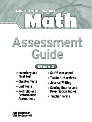
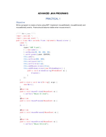
DYNAMIC NEW ART PROGRAMEvery piece of art has been updated to make it more vibrant, three-dimensional,and instructional. The authors examined every figure to ensure it was engaging and accurate. The fourteenth edition’sart program willhelp with understanding the key concepts of anatomy and physiology.11p 12n0 17p 18n0 14th Edition Cranial cavity Chlorine atom (Cl) atom (Na) Sodium Separate atoms (a)If a sodium atomloses an electron to a chlorine atom, the sodium Vertebral canal17p a sodium11p ion (Na ), and the chlorine atom becomes atom becomes 00 12n18na chlorideion (Cl ). Thoracic cavity Diaphragm Sodium atom (Na)Chlorine atom (Cl) 11p17pAbdominal 1 1 (a) Separate atomscavity12n018n0If a sodium atom loses an electron to a chlorine atom, the sodiumAbdominopelvicth atomatom becomes a sodium ion (Na ), and the chlorinebecomes cavitya chloride ion (Cl ). Pelvic cavity (a) )Chloride ion (Cl ) Sodium ion (Na Sodium chloride11p 17p (b) Bonded 1 1 12n018n0ionsThese oppositely charged particles attract electrically and join by an ionic bond. 13th Edition Realistic, threedimensional figuresprovide depth andorientation.14 EditionChloride ion (Cl )Sodium ion (Na )Na 13th EditionCl Sodium chloride(b) Bonded ionsThese oppositely charged particles attract electrically and join byan ionic bond.Colors highlighting atomicSalt crystalnuclei complement the(c)atomIonically bonded substances form arrays such as a crystal of NaCl.colors in molecular models.Na 14th EditionCl (c) Salt crystalIonically bonded substances form arrays suchas a crystal of NaCl.Primordialfollicle13th ntralfollicle3Matureantral follicleOvulation45Corpusluteum6Corpusalbicans1 At puberty, in response to hormonal stimulation, primordialfollicles (0.02 mm) begin maturing into primary follicles (0.1 mm).4 The antral follicle will grow quite rapidly and mature in about15 days prior to ovulation (20 mm).2 It takes two to three months for the primary follicles to reachthe pre-antral stage. It takes another 70 additional days for apre-antral follicle to reach a size of 0.2 mm.5 Following ovulation, the remants of the antral follicle becomethe corpus luteum.Process portrayed more accurately.xii3 In another 65 to 70 days the pre-antral follicle will have becomean antral follicle.(b)6 If fertilization of the ovulated secondary oocyte does not occur,the corpus luteum degenerates to become the corpus albicans.
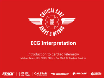
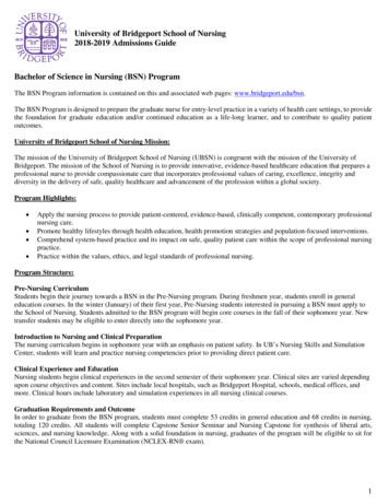
14th EditionStriationsNuclei (nearperiphery of cell)13th EditionPortion of askeletal musclefiber(a)Line art for micrographs is three-dimensionalto help visualize more than just the flatmicroscopic sample.(b)14th Edition13th eticulumCisternae ofsarcoplasmic reticulumNucleusTriadTransverse tubuleOpenings hickfilamentsThinfilamentsNucleusThis longitudinal section shows theinterior structures of a muscle fiberrevealing more detail of the myofibrils,and thick and thin filaments.14th EditionRegion ofaction potentialColor follows the movementof the action potential. –––––––– – –––––––– – –––––– –––––– 13th Edition (a) ––– Direction of conduction––– (b)(c) –––––– –– ––––––– –– – xiii
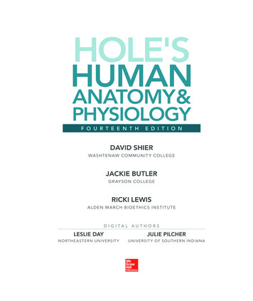
SS LERACTIEMuscular SystemE ASSLearnCLEARN, PRACTICE, ASSESSN P9ARLearning OuTcOMeSlearning tools to help the student succeed. . .Check out the Chapter Preview, Foundations for Success,on page 1. The Chapter Preview was specifically designedto help the student LEARN how to study. It provideshelpful study tips.After you have studied this chapter, you should be able to:9.1 Structure of a Skeletal Muscle1 Describe the structure of a skeletal muscle. (pp. 292–293)2 Name the major parts of a skeletal muscle fiber anddescribe the functions of each. (pp. 293–296)9.2 Skeletal Muscle contraction3 beginsDescribeneural controlof skeletallactic acidtotheaccumulatein musclesandmuscleto appear in old).Inthebody,lacticacid dissoLearning Outcomes open chapters, and are closely linked to Chapterassessments4 Identifythe major events of skeletal muscle fiberciates rapidlyinto lactateion(lactate) and hydrogen ion. Recentcontraction.(pp.298–299)and integrative assessments/Critical thinking questions found at the end of each chapter.studies onof bothhydrogenion andmusclelactatefibersuggest that5 theListeffectthe energysourcesfor skeletallearning outcomes are also tied to Connect content.contraction.299–302)the relationshipbetween(pp.lacticacid and muscle fatigue may not beEnergy toEnergyGlucose9.2 synthesizeSkeletalMuscleContraction6 Describeoxygen debt.(pp. in302–303)as clear-cutas once thought.Researchthis area is Synapticongoing.fromvesiclesFalsely colored scanning electron micrograph (SEM)of a normal human7Describehowamusclemaybecomefatigued.(p. enuousexerciseofaerobictrainingstimulatesnewA alcelskeletal muscleATPfiber reveals the characteristic bandingATP pattern of ndinto muscles,supplyingmyofibrils(3,000 ).constituents. The result is a movementlularand chemicalthewithinchapter. Itanswersto thequestion:“Whatis themorebig oxygen and8 Distinguish between a twitch and a sustained contraction.nutrientsto the musclefibers.physicaltraining also addsthe myofibrils inPyruvicwhichacidthe filaments of actin and myosinslide of howpicturethis chapterrelatestoSuchHumanAnatomy(pp. 304–306)past one another, shortening the sarcomeres. When this easetheir tionsproducePhysiology”?the muscle fiber shortens and pulls on its attachments.Both the adaptationsof increasedbloodmaintainsupply andmore(p.mitochonbody movementsand helpposture.306)Muscular System10 rs.driaPhysiologyboostability Revealedofbetweenmuscle fibersto produceATPaerobically.Anatomy andatLactic acidMotor(pp. 306–307)Learning ametime.AcetylcholineSynapticthe beginning of each chaptertells which system inneuron axoncleftEverything we do to express ourselves uses muscles. Consciously A cramp9.4is ontraction.APRin appliesto this11chapter.Actin, myosin,troponin,and tropomyosinare g The causeGlycolysisandSynthesisof glucoseofmuscle crampsis thenot structuresfully understood.One hypothmuscle lacticraisingacid formationacida question, and evenmultiunit smooth muscleand visceral sarcolemmasmooth muscle.or frowning,a hand in fromclasslacticto askMotoresis suggeststhatcramps may result from changes in electrolyteUnderstandingWordshelpsA rod-shapedmuscle protein calleddystrophin, for example,(in muscle)(in liver)(p. 307)end plateraising an eyebrow.concentrationin thetheextracellularsurroundingtheof andmuscleMuscle fiberMyofibrilaccounts for only 0.002% of total muscle protein in skeletalthe student rememberscientificcontraction fluid12 ntrolledstimulationmuscle, but its absence causes the devastating inheriteddis- meanings. Examinesmooth muscle fibers. (p. oo.All muscles of the muscle.anaerobically,to produceglucose.order Duchennemusculardystrophy,whichaffectsmales.9.5 cardiac MuscleGlycogen9The WhOLe PicTureAfter you have studied this chapter, you should be able to:9.1 Structure of a Skeletal Muscle1 Describe the structure of a skeletal muscle. (pp. 292–293)2 Name the major parts of a skeleta
IV. Title: Hole’s human anatomy and physiology. V. Title: Human anatomy & physiology. QP34.5.S49 2016 612–dc23 2014021876 The Internet addresses listed in the text were accurate at the time of publication. The inclusion of a website does not indicate an endorsement by the authors or McGraw-Hill Education, an