Transcription
1An Introductionto Anatomyand PhysiologyLearning OutcomesThese Learning Outcomes correspond by number to this chapter’s sectionsand indicate what you should be able to do after completing the chapter.1-1 1-2 1-3 1-4 1-5 1-6 1-7 Describe how to use the text and art to master learning. p. 2Define anatomy and physiology, explain the relationship betweenthese sciences, and describe various specialties of eachdiscipline. p. 3Identify the major levels of organization in organisms, from thesimplest to the most complex, and identify the major componentsof each organ system. p. 6Describe the origins of anatomical and physiological terms,and explain the significance of Terminologia Anatomica. p. 7Use anatomical terms to describe body regions, bodysections, and relative positions. p. 7Identify the major body cavities of the trunkand their subdivisions, and describethe functions of each. p. 14Explain the concept of homeostasis.p. 181-8 Describe how negative feedbackand positive feedback areinvolved in homeostaticregulation. p. 19M01 MART6026 11 SE C01 pp001-026.indd 105/11/16 12:01 am
CLINICAL CASE Using A&P to Save a LifeAn emergency medical technician (EMT) is onthe way to the emergency department with ayoung victim of street violence. A knife with1a 6-inch blade had been found next to thebleeding, unconscious man.“We have a young male with multiplestab wounds. He has lost a lot of blood andwe can barely get a blood pressure,” theEMT radios to the triage nurse in the emergency department as the ambulance squealsthrough traffic. “We started an IV and we arepouring in fluid as fast as we can.”“Where are the wounds?” asks the receiving nurse.“He has a deep wound in his right upper quadrant, justinferior to the diaphragm. I can see bruising from the hub of theknife around the wound, and there is another wound in his anteriorAn Introduction to Studyingthe Human BodyWelcome to the field of human anatomy and physiology—known simply as A&P! In this textbook we will introduce youto the inner workings of the human body, giving informationabout both its structure (anatomy) and its function (physiology). Many students who use this book are preparing for jobsin health-related fields; but regardless of your career choice,you will find the information within these pages relevant toyour future.We will focus on the human body, but the principles youwill learn apply to other living things as well. Our world contains an enormous diversity of living organisms, which varywidely in appearance and lifestyle. One aim of biology—thestudy of life—is to discover the unity and the patterns thatunderlie this diversity. As we study human anatomy and physiology, three main concepts will emerge: (1) the principle ofcomplementarity of structure and function, (2) the hierarchyof structural relationships, and (3) homeostasis, the tendencytoward internal balance. These principles are the foundation forlearning about the human organism.Before we begin with the science of human anatomy andphysiology, let’s turn our attention to the science of learningand learning strategies. To make the most of your learningexperience, apply these strategies, which were collected fromacademic research.1-1 To make the most of your learning,read the text and view the art togetherLearning Outcome Describe how to use the text and art tomaster learning.right thigh. His pulse is 120 and thready(weak). His blood pressure is 60 over 30.”“How long has he been down?” questions the nurse.“Less than a half hour. We intubatedhim (inserted a breathing tube) and starteda large-bore IV as soon as we got there. Weare 10 minutes out now.”“Keep the fluids going wide open, keeppressure on the thigh, and take him directlyto Trauma Room 1,” come the instructions.Meanwhile, the nurse orders the trauma team to Trauma Room 1,orders X-Ray to be on standby in the room, and requests 4 units oftype O negative whole blood—the universal donor blood—from theblood bank. Will the team be ready to save this young man?To find out, turn to the Clinical Case Wrap-Up on p. 26.Getting to Know Your TextbookThis first section of the book sets the stage for your success inthis course and introduces you to the basic principles of learning. Just as there are three underlying concepts in A&P, there aretwo basic principles to using your textbook effectively to learnA&P. Practicing these principles will help you throughout yourcollege career.Let’s start. Think back to your first childhood book. Youmost likely began with a “picture book.” Then, as you learnedthe alphabet and developed speech, you progressed to “wordbooks.” The next step was “chapter books.” Somewhere alongthe way, you quit looking at pictures and focused solely on thewords (text). Maybe the shift in focus to text-based readingwithout looking at the pictures happened in high school. Youbegan reading words—paragraph upon paragraph, page uponpage of words. Now, you are in college, and we need to realignyour focus.In college, you are faced with lots of new terms, abstractconcepts, and unfamiliar images. That’s great, because collegeis intended to increase your knowledge and expand your horizons. However, research has shown that undergraduate students have a tendency to simply read the text (also called thenarrative), without paying attention to the pictures (referred toas visuals, art, diagrams, illustrations, figures, or images). Whileyou can certainly learn from this approach, further researchdemonstrates that when students read the text and then look atthe corresponding picture, they actually learn the material better!Although this may sound quite intuitive, most students donot do that. So, we wrote a book that truly integrates text withart to help you learn A&P. Please continue reading as we walkyou through the process of using a textbook to enhance yourlearning. Much of what we’re about to tell you applies to most2M01 MART6026 11 SE C01 pp001-026.indd 205/11/16 12:01 am
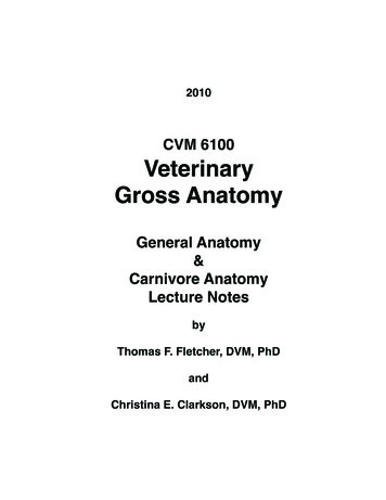
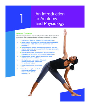
Chapter 1 An Introduction to Anatomy and Physiology 3of your college textbooks, but we’ll focus on this book, since itwas designed for students such as you.Anatomy of a ChapterYour book is broken down into sections with text–art integration, and specific learning outcomes for each section based on alearning classification scheme. A section is a unit about a topicthat continues to build on previously learned topics. The sectional layout promotes logical, efficient navigation through thematerial, while callouts to figures integrate the text with theart. Text–art integration implies that the figures are close tothe lines of text and that the figure legends are adjacent to theart. Look at that figure when you see a callout for it. The figurecallouts look like this: (Figure 1–1). They are color-coded onpurpose so you can stop reading, look at the figure, and thenfind your place again when you go back to reading the text. So,strategy #1 is to read the text and then study the image that goesalong with the narrative.Learning outcomes are educational objectives that usekey verbs and target specific skills, goals, aims, and achievements. The learning outcomes appear at the beginning of eachchapter and within the chapter under the sentence-based headings. Strategy #2 is to pay attention to these learning outcomesbecause they are tied directly to testing and tell you what you shouldbe able to do after reading that specific section and studying theimages. These learning outcomes are based on a learning classification scheme, which identifies the fundamental levels oflearning from lower order skills to those of higher-order skills.Figure 1–1A Conceptual Framework for Learning.ngowiKnand rememberingand undndingerstaneheprdimngoCApplying anddescribingSynthesizingandcreatingEvaluatingand measuringM01 MART6026 11 SE C01 pp001-026.indd 3AnalyzingandexplainingYou’ll see key verbs in your learning outcomes and you’ll noticesome overlap among them within the levels of learning. Fromlower to higher, these levels are (1) knowing and remembering,(2) comprehending and understanding, (3) applying and1describing, (4) analyzing and explaining, (5) evaluating andmeasuring, and (6) synthesizing and creating (Figure 1–1).(Here is where you can practice using what you just learned:Look at that figure, think about it, and then return to this text.)If you practice these basic strategies—(1) read the narrativeand study the image and (2) pay attention to the learning outcomes—you are well on your way to success!Checkpoint1. Describe a learning outcome.2. Explain how to use your textbook most effectively toenhance your learning.See the blue Answers tab at the back of the book.1-2 Anatomy (structure) and physiology(function) are closely integratedLearning Outcome Define anatomy and physiology, explainthe relationship between these sciences, and describe variousspecialties of each discipline.Anatomy is the study of internal and external body structuresand their physical relationships among other body parts. Incontrast, physiology is the study of how living organisms perform their vital functions. Someone studying anatomy might,for example, examine where a particular muscle attaches to theskeleton. Someone studying physiology might consider how amuscle contracts or what forces a contracting muscle exerts onthe skeleton. You will be studying both anatomy and physiology in this text, so let’s look at the relationships between thesesciences.Anatomy and physiology are closely integrated, both intheory and practice. Anatomical information provides cluesabout functions, and physiological processes can be explainedonly in terms of the underlying anatomy. This is a very important concept in living systems:All specific functions are performed by specific structures,and the form of a structure relates to its function. This isknown as the principle of complementarity of structure andfunction.The link between structure and function is always present,but not always understood. For example, the anatomy of theheart was clearly described in the 15th century, but almost200 years passed before the heart’s pumping action wasdemonstrated.Anatomists and physiologists approach the relationshipbetween structure and function from different perspectives. Tounderstand the difference, suppose you asked an anatomist and05/11/16 12:01 am
4 UNIT 1 Levels of Organizationsa physiologist to examine a car and report their findings. Theanatomist might begin by measuring and photographing thevarious parts of the car and, if possible, taking it apart and putting it back together. The anatomist could then explain its key1structural relationships—for example, how the pistons are seatedin the engine cylinders, how the crankshaft is connected to thepistons, and how the transmission links the drive shaft to theaxles and, thus, to the wheels. The physiologist also would notethe relationships among the car’s parts, but he or she wouldfocus mainly on its functional characteristics, such as how thecombustion of gasoline in the cylinders moves the pistons upand down and makes the drive shaft rotate, and how the transmission conveys this motion to the axles and wheels so that thecar moves. Additionally, he or she might also study the amountof power that the engine could generate, the amount of forcetransmitted to the wheels in different gears, and so forth.Our basic approach in this textbook will be to start withthe descriptive anatomy of body structures (appearance, size,shape, location, weight, and color) before considering therelated functions. Sometimes the groups of organs that makeup an organ system perform very diverse functions, and in thosecases we consider the functions of each individual organ separately. A good example is our discussion of the digestive systemand its organs. You will learn about the functions of the salivary glands in one section, and the functions of the tongue inanother. In other systems, the organs work together so extensively that we present an overall discussion of their physiology,after we describe the system’s anatomy. The lymphatic system(which contains a network of vessels) and the cardiovascularsystem, for example, are treated using this approach.AnatomyWhen you look at something, how far away you are from itoften determines what you see. You get a very different view ofyour neighborhood from a satellite photo than from your frontyard. Similarly, your method of observation has a dramatic effect on your understanding of the structure of the human body.Based on the degree of structural detail being considered, we divide human anatomy, the study of the structure of the humanbody, into gross (macroscopic) anatomy and microscopic anatomy.Gross AnatomyGross anatomy, or macroscopic anatomy, involves examiningfairly large structures. Gross anatomy (from the Latin term grossus, meaning “thick” or “massive”) can be conducted withoutusing a microscope and can involve the study of anatomy bydissecting a cadaver. There are many different forms of grossanatomy: Surface anatomy, or superficial anatomy, is the study of thegeneral form of the body’s surface, especially in relation toits deeper parts.M01 MART6026 11 SE C01 pp001-026.indd 4 Regional anatomy focuses on the anatomical organizationof specific areas of the body, such as the head, neck, ortrunk. Many advanced courses in anatomy stress a regionalapproach, because it emphasizes the spatial relationshipsamong structures already familiar to students.Sectional anatomy is the study of the relationship of thebody’s structures by examining cross sections of the tissueor organ.Systemic anatomy is the study of the structure of organ systems,which are groups of organs that function together in acoordinated manner. Examples include the skeletal system,composed primarily of bones; the muscular system, made upof skeletal muscles; and the cardiovascular system, consistingof the heart, blood, and vessels. We take a systemic anatomyapproach in this book because this format works better toclarify the functional relationships among the componentorgans. We introduce the 11 organ systems in the humanbody later in the chapter.Clinical anatomy includes a number of subspecialtiesimportant in clinical practice. Examples include pathologicalanatomy (anatomical features that change during illness),radiographic anatomy (anatomical structures seen usingspecialized imaging techniques), and surgical anatomy(anatomical landmarks important in surgery).Developmental anatomy describes the changes in form thattake place between conception and adulthood. The techniques of developmental anatomists are similar to thoseused in gross anatomy and in microscopic anatomy (discussed next) because developmental anatomy considersanatomical structures over a broad range of sizes—from asingle cell to an adult human. The most extensive structuralchanges take place during the first two months of development. The study of these early developmental processes iscalled embryology (em-bre-OL-o-je).Microscopic AnatomyMicroscopic anatomy deals with structures that we cannotsee without magnification. The boundaries of microscopicanatomy are set by the limits of the equipment we use. With adissecting microscope you can see tissue structure. With a lightmicroscope, you can see basic details of cell structure. And withan electron microscope, you can see individual molecules thatare only a few nanometers (billionths of a meter) across.Microscopic anatomy includes two major subdivisions:cytology and histology. Cytology (sı-TOL-o-je) is the studyof the internal structure of individual cells, the simplest unitsof life. Cells are made up of chemical substances in variouscombinations, and our lives depend on the chemical processesthat take place in the trillions of cells in the body. For thisreason, we consider basic chemistry (Chapter 2) before weexamine cell structure (Chapter 3). Histology (his-TOL-o-je).05/11/16 12:01 am
Chapter 1 An Introduction to Anatomy and Physiology 5 Clinical Note Habeas Corpus (“You Shall Have the Body”)It is the first day of Anatomy. Students await the arrival oftheir white-coated and gloved professor. Anxiety mounts as astretcher covered in surgical drapes is wheeled in. This is thecadaver. Who will faint? Will it be me? For many students inthe health professions, cadaver dissection is a cornerstone oftheir training. These students are following in a revered tradition that began 2300 years ago with the first examinations ofthe body after death by Greek royal physicians. The expression “a skeleton in your closet” dates from a later era whenmedical students had to procure bodies on their own forstudy (and keep them hidden in the closet). There is much tobe learned from death. Cadaver dissections also reveal muchabout life. After working closely on a cadaver for months,is the examination of tissues—groups of specialized cells thatwork together to perform specific functions (Chapter 4). Tissues combine to form organs, such as the heart, kidney, liver,or brain, each with specific functions. Many organs are easy toexamine without a microscope, so at the organ level we crossthe boundary from microscopic anatomy to gross anatomy. Aswe proceed through the text, we will consider details at all levels, from microscopic to macroscopic.PhysiologyHuman physiology is the study of the functions, or workings,of the human body. These functions are complex processes andmuch more difficult to examine than most anatomical structures. As a result, there are even more specialties in physiologythan in anatomy. Examples include the following: Cell physiology, the study of the functions of cells, is thecornerstone of human physiology. Cell physiology looks atthe chemistry of the cell. It includes both chemical processeswithin cells and chemical interactions among cells.Organ physiology is the study of the function of specificorgans. An example is cardiac physiology, the study of heartfunction—how the heart works.Systemic physiology includes all aspects of the functioning of specific organ systems. Cardiovascular physiology,respiratory physiology, and reproductive physiology areexamples.Pathological physiology is the study of the effects of diseaseson organ functions or system functions. Modern medicinedepends on an understanding of both normal physiologyand pathological physiology.Physicians normally use a combination of anatomical,physiological, chemical, and psychological information whenM01 MART6026 11 SE C01 pp001-026.indd 5students develop an attachment to “their” body, oftennaming it. The intimate revelations of the scalpel, the highlypersonal variations of humananatomy, the Rubik’s cube ofdisease, and the stark reality of death combine to leavea deep intellectual and emotional mark on the student.Students and faculty may end the course with a ceremony topay their respects to this human body and to this privilegedexperience.1they evaluate patients. When a patient presents with signs (anobjective disease indication like a fever) and symptoms (asubjective disease indication, such as tiredness), the physicianwill look at the structures affected (gross anatomy), perhapscollect a fluid or tissue sample (microscopic anatomy) foranalysis, and ask questions to find out what changes from normal functioning the patient is experiencing. Think back to yourlast trip to a doctor’s office. Not only did the physician examineyour body, noting any anatomical abnormalities, but he or shealso evaluated your physiological processes by asking questions,observing your movements, listening to your body sounds, taking your temperature, and perhaps requesting chemical analysesof fluids such as blood or urine.In evaluating all these observations to reach a diagnosis,physicians rely on a logical framework based on the scientificmethod. The scientific method is a system of advancingknowledge that begins by proposing a hypothesis to answer aquestion, and then testing that hypothesis with data collectedthrough observation and experimentation. This methodis at the core of all scientific thought, including medicaldiagnosis.Checkpoint3. Define anatomy.4. Define physiology.5. Describe how anatomy and physiology are closelyrelated.6. What is the difference between gross anatomy andmicroscopic anatomy?7. Identify several specialties of physiology.8. Why is it difficult to separate anatomy from physiology?See the blue Answers tab at the back of the book.05/11/16 12:01 am
6 UNIT 1 Levels of Organizations1-3 Levels of organization progressfrom chemicals to a complete organismLearning Outcome Identify the major levels of organization in1 organisms, from the simplest to the most complex, and identifymajor components of each organ system.Our understanding of how the human body works is basedon investigations of its different levels of organization. Higherlevels of organization are more complex and more variablethan lower levels. Chapters 2, 3, and 4 consider the chemical, cellular, and tissue levels of organization of the humanbody. These levels are the foundations of more complex structures and vital processes, as we describe in Chapters 5–29. Thesix levels of organization of the human body are shown inSpotlight Figure 1–2 and include: The Chemical Level. Atoms are the smallest stable units ofmatter. They can combine to form molecules with complex shapes. The atomic components and unique threedimensional shape of a particular molecule determine itsfunction. For example, complex protein molecules form filaments that produce the contractions of muscle cells in theheart. We explore this level of organization in Chapter 2.The Cellular Level. Cells are the smallest living units in thebody. Complex molecules can form various types of largerstructures called organelles. Each organelle has a specificfunction in a cell. Energy-producing organelles provide theenergy needed for heart muscle cell contractions. We examine the cellular level of organization in Chapter 3.The Tissue Level. A tissue is a group of cells workingtogether to perform one or more specific functions. Heartmuscle cells, also called cardiac muscle cells (cardium,heart), interact with other types of cells and with materialsoutside the cell to form cardiac muscle tissue. We considerthe tissue level of organization in Chapter 4.The Organ Level. Organs are made of two or more tissuesworking together to perform specific functions. Layers ofcardiac muscle tissue, in combination with another type oftissue called connective tissue, form the bulk of the wall ofthe heart, which is a hollow, three-dimensional organ.The Organ System Level. A group of organs interacting toperform a particular function forms an organ system.Each time the heart contracts, for example, it pushes bloodinto a network of blood vessels. Together, the heart, blood,and blood vessels make up the cardiovascular system, oneof 11 organ systems in the body. This system functions todistribute oxygen and nutrients throughout the body.The Organism Level. An individual life form is an organism.In our case, an individual human is the highest level oforganization that we consider. All of the body’s organsystems must work together to maintain the life and healthof the organism.M01 MART6026 11 SE C01 pp001-026.indd 6The organization at each level determines not only thestructural characteristics but also the functions of higher levels. For example, the arrangement of atoms and molecules atthe chemical level creates the protein filaments and organelles at the cellular level that give individual cardiac musclecells the ability to contract. At the tissue level, these cells arelinked, forming cardiac muscle tissue. The structure of thetissue ensures that the contractions are coordinated, producing a powerful heartbeat. When that beat occurs, the internalanatomy of the heart, an organ, enables it to function as apump. The heart is filled with blood and connected to theblood vessels, and its pumping action circulates blood throughthe vessels of the cardiovascular system. Through interactionswith the respiratory, digestive, urinary, and other systems, thecardiovascular system performs a variety of functions essentialto the survival of the organism.Something that affects a system will ultimately affect eachof the system’s parts. For example, after massive blood loss, theheart cannot pump blood effectively. When the heart cannotpump and blood cannot flow, oxygen and nutrients cannotbe distributed to the heart or around the body. Very soon,the cardiac muscle tissue begins to break down as individualmuscle cells die from oxygen and nutrient starvation. Thesechanges will not be restricted to the cardiovascular system.All cells, tissues, and organs in the body will be damaged.Spotlight Figure 1–2 illustrates the levels of organization andintroduces the 11 interdependent, interconnected organ systems in the human body.The cells, tissues, organs, and organ systems of the bodycoexist in a relatively small, shared environment, much like theresidents of a large city. Just as city dwellers breathe the sameair and drink the water supplied by the local water company,cells in the human body absorb oxygen and nutrients from thefluids that surround them. If a city is blanketed in smog or itswater supply is contaminated, its inhabitants will become ill.Similarly, if the body fluid composition becomes abnormal,cells will be injured or destroyed. For example, suppose thetemperature or salt content of the blood changes. The effecton the heart could range from the need for a minor adjustment (heart muscle tissue contracts more often, raising theheart rate) to a total disaster (the heart stops beating, so theindividual dies).Checkpoint9. Identify the major levels of organization of the humanbody from the simplest to the most complex.10. Identify the organ systems of the body and cite somemajor structures of each.11. At which level of organization does a histologistinvestigate structures?See the blue Answers tab at the back of the book.05/11/16 12:01 am
Chapter 1 An Introduction to Anatomy and Physiology 71-4 Medical terminology is importantto understanding anatomy andphysiologyLearning Outcome Describe the origins of anatomical andphysiological terms, and explain the significance of TerminologiaAnatomica.Early anatomists faced serious problems when trying to communicate. Saying that a bump is “on the back,” for example,does not give very precise information about its location. Soanatomists created illustrated maps of the human body andgave each structure a specific name. They used prominent anatomical structures as landmarks, measured distances in centimeters or inches, and discussed these subjects in specializeddirectional terms. Modern anatomists continue and build onthese practices. In effect, anatomy uses a special language thatyou must learn almost at the start of your study.That special language, called medical terminology,involves using word roots, prefixes, suffixes, and combiningforms to build terms related to the body in health and disease. Many of the anatomical and physiological terms you willencounter in this textbook are derived from Greek or Latinroots that originated more than 1500 years ago. In fact, the termanatomy is derived from Greek roots that mean “a cutting open”;the term physiology also comes from Greek. Learning the wordparts used in medical terminology will greatly assist in yourstudy of anatomy and physiology and in your preparation forany health-related career.There are four basic building blocks—or word parts—ofmedical terms. Word roots are the basic, meaningful parts ofa term that cannot be broken down into another term withanother definition. Prefixes are word elements that are attachedto the beginning of words to modify their meaning but cannot stand alone. Suffixes are similar to prefixes, except they areword elements or letters added to the end of a word or wordpart to form another term. Combining forms are independentwords or word roots that are used in combination with words,prefixes, suffixes, or other combining forms to build a newterm. As we introduce new terms, we will provide notes onpronunciation and relevant word parts. In addition, the tableinside the back cover of your textbook lists many commonlyused word roots, prefixes, suffixes, and combining forms.To illustrate the building of medical terms, consider theword pathology (puh-THOL-o-je). Breaking this word into itsbasic parts reveals its meaning. The prefix path- refers to disease(the Greek term for “disease” is pathos). The suffix -ology means“study of.” So pathology is the study of disease.Latin and Greek terms are not the only ones that have beenimported into the anatomical vocabulary over the centuries,and this vocabulary continues to expand. Many anatomicalstructures and clinical conditions were first named after eitherthe discoverer or, in the case of diseases, the most famous.M01 MART6026 11 SE C01 pp001-026.indd 7victim. During the past 100 years, most of these commemorative names, or eponyms (EH-po-nimz), have been replaced bymore precise terms. Where appropriate, we will give both theeponym and the more precise term, because in clinical medi1cine, both terms may be used.To avoid the miscommunication that plagued the earlyanatomists, it is important for scientists throughout the worldto use the same name for each body structure. In 1998, twoscientific organizations—the Federative Committee on Anatomical Terminology (FCAT) and the International Federationof Associations of Anatomists (IFAA)—published TerminologiaAnatomica (TA). Terminologia Anatomica established the worldwide standard for human anatomical terminology. The successor of FCAT is the Federative International Programme onAnatomical Terminologies (FIPAT). In April 2011, FIPAT published TA online. Latin continues to be the language of anatomy,but this reference provides an English equivalent term for eachanatomical structure. For example, the tendo calcaneus (Latin) isalso called the c
1-2 Anatomy (structure) and physiology (function) are closely integrated Learning Outcome Define anatomy and physiology, explain the relationship between these sciences, and describe various specialties of each discipline. Anatomy is the study of internal and external body structures and their physical relationships among other body parts. In