Transcription
REVISTA DE ODONTOLOGIA DA UNESPORIGINAL ARTICLERev Odontol UNESP. 2017 Sept-Oct; 46(5): 267-272 2017 - ISSN 1807-2577Doi: otometric assessment of tooth bleaching underorthodontic braquets bonded with different materialsAvaliação espectrofotométrica do clareamento dental sob braquete ortodôntico fixadocom diferentes materiaisCamila Maria Lima de CASTROa*, Higor Catta Preta BORGESa, Karoline Cardoso Ferro BARROSa,Natanael Barbosa dos SANTOSa, Larissa Silveira de Mendonça FRAGOSOaaFaculdade de Odontologia, UFAL – Universidade Federal de Alagoas, Maceió, AL, BrasilResumoIntrodução: pacientes estão sendo submetidos ao clareamento durante tratamento ortodôntico com finalidadeestética ou para antecipar a troca de restaurações após finalização do tratamento ortodôntico. Objetivo: avaliar aefetividade do clareamento dentário sob braquete ortodôntico fixado com diferentes materiais. Material e método: cemblocos de esmalte bovino foram divididos em dois grupos, clareamento de consultório e clareamento caseiro. O declareamento de consultório foi subdividido em cinco (n 10): HP (controle – sem braquetes); SA(t) – braquetes fixadoscom Transbond XT, sem clareamento; SA(fm) – braquetes fixados com Fill Magic Ortodôntico, sem clareamento;HP(t) – braquetes fixados com Transbond XT submetidos ao clareamento e HP(fm) – braquetes fixados comFill Magic Ortodôntico submetidos ao clareamento. O clareamento caseiro seguiu a mesma divisão descrita parao clareamento de consultório substituindo-se o agente clareador. Foram realizadas avaliações da cor através doespectrofotômetro nos tempos: 1) antes do manchamento (baseline); 2) após o manchamento; 3) após fixação dosbraquetes e procedimentos clareadores. Os dados foram submetidos à ANOVA e diferenças analisadas através do testede Tukey (p 0,05). Resultado: Para os dois tipos de clareamento o grupo controle teve ação clareadora efetiva. Gruposfixados com Transbond XT apresentaram maior potencial clareador dentre os grupos que apresentaram acessórioortodôntico, mas com ação clareadora diferente do controle positivo. Grupos fixados com Fill Magic Ortodônticonão apresentaram ação clareadora, assemelhando-se aos grupos controle negativo (saliva artificial). Conclusão: obraquete ortodôntico prejudicou a efetividade do tratamento clareador caseiro e de consultório, independente daresina utilizada para a fixação.Descritores: Braquetes ortodônticos; clareamento dental; colagem dentária.AbstractIntroduction: patients have been submitted to tooth bleaching during orthodontic treatment for aesthetic purposes orto anticipate the replacement of restorations after completion of the treatment. Objective: to evaluate the effectivenessof tooth bleaching under orthodontic brackets bonded with different materials. Material and method: a hundredbovine enamel blocks were divided into two groups, at-home and in-office tooth bleaching. In-office bleaching wassubdivided into five groups (n 10): HP (control - without brackets); SA(t) - brackets bonded with Transbond XT,without bleaching; SA(fm) - brackets bonded with Orthodontic Fill Magic, without bleaching; HP(t) - brackets bondedwith Transbond XT subjected to bleaching; and HP(fm) - brackets bonded with Orthodontic Fill Magic subjected tobleaching. At-home bleaching followed the same treatments, only replacing the bleaching agent. Spectrophotometricassessment was used for tooth color determination at three moments: 1) before staining (baseline); 2) after staining;3) after bonding the brackets and bleaching procedures. Data were submitted to ANOVA and analyzed by Tukey’stest (p 0.05). Result: For both types of bleaching, the control group had an effective bleaching action. Groupsusing Transbond XT presented greater bleaching potential among the groups with orthodontic accessory, but thebleaching action differed from the positive control. Groups using Orthodontic Fill Magic presented no bleachingaction, resembling the negative control groups (artificial saliva). Conclusion: the orthodontic bracket impaired theeffectiveness of the at-home and in-office bleaching treatment, regardless of the resin used for bonding.Descriptors: Orthodontic brackets; tooth whitening; dental bonding.
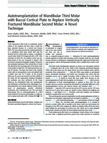
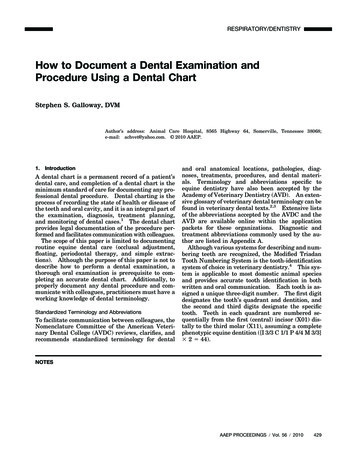
268Castro, Borges, Barros et al.INTRODUCTIONOne of the reasons of discontentment among orthodonticpatients is the enamel color change that occurs during treatment1,2.The staining of the teeth in this period may be associated with thecolor instability of the resinous material used to bond the bracket3,or even by demineralization of the enamel4 or the direct absorptionof food colorants2.The mechanism of action of tooth whitening involves anoxidation‐reduction reaction where the hydrogen peroxide reducesorganic pigments impregnated in the enamel and dentin, thusallowing their elimination. When hydrogen peroxide gets in contactwith the enamel surface, it releases unstable oxygen, which bindsto other substances that are free or weakly bound to a particularsubstrate, for stabilization5.Dental enamel is a highly mineralized crystalline tissue thatpresents some permeability, allowing the diffusion of substances andthe ionic exchange with the buccal environment6. Tooth whiteningis only possible because of this permeability and the low molecularweight of some active chemical components of bleaching agents,such as hydrogen peroxide7.Dental whitening can be performed by two techniques: at-homeand in-office6,8. At home, bleaching agents of low concentration inflexible trays are used in daily basis and supervised by the dentist.The in-office technique uses high concentrations of bleaching agentsfor short periods of exposure6,9.Hydrogen peroxide is the active compound of the whiteningprocess. Thus, when bleaching is required, hydrogen peroxide‑basedbleaching agents must be used, an option widely used in the in‑officetechnique. Carbamide peroxide-based products provide hydrogenperoxide in a gradual and continuous way; however, 10% of carbamideperoxide results in approximately 3.6% hydrogen peroxide9.Aware of the diffusion capacity of bleaching agents in dentaltissues, orthodontists have subjected their patients to dentalbleaching during orthodontic treatment for aesthetic purposes orto anticipate the exchange of dental restorations after orthodontictreatment is completed. However, the real effectiveness of thediffusion of the bleaching agent under the orthodontic accessoryis not fully understood so far, and more, if after whitened, there isany difference in the effectiveness of the treatment depending onthe material used to bond the brackets.In order to address these questions, the present study aimed toevaluate through reflectance spectrophotometry the effectivenessof tooth whitening under orthodontic brackets bonded withdifferent materials.MATERIAL AND METHODPreparation of SpecimensOne hundred bovine incisor teeth were extracted in aslaughterhouse and refrigerator in Alagoas (MAFRIAL), storedin 1% thymol solution and kept under refrigeration until use.The teeth were cleaned with a scalpel blade, in order to remove theperiodontal ligament. They then had the coronal portion separatedRev Odontol UNESP. 2017 Sept-Oct; 46(5): 267-272from the root (Figure 1A) with the double-sided diamond disk(KG Sorensen, Barueri, SP, Brazil) at low rotation under constantirrigation, removing the coronal pulp and discarding the root.A high concentration diamond wafering blade (4” x 0.12 x 0.5,Extec, Enfield, USA) coupled to a metallographic cut-off machine(Figure 1B) (South Bay Technology Inc. San Clemente, CA,USA) was used to perform four cuts in the coronal portion, inthe mesio‑distal and incisor-cervical directions, obtaining dentalblocks with dimensions of 8.0 x 8.0 mm (Figure 1C) with avariation of 0.3 mm. The size of the blocks was checked with adigital micrometer (Mitutoyo, Aurora, Illinois, USA) (Figure 1D).The dental blocks were kept in distilled water.The blocks were bonded with sticky wax in acrylic bases tofacilitate handling. For this, the vestibular enamel surfaces wereplaced in contact with the wax, that is, facing downwards, and thedentin facing upwards, so that the dentin was exposed. Dentinplaning was done with 320 grit silicon carbide sandpaper in ametallographic polish (APL 4, Arotec, Cotia, SP, Brazil) (Figure 1E)for due evenness, at low speed for 15 seconds. Then, the dentalblocks were repositioned on the acrylic bases, this time with thesurfaces of the buccal enamel facing upwards and pressed withthe aid of a parallelogram (Figure 1F), constituting the specimens.Whitening TestThe acrylic bases were painted with matte black paint (Figure 1G)to prevent the passage of external light during the color readingprocess, which could interfere with the final result of the assessment.The specimens showing cracks, hypoplasia and spotting wereexcluded from the sample and replaced.The specimens were randomly assigned to two groups: in-officebleaching (Whintness HP Blue, FGM, Joinville, Santa Catarina,Brazil) and at-home bleaching (Whitness Perfect, FGM, Joinville,Santa Catarina, Brazil). Each group was divided into five subgroups(n 10).In-office bleaching:1. HP - positive control: no brackets;2. SA(t) - negative control: brackets bonded with TransbondXT (3M Unitek, São José do Rio Preto, SP, Brazil), withoutbleaching;3. SA(fm) - negative control: brackets bonded with OrthodonticFill Magic (Vigodent, Rio de Janeiro, RJ, Brazil), withoutbleaching;4. HP(t) - brackets bonded with Transbond XT subjected tobleaching;5. HP(fm) - brackets bonded with Orthodontic Fill Magicsubjected to bleaching.At-home bleaching:1. WP - positive control: no brackets;2. SA(t) - negative control: brackets bonded with TransbondXT, without bleaching;
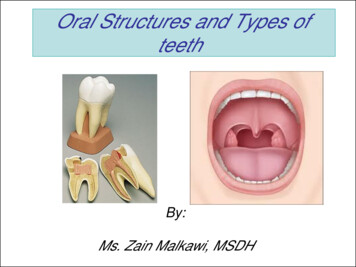
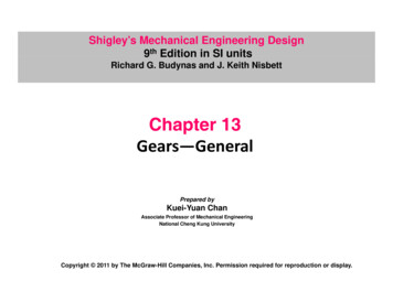
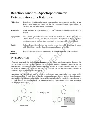
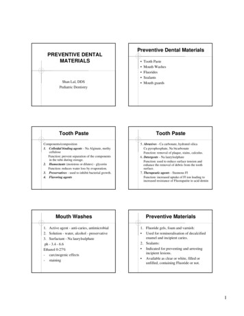
Rev Odontol UNESP. 2017 Sept-Oct; 46(5): 267-272Spectrophotometric assessment of tooth.269Figure 1. Sequence of methodology. (A) Coronal portion of the teeth after root excision; (B) Metallographic cut-off machine used for all dentalcuts; (C) Dental blocks with dimensions of 8.0 x 8.0 mm; (D) Size checked with digital micrometer; (E) Metallographic polishing machine used forpolishing; (F) Teeth pressed with the paralellometer; (G) Acrylic bases painted with matte black paint; (H) Specimens after staining; (I) Bondingof metal brackets; (J and K) Application of the respective bleaching treatments.3. SA(fm) - negative control: brackets bonded with OrthodonticFill Magic, without bleaching;4. WP(t) - brackets bonded with Transbond XT subjected tobleaching;5. WP(fm) - brackets bonded with Orthodontic Fill Magicsub‑ected to bleaching.Then the specimens were submitted to the staining stage(Figure 1H) with black tea (Maratá, Itaporanga D’Ajuda, SE, Brazil),prepared in the proportion of 10 g of tea to 1 L of distilled waterboiled for two minutes) for five consecutive days with two dailyexchanges, being kept in an orbital shaker table (TermoagitatorTE-420, Tecnal, Piracicaba, SP, Brazil) at 37 C and 100 rpm. Beforeeach new exchange, the specimens were washed in distilled waterand immersed in artificial saliva for one hour, until they werere‑immersed in the tea.After this, stainless steel brackets (Morelli, SP, Brazil) (Figure 1I),Roth, for lower incisors, with slot 022” x .030” in 1.7 mm x 2.3 mmbase mesh, were bonded with the resins corresponding to eachgroup, according to the manufacturers’ guidelines. The applicationof bleaching treatments began (Figures 1J and 1K) following theindications of the manufacturers. Before the application of all thetreatments, the specimens were washed in distilled water, placedin artificial saliva and kept in an orbital table. The groups thatdid not undergo bleaching remained immersed in artificial salivathroughout the experiment.Color assessment was performed using a digital spectrophotometer(CR-321, MINOLTA Co., Tokyo, Honshu, Japan) at three moments:Initial - before staining; Intermediate - after staining; and Final - afterbleaching. The spectrophotometer describes the three-dimensionalcolor through the CIELAB scale, which makes use of a mathematicalsystem where all colors are defined by the coordinates of the threeaxes: L*, a* and b*. The L* axis corresponds to the brightness andvaries from 0 (black) to 100 (white). The a* and b* axes are relatedto color and saturation, but a* varies from red (positive) to green(negative) and b* from yellow (positive) to blue (negative). The colorreading was always performed at the central point of the block,in the same environment and with the same type of illumination.Statistical AnalysisThe values of the parameters L*, a* and b* obtained byspectrophotometric analysis were submitted to one-factor analysisof variance (ANOVA) and any differences were evaluated throughthe Tukey test (p 0.05).RESULTAfter the staining stage, no discrepancy in the total colorvariation (ΔE) was seen between the experimental groups, eitherin the in-office bleaching (ANOVA, p 0.31) Figure 2A, or in theat-home bleaching (ANOVA, p 0.28) Figure 2B.
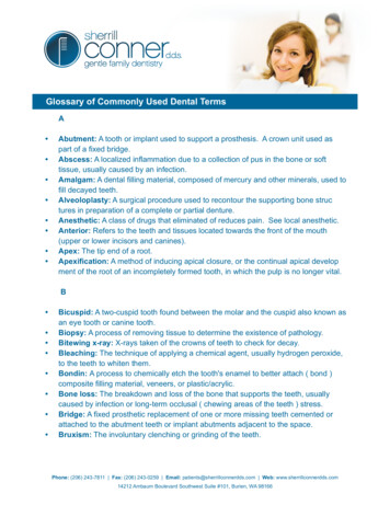
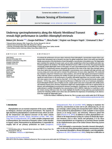
270Castro, Borges, Barros et al.Rev Odontol UNESP. 2017 Sept-Oct; 46(5): 267-272Figure 3. Total color variation (ΔE) between the staining and in-officebleaching stages of the experimental groups. There was evidence ofheterogeneity among groups (ANOVA, p 0.0007).Figure 2. Total color variation (ΔE) between the baseline and stainingphases. (A) Experimental groups submitted to in-office bleaching;(B) Experimental groups submitted to at-home bleaching. Both resultsindicate homogeneity.IN-OFFICE BLEACHINGWhen comparing ΔE after the bleaching stage Figure 3, the groupswere found to be heterogeneous, and therefore statistically differentfrom each other (ANOVA, p 0.0007). Thus, it was necessary toapply the Tukey test to identify the relationship between the groups.The HP group diverged from the others and triggered the greatestvariation among all groups tested. The HP(t) group was statisticallydifferent from all groups and caused a significant change in totalcolor variation. The HP(fm) group was statistically different fromthe groups that presented the highest bleaching action, i.e. thegroup with brackets HP(t) and without brackets (HP), and wassimilar to the negative controls SA(t) and SA(fm).AT-HOME BLEACHINGWhen comparing ΔE after the bleaching stage Figure 4, thegroups were found to be heterogeneous, and therefore statisticallydifferent from each other (ANOVA, p 0.0000001). Thus, it wasnecessary to apply the Tukey test to identify the relationship betweenthe groups. The WP group diverged from the others and triggeredthe greatest variation among all groups tested. The WP(t) groupwas statistically different from all groups and caused a significantchange in total color variation. The WP(fm) group was statisticallydifferent from the groups that presented the highest bleachingaction, i.e. the group with brackets WP(t) and without brackets(WP), and was similar to the negative controls SA(t) and SA(fm).DISCUSSIONIn order for the study to be carried out in an adequate manner, itwas essential that the specimens behave homogeneously in relationto the ΔE parameter (in the staining stage), which was confirmedin both at-home and in-office bleaching.Figure 4. Total color variation (ΔE) between the staining and at-homebleaching stages of the experimental groups. There was evidence ofheterogeneity among groups (ANOVA, p 0.0000001).As the main objective of the research was to analyze the chemicalbleaching potential in the presence of orthodontic brackets bondedwith different materials, it was necessary that the specimens werestained to be then subjected to bleaching. The stain is intendedto simulate the accumulation of pigments adhered to the surfaceof the teeth, usually from food, the so-called extrinsic stains.According to Téo et al.10, of all the substances that have potentialof pigmentation, the one that promoted greater staining was blacktea. For this reason we used this substance in the present study.The staining was effective in both at-home and in-office bleachinggroups, since the L* value decreased in all groups. Furthermore,after staining, the experimental groups were homogeneous whencompared to each other.The spectrophotometric assessment showed that althoughthere was bleaching in the groups where the orthodontic bracketswere bonded, these groups differed statistically from their positivecontrol groups (HP and WP).The greater bleaching action of the positive control groups isexplained by the fact that the hydrogen peroxide is in direct contactwith the surface to be cleared, which was not the case with theother groups. Therefore, the former was used as a positive controlbecause it has clinically and scientifically proven whitening action.The HP(t) and WP(t) groups were statistically different fromboth their negative and positive controls. This can be explainedby the fact that the exposure to the whitening agent in the poresand periquimacies of the enamel surface did not occur uniformly
Rev Odontol UNESP. 2017 Sept-Oct; 46(5): 267-272Spectrophotometric assessment of tooth.in the areas below the bracket, what is the case of bleaching in theabsence of brackets. However, the bleaching action observed inthe HP(t) and WP(t) groups showed that the bleaching agent wasable to infiltrate into the bonding material, thus allowing bleaching,although statistically different from the positive controls11,12.In turn, the groups HP(fm) and WP(fm) were statistically differentfrom the groups that had bleaching action with brackets - HP(t)and WP (t) - and without brackets - HP and WP. The similaritywith its negative controls show that the Fill Magic Orthodonticresin negatively influenced the potential of the bleaching agentsin the in-office and at-home methods, preventing it to infiltrate,resulting in less bleaching.The results of this study agree with those found by Lunardi et al.13,who showed in their study that the resin used to bond the orthodonticbrackets in contact with the enamel affects the effectiveness of thedental bleaching, as if the bleaching gel were unable to penetrateuniformly throughout the sample, resulting in a poorly lit areaunder the orthodontic bracket.Hintz et al. observed in their study the difficult diffusion ofthe bleaching agent where the bracket had been placed and thenremoved. According to these authors, it is possible that the tags of14271the resin remaining after the removal of the brackets act preventingthe diffusion and action of the bleaching agent.The results of this study lead the authors to agree Consolaro et al.12who stated that bleaching during orthodontic treatment shouldonly be performed in indispensable cases, such as for professionalreasons, and that this procedure must be a artifice and not a generalrule, since it is known to which extent bleaching in the presence oforthodontic brackets can lead to color irregularities.It was found, therefore, that the bonding of the brackets and thelack of direct contact of the enamel with the bleaching gels affectsthe result of the bleaching treatment, as the bleaching agent doesnot penetrate uniformly throughout the specimen, causing the areaunder the bracket to be less effectively cleared.CONCLUSIONConsidering the limitations of this in vitro study and basedon the results, we can conclude that orthodontic brackets impairthe effectiveness of at-home and in-office bleaching treatment,regardless of the resin used for bonding.REFERENCES1. Karamouzos A, Athanasiou AE, Papadopoulos MA, Kolokithas G. Tooth-color assessment after orthodontic treatment: a prospectiveclinical trial. Am J Orthod Dentofacial Orthop. 2010 Nov;138(5):537.e1-8; discussion 537-9. PMid: 21055582. http://dx.doi.org/10.1016/j.ajodo.2010.03.026.2. Trakyali G, Özdemir FI, Arun T. Enamel colour changes at debonding and after finishing procedures using five different adhesives. Eur JOrthod. 2009 Aug;31(4):397-401. PMid:19460855. http://dx.doi.org/10.1093/ejo/cjp023.3. Faltemeier A, Rosentrit M, Reicheneder C, Behr M. Discolouration of orthodontic adhesives caused by food dyes and ultraviolet light. EurJ Orthod. 2008 Feb;30(1):89-93. PMid:17873146. http://dx.doi.org/10.1093/ejo/cjm058.4. Knösel M, Attin R, Becker K, Attin T. External bleaching effect on the color and luminosity of inactive white-spot lesions after fixedorthodontic appliances. Angle Orthod. 2007 Jul;77(4):646-52. PMid:17605483. http://dx.doi.org/10.2319/060106-224.5. Lia Mondelli RF, Garrido Gabriel TR, Piola Rizzante FA, Magalhães AC, Soares Bombonatti JF, Ishikiriama SK. Do different bleaching protocolsaffect the enamel microhardness? Eur J Dent. 2015 Jan-Mar;9(1):25-30. PMid:25713480. http://dx.doi.org/10.4103/1305-7456.149634.6. Patel A, Louca C, Millar BJ. An in vitro comparison of tooth whitening techniques on natural tooth colour. Br Dent J. 2008 May 10;204(9):E15;discussion 516-7. PMid: 18408707. http://dx.doi.org/10.1038/sj.bdj.2008.291.7. Arwill T, Myrberg N, Söremark R. Penetration of radioactive isotopes through enamel and dentin. II. Transfer of 22Na in fresh and chemicallytreated dental tissues. Odontol Revy. 1969;20(1):47-54. PMid:5257991.8. Joiner A. The bleaching of teeth: a review of the literature. J Dent. 2006 Aug;34(7):412-9. PMid:16569473. http://dx.doi.org/10.1016/j.jdent.2006.02.002.9. Silva FMM, Nacano LG, Pizi ECG. Avaliação clínica de dois sistemas de clareamento dental. ROBRAC: Rev Odontol Brasil Central. 2012Out;21(56):473-9.10. Téo TB, Takahashi MK, Gonzaga CC, Lopes MGK. Avaliação após o clareamento, da alteração de cor de dentes bovinos imersos em soluçõescom elevado potencial de pigmentação. Rev Sul-Bras Odontol. 2010 Out-Dez;7(4):401-5.11. Jadad E, Montoya J, Arana G, Gordillo LA, Palo RM, Loguercio AD. Spectrophotometric evaluation of color alterations with a new dentalbleaching product in patients wearing orthodontic appliances. Am J Orthod Dentofacial Orthop. 2011 Jul;140(1):e43-7. 10.11.021.12. Consolaro A, Consolaro RB, Francischone L. Clareação dentária e o tratamento ortodôntico: esclarecimentos e orientações. Rev Clín OrtodonDental Press. 2013 Ago-Set;12(4):114-9.13. Lunardi N, Correr AB, Rastalli ANS, Lima DANL, Consani RLX. Spectrophotometric evaluation of dental bleaching under orthodonticbracket in enamel and dentin. J Clin Exp Dent. 2014 Oct;6(4):e321-6. PMid:25593650. http://dx.doi.org/10.4317/jced.51168.14. Hintz JK, Bradley TG, Eliades T. Enamel colour changes following whitening with 10 per cent carbamide peroxide: a comparison oforthodontically-bonded/debonded and untreated teeth. Eur J Orthod. 2001 Aug;23(4):411-5. PMid:11544791. http://dx.doi.org/10.1093/ejo/23.4.411.
272Castro, Borges, Barros et al.Rev Odontol UNESP. 2017 Sept-Oct; 46(5): 267-272CONFLICTS OF INTERESTSThe authors declare no conflicts of interest.*CORRESPONDING AUTHORCamila Maria Lima de Castro, Faculdade de Odontologia de Alagoas, UFAL – Universidade Federal de Alagoas, TravessaProfessor José da Silveira Camerino, 635, Condomínio Ilha Vitória, Bloco B, Apartamento 02, Farol, 57057-420 Maceió - AL,Brasil, e-mail: camilalc20@gmail.comReceived: May 26, 2017Accepted: September 1, 2017
Hydrogen peroxide is the active compound of the whitening process. Thus, when bleaching is required, hydrogen peroxide-based bleaching agents must be used, an option widely used in the in-office technique. Carbamide peroxide-based products provide hydrogen peroxide i