Transcription
RESPIRATORY/DENTISTRYHow to Document a Dental Examination andProcedure Using a Dental ChartStephen S. Galloway, DVMAuthor’s address: Animal Care Hospital, 8565 Highway 64, Somerville, Tennessee 38068;e-mail: achvet@yahoo.com. 2010 AAEP.1.IntroductionA dental chart is a permanent record of a patient’sdental care, and completion of a dental chart is theminimum standard of care for documenting any professional dental procedure. Dental charting is theprocess of recording the state of health or disease ofthe teeth and oral cavity, and it is an integral part ofthe examination, diagnosis, treatment planning,and monitoring of dental cases.1 The dental chartprovides legal documentation of the procedure performed and facilitates communication with colleagues.The scope of this paper is limited to documentingroutine equine dental care (occlusal adjustment,floating, periodontal therapy, and simple extractions). Although the purpose of this paper is not todescribe how to perform a dental examination, athorough oral examination is prerequisite to completing an accurate dental chart. Additionally, toproperly document any dental procedure and communicate with colleagues, practitioners must have aworking knowledge of dental terminology.Standardized Terminology and AbbreviationsTo facilitate communication between colleagues, theNomenclature Committee of the American Veterinary Dental College (AVDC) reviews, clarifies, andrecommends standardized terminology for dentaland oral anatomical locations, pathologies, diagnoses, treatments, procedures, and dental materials. Terminology and abbreviations specific toequine dentistry have also been accepted by theAcademy of Veterinary Dentistry (AVD). An extensive glossary of veterinary dental terminology can befound in veterinary dental texts.2,3 Extensive listsof the abbreviations accepted by the AVDC and theAVD are available online within the applicationpackets for these organizations. Diagnostic andtreatment abbreviations commonly used by the author are listed in Appendix A.Although various systems for describing and numbering teeth are recognized, the Modified TriadanTooth Numbering System is the tooth-identificationsystem of choice in veterinary dentistry.4 This system is applicable to most domestic animal speciesand provides accurate tooth identification in bothwritten and oral communication. Each tooth is assigned a unique three-digit number. The first digitdesignates the tooth’s quadrant and dentition, andthe second and third digits designate the specifictooth. Teeth in each quadrant are numbered sequentially from the first (central) incisor (X01) distally to the third molar (X11), assuming a completephenotypic equine dentition ([I 3/3 C 1/1 P 4/4 M 3/3] 2 44).NOTESAAEP PROCEEDINGS Ⲑ Vol. 56 Ⲑ 2010429
RESPIRATORY/DENTISTRY101–111: Maxillary right quadrant, permanentdentition.201–211: Maxillary left quadrant, permanentdentition.301–311: Mandibular left quadrant, permanentdentition.401– 411: Mandibular right quadrant, permanent dentition.501–508, 601– 608, 701–708, and 801– 808: deciduous 100, 200, 300, 400 dentition, respectively.The typical domestic male horse is missing his mandibular wolf teeth, and many domestic mares areadditionally missing all canine teeth; therefore, thedental formulae for male and female equids are ([I3/3 C 1/1 P 4/3 M 3/3] 2 42) and ([I 3/3 C 0/0 P4/4 M 3/3] 2 38), respectively. In the ModifiedTriadan System, “The Rule of Four and Nine” isused to simplify annotation among various speciesand variations within a species. Tooth X04 is always the canine tooth (104, 204, 304, 404), and toothX09 is always the first molar (109, 209, 309, 409).Applying this rule, the first molarized cheek tooth(the second premolar) in domestic horses is toothX06 (106, 206, 306, 406).The Dental ChartThe dental chart is a record of the condition of thepatient’s dentition and oral cavity. It should include a dental history, oral-examination findings,proposed and completed dental procedures, proposed future dental care, and home-care instructions.5 Although many small animal and humandentists prefer a two-chart system (one chart forrecording examination findings, diagnoses, and proposed treatment planning and a second chart forrecording the treatment performed), most equinedental practitioners use a combined report for boththe examination and treatments. The most commonly accepted chart format is an anatomical dentaldiagram supplemented by brief descriptions to clarify the examination findings, diagnoses, and procedure performed. Most dental charts are designedwith a fill-in-the-blank and check-off format to ensure consistent documentation. The dental chartshould include a legend for non-standardized symbols and abbreviations; however, the use of approvedAVDC/AVD abbreviations should minimize this requirement. To meet the legal requirements of medical documentation, most state veterinary-practiceacts require that the following information be included in the medical record6:1.2.3.4.5.6.7.430DatePrimary complaintHistoryPhysical examination findingsPreliminary diagnosis with rule-outsTests performed and resultsDiagnosis2010 Ⲑ Vol. 56 Ⲑ AAEP PROCEEDINGS8.Treatment plan, implementation, drugs administered, and procedures performed9. Prognosis10. Patient progress11. Client communication2.Materials and MethodsThe following outline describes the steps in documenting a dental procedure using the author’s combined format (examination and treatment) equinedental chart (Appendix B):I. Documentation of all veterinary cases begins with recording the owner information,patient’s signalment, and primary complaint for the visit.II. The patient’s history is taken with particular emphasis on the horse’s use, bit and bridle, diet, and masticatory and performanceproblems.III. A thorough physical examination is performed and documented. The clinicianmust first rule out sources of systemic disease before any elective dental procedure isperformed. Because sedative restraint isrequired for a thorough dental examination,emphasis during the physical examinationshould be placed on the horse’s body condition and cardiovascular system.IV. After diseases of other body systems areruled out, the horse’s head is examined, andabnormalities are recorded.V. On completion of the external examination,the horse is sedated for oral examination. Sedative and other medications arerecorded on the dental chart as they aregiven during the procedure.VI. Oral examination includes the examinationof all tissues in the mouth. The soft-tissuefindings are documented in the appropriatefill-in-the-blank section of the chart (e.g., acheek laceration caused by a hard enamelpoint on the maxillary right first molar isabbreviated LAC/B 110).VII. Dental abnormalities are documented on thedental diagram and explained in the examfindings section of the chart using the appropriate diagnostic abbreviation followed bythe affected tooth’s Triadan number and theaspect of the tooth, when appropriate. Thetooth aspects are apical, coronal, occlusal,mesial (M), distal (D), palatal (P), lingual(L), and vestibular (V)7. A forward slash (/)or a space is often used between abbreviations for clarity. For example, a hook onthe maxillary right first cheek tooth is abbreviated HK 106.A. Clinically missing teeth are circled onthe diagram and annotated by the toothnumber and abbreviation O (e.g., an absent maxillary left second incisor is ab-
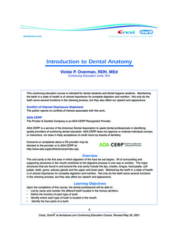
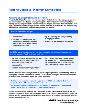
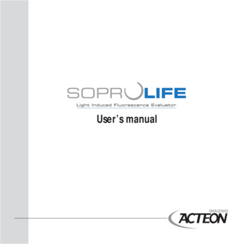
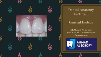
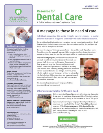
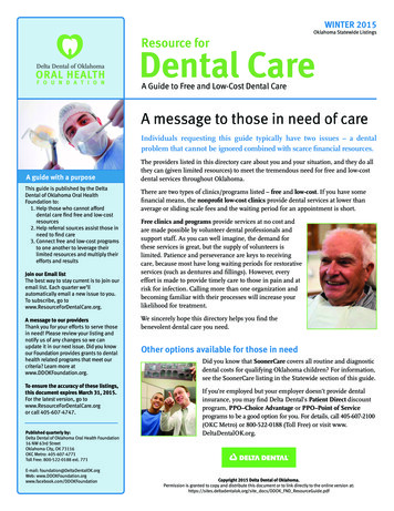
RESPIRATORY/DENTISTRYFig. 1.Dental diagram charting a diagonal bite 4 (DGL/4).breviated 0/202). During the mixeddentition period, unerupted molars arerecorded by circling the adult molar onthe dental diagram.B. The presence of deciduous dentition isannotated on the dental diagram by placing a single line through the adult toothnumber and writing in the appropriatedeciduous tooth number (e.g., 508).C. Supernumerary teeth and retained deciduous teeth are drawn on the diagramand appropriately annotated (e.g., SN111, not 112, and RD 503).D. An unerupted or partially erupted toothis usually impacted; therefore, a blindmaxillary right wolf tooth is abbreviatedTI 105.E. Dental malocclusions, fractures, cavities,and periodontal pockets are drawn onthe chart to approximate the outline ofactual finding and annotated in the exam-findings section.VIII. Malocclusions and other abnormal dentalfindings commonly effecting the incisors include the following:A. Diagonal bites are defined with respectto the mandibular incisors. DGL/3 is adiagonal bite in which the mandibularleft incisors are longer than the mandibular right incisors (Fig. 1). DGL/4 is adiagonal bite in which mandibular rightincisors are longer.B. Ventral curvature (CV) and dorsal curvature bites (CD) are the dental termsfor a smile and frown bite, respectively.C. Although overbites and underbites usually affect the entire dentition of a patient, these malocclusions are typicallyrecorded in the incisor part of the examfindings section as MAL2 or MAL3, respectively.D. Hooks on the maxillary third incisors area common finding (HK 103/203) (Fig. 2).E. Abnormal wear patterns or attritionsuch as that seen in cribbers is recordedFig. 2. Dental diagram charting hook malocclusions on the maxillary third incisors (HK 103/203).by describing the affected aspect of thetooth (e.g., cribbing attrition on the vestibular aspect of the maxillary first incisors is abbreviated AT 101V/201V).F. Crown fractures of the incisors should bedrawn on the dental chart and described(e.g., a crown fracture of the maxillaryright third incisor is abbreviated T/FX403CR). The extent of the fracture canbe further described using the tooth-fracture abbreviations (T/FX/) in AppendixA.G. Iatrogenic pulp damage secondary tooverreduction of the incisors with powerinstrumentation is a common finding. Exposed pulp is differentiatedbased on its vitality and recorded (e.g., aliving, bleeding pulp in the mandibularright third incisor is abbreviated T/PE/V203, whereas a necrotic, non-vital pulp inthe same tooth is abbreviated T/PE/NV203).H. Cavities (CA) should be staged accordingto severity.1. Stage 1: cavities in the cementumonly (CA1).2. Stage 2: cavities through the cementum and into the enamel (CA2).3. Stage 3: cavities involving the cementum, enamel, and dentin (CA3).4. Stage 4: cavities exposing pulp(CA4).I. Tooth resorption (TR; equine odontoclastic tooth resorption and hypercementosis[EOTRH]) should be classified using theAVDC classification (see TR in AppendixA).IX. Dental findings commonly affecting the canine teeth include:A. Tartar (calculus [CAL]) that may be associated with periodontal disease (discussed below).B. Blind canines in young males and mares(TI).AAEP PROCEEDINGS Ⲑ Vol. 56 Ⲑ 2010431
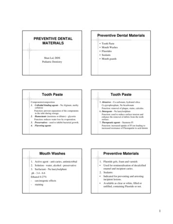
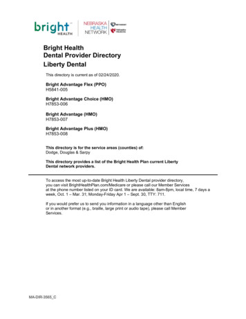
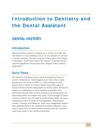
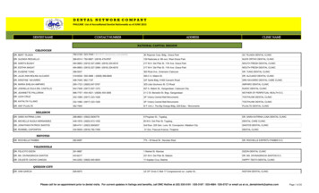
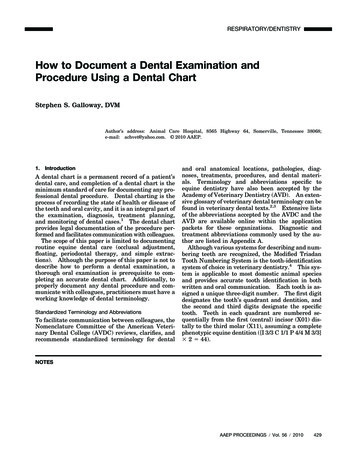
RESPIRATORY/DENTISTRYFig. 3. Dental diagram charting a typical cheek-tooth malocclusion pattern (HK 206/311, WV 308 –9, ATR 210/311).C. Vestigial canines commonly seen inmares. No dental abbreviation is recognized for this finding; therefore, the author uses a check-the-box format in theexam-findings section of the dental chartto record this finding.D. Cavities and tooth resorption are annotated as described for incisors.X. Dental findings commonly involving the wolfteeth include missing (0) and blind teeth(TI).XI. Dental malocclusions and findings commonly affecting the cheek teeth includehooks (HK), ramps (RMP), waves (WV),steps (STP), abnormal transverse ridges(ATR), hard enamel points (PTS), cuppedteeth (CUPD), and expired teeth (EXP).These findings are documented on the dental diagram by drawing the lateral profileof the cheek-tooth arcade onto the diagram. The individual tooth malocclusionsare clarified in the exam-findings section ofthe dental chart (Fig. 3).XII. Dental abnormalities affecting the occlusalaspect of the cheek teeth, such as fracturesand infundibular cavities, are best documented on an occlusal diagram such as theDuToit Equine Endodontic Numbering System Chart (Appendix C). The lesion isdrawn onto the chart and described both inthe occlusal chart margin and in the examfindings section of the dental chart.A. Occlusal fractures often fit into one of thefollowing categories:1. Chip: fracture involving only the occlusal margin (T/FX/CHIP).2. Wedge: fracture outside the infundibulae, involving one or more pulphorns (T/FX/WDG).3. Sagittal: fracture through the infundibulum; classically, through both infundibulae (T/FX/SAG) (Fig. 4).4. The AVDC has further divided toothfractures into seven classifications(See T/FX/ in Appendix A).B. Infundibular cavities (INF/CA) should bestaged according to severity:1. Stage 1: cavities in the infundibularcementum only (INF/CA1).2. Stage 2: cavities involving the infundibular cementum and infundibularenamel ring (INF/CA2).3. Stage 3: cavities involving the infundibular cementum, enamel, and dentin (INF/CA3).4. Stage 4: cavities through the infundibulae resulting in tooth fracture(INF/CA4). This staging is rarelyused, because the pathology is usuallydocumented as a sagittal fracture (T/FX/SAG).XIII. Periodontal disease should be noted on thedental diagram and described in the examination findings.A. Periodontal pockets should be probed,and their depths should be recorded (e.g.,a 15-mm deep periodontal pocket on thedistopalatal interproximal aspect of themaxillary left fourth premolar is abbreviated PP15 208IPD/P (Fig. 5).B. Teeth affected by periodontal diseaseshould be checked for mobility, and theindex should be recorded:1. M1: less than 1 mm movement inany direction.Fig. 4. Dental diagram charting a sagittal fracture of tooth 109 with a missing (vestibular) buccal slab, a grade 2 infundibular cavityin the mesial infundibulum of tooth 110, and cupping in tooth 111 (T/FX/SAG 109, 0 109/V, INF/CA2 110, CUPD 111).4322010 Ⲑ Vol. 56 Ⲑ AAEP PROCEEDINGS
RESPIRATORY/DENTISTRYFig. 5. Dental diagram charting a diastema and periodontalpocketing between the maxillary left third and fourth cheek teeth(DIA/PP15 208IPD).2. M2: less than 2 mm movement inany direction.3. M3: mov
4. Stage 4: cavities exposing pulp (CA4). I. Tooth resorption (TR; equine odontoclas-tic tooth resorption and hypercementosis [EOTRH]) should be classified using the AVDC classification (see TR in Appendix A). IX. Dental findings commonly affecting the ca-nine teeth include: A. Tartar (calculus [CAL]) that may be as-sociated with periodontal disease (dis-cussed below). B. Blind canines in .