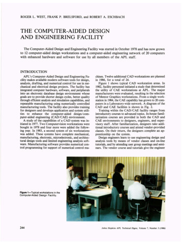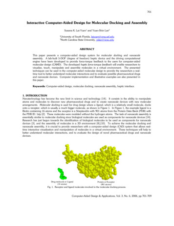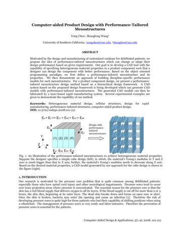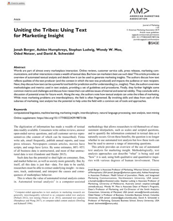
Transcription
Computer AidedDrug DesignDocking and other Virtual ScreeningMethods
Drug Design Lingo: Target: any macromolecule whose function can bemanipulated/altered to result in diseasetreatment! Ligand: compound (typically small molecule) thatmay bind to a target, with hopes of it serving as atreatment HTS: High Throughput Screening; experimentalmethod of assaying many (thousands) ofcompounds for activity Binding Mode: orientation of ligand in a bindingsite
Goals/objectives of CADD: Find/design ligands to bind/regulate targetmacromolecules
Goals/objectives of CADD: Find/design ligands to bind/regulate targetmacromolecules Virtual High Throughput Screening Thousands of ligands/1-2 targets Target Structure/Binding Site Prediction 1-2 targets Off-path Target Screening 10-20 ligands/10-20 targets Binding Mode Analysis/Prediction 10-20 ligands/1-2 targets
Goals/objectives of CADD: Find/design ligands to bind/regulate targetmacromolecules Virtual High Throughput Screening Thousands of ligands/1-2 targetsDocking,Pharmacophore Modeling Target Structure/Binding Site Prediction 1-2 targetsHomology Modeling, Binding Site Prediction Off-path Target ScreeningBinding Site Comparison, 10-20 ligands/10-20 targets Cross-Docking Binding Mode Analysis/Prediction 10-20 ligands/1-2 targetsFlexible Docking
An Example of CADD Success!Doman, T. N., et al. Molecular Docking and HighThroughput Screening for Novel Inhibitors of ProteinTyrosine Phosphatase-1B J. Med. Chem. 2002. 45,2213-2221
What functionalityare you lookingfor? What information do you have? What information do you want? Directory of Computer Aided DrugDesign Tools (very great link! cross referencesfunctionality by software!)c
What functionalityare you lookingfor? What information do you have? What information do you want? Directory of Computer Aided DrugDesign Tools (very great link! cross referencesfunctionality by software!)cYou know the structure of thetarget binding site!You know a ligandconfirmed to bind target!Saturday Morning!!
What functionalityare you lookingfor? What information do you have? What information do you want? Directory of Computer Aided DrugDesign Tools (very great link! cross referencesfunctionality by software!)cLet’s take 5 minutes, to all follow this link separately youmight find something that interests you!
Molecular Docking Simulations:Introduction and Tutorial
Docking What is it?
“In the field of molecular modeling, docking is a method which predicts the preferred orientation ofone molecule to a second when bound to each other to form a stable complex. Knowledge of thepreferred orientation in turn may be used to predict the strength of association or bindingaffinity between two molecules using, for example, scoring functions.” (from king (molecular)Lengauer T, Rarey M. Computational methods for biomolecular docking. 1996. 6(3), 402-406
“In the field of molecular modeling, docking is a method which predicts the preferred orientation ofone molecule to a second when bound to each other to form a stable complex. Knowledge of thepreferred orientation in turn may be used to predict the strength of association or bindingaffinity between two molecules using, for example, scoring functions.” (from Wikipedia)fun sunshineget into Docking (molecular)Lengauer T, Rarey M. Computational methods for biomolecular docking. 1996. 6(3), 402-406
Docking Goals:1. Identify false positives and falsenegatives before experimentalscreening (narrow compoundlibrary)2. Predict binding modes & relativebinding affinity3. Suggest possible successfulcompounds
Docking can predict binding modes as well as possible binders!Number ofCompoundLigand PubChem IDNumberDocking 09909190104361206398761 (Maxacalcitol)ChemSpider ID: 1466931135253652895489935197446912452895015288670 504-10.49947-10.07298-9.211291-8.99854-7.881713
There are many docking programs .1-Click DockingAADSADAMAutoDockAutoDock d ODOCKSOFTDockingSurflex-DockSwissDockVoteDockYUCCA
Why so many different docking programs? Protein, Ligands, etc. Ligand Whole molecule Fragment Based Protein Rigid DockingFlexible Receptor DockingSemi-flexible dockingFull Protein Flexible Docking Water / co-factors / metals Explicit Implicit Scoring Function EmpiricalForce FieldKnowledge BasedConsensus
The Specifics: What do we need to predict a binding mode?Ligand ofInterestConformationalSearch AlgorithmEnergetic ScoringFunctionPredictedBinding Mode
The Specifics: What do we need to predict a binding mode?may be a database ofligands .Ligand ofInterestConformationalSearch Algorithm Molecular DynamicsSimulations Genetic Algorithm Systematic Searching (i.e.,rotamer libraries and such)Energetic ScoringFunctionPredictedBinding Mode
AutoDock Vina: A Rigid, Grid-based Docking ProcedureVina represents shape and properties of the receptor as a gridof points, where each point in space is assigned a value in afield! (ligand flexible, protein rigid)(draw grid here on board)The non-bonded energetic terms of a docked ligand are then minimizedwithin this grid/field, rather minimized via explicit atom-atom calculations.
Is “rigid” the best model forreceptor/ligand interaction?
Is “rigid” the best model forreceptor/ligand interaction?
Results: “Canonical” Cross Docking Test Set
“Lock and key” implies some element of rigidity“Hand and glove” model implies receptor and ligand are both flexibleThus we need:
“Lock and key” implies some element of rigidity“Hand and glove” model implies receptor and ligand are both flexibleThus we need:
“Lock and key” implies some element of rigidity“Hand and glove” model implies receptor and ligand are both flexibleThus we need:Flexible ligand/flexible receptor docking!
Induced Fit Docking (IFD) in Schrödinger:Initial ligand docking with Glide SP (using reducedvdW radii, can mutate large side-chains to alanine)Prime (protein structure prediction tool) used foreach initial pose to predict multiple receptorconformationsGlide XP “redocking” into different receptorconformersGlideScore calculated and complexes ranked, XPdescriptors written
CHARMM-based Flexible Receptor DockingO’Boyle, N. M.; Vandermeersch, T.; FlProtein 2acr contrast 900dpi.pngynn, C. J.; Maguire, A. R.;Hutchison, G. J. Cheminf., 2011, 3, 8-15.Lee M. S.: Feig, M.; Salsbury, Jr. F. R.; and Brooks III. C. L. J. Comp. Chem., 2003, 24, 1348-1356.Suárez, M, P. T.; and Alfonso J. Syst. Synth. Biol., 2008, 2.3, 105-113.Wu, X., Brooks, B.R. J. Chem. Phys., 2011, 135, 204101.
CHARMM-based Flexible Receptor Docking
So is rigid docking useless? NO! Use it to identify falsepositives and false negativesbefore further screening! rigid docking is computationallyinexpensive narrow the library before usingexpensive tools Use flexible docking to predictbinding modes and affinities
Tutorial:Docking with AutoDock VinaAutoDock Vina Publication
1. Navigate to the PDBwebsite(http://www.rcsb.org/pdb)and search for the PDB structureId 1MVC)2. Click “Download Files PDBFormat”, this will downloadthe 1MVC structure (a humanRxR) with bound BMS649agonist.
This is PyRx, a GUI for AutoDock Vina!The Navigator Panel is where you canload and organize molecules for jobs.The View Panel is where you can viewmolecules, documents, plots andcharts! You can also make plots,documents and charts. The Controlssection has a Vina wizard, an AutoDockWizard, a Babel wizard, and a pythonshell, as well as an error log.1.From the top toolbar in PyRx, clickthe “Load Molecule” icon.
This is PyRx, a GUI for AutoDock Vina!The Navigator Panel is where you canload and organize molecules for jobs.The View Panel is where you can viewmolecules, documents, plots andcharts! You can also make plots,documents and charts. The Controlssection has a Vina wizard, an AutoDockWizard, a Babel wizard, and a pythonshell, as well as an error log.1.From the top toolbar in PyRx, clickthe “Load Molecule” icon.2.A ‘Finder’ window (or windowsequivalent) will open. Navigate tothe downloads folder, select“1mvc.pdb” to open.
This is PyRx, a GUI for AutoDock Vina!The Navigator Panel is where you canload and organize molecules for jobs.The View Panel is where you can viewmolecules, documents, plots andcharts! You can also make plots,documents and charts. The Controlssection has a Vina wizard, an AutoDockWizard, a Babel wizard, and a pythonshell, as well as an error log.1.From the top toolbar in PyRx, clickthe “Load Molecule” icon.2.A ‘Finder’ window (or windowsequivalent) will open. Navigate tothe downloads folder, select“1mvc.pdb” to open.3.The macromolecule is now loadedin the 3D Scene!
Now we need to modify the 1mvc.pdbfile so that we can have themacromolecule (1mvc) and the ligand(bms649) in separate pdb files.1.In the “View” Panel, select the“Documents” Tab.
Now we need to modify the 1mvc.pdbfile so that we can have themacromolecule (1mvc) and the ligand(bms649) in separate pdb files.1.In the “View” Panel, select the“Documents” Tab.2.Click the “Open” icon (a Folder),again a “Finder” window will open,select “1mvc.pdb”.
Now we need to modify the 1mvc.pdbfile so that we can have themacromolecule (1mvc) and the ligand(bms649) in separate pdb files.1.In the “View” Panel, select the“Documents” Tab.2.Click the “Open” icon (a Folder),again a “Finder” window will open,select “1mvc.pdb”.3.This is what it should look likeafter opening the 1mvc.pdf file indocuments!
Now we need to modify the 1mvc.pdbfile so that we can have themacromolecule (1mvc) and the ligand(bms649) in separate pdb files.1.In the “View” Panel, select the“Documents” Tab.2.Click the “Open” icon (a Folder),again a “Finder” window will open,select “1mvc.pdb”.3.This is what it should look likeafter opening the 1mvc.pdf file indocuments!4.Scroll to nearly the bottom of1mvc.pdb, looking for lines thatstart with the word “HETATM”The lines of interest are:HETATM 1773 O1 BM6 A HETATM 1800 C24 BM6 A Ctrl C (copy) these lines of thepdb file!
Modifying 1mvc.pdb (cont.)5. Make a new document, this will bethe BMS649 ligand file, by clicking the“New” Icon (looks like a piece ofpaper).
Modifying 1mvc.pdb (cont.)5. Make a new document, this will bethe BMS649 ligand file, by clicking the“New” Icon (looks like a piece ofpaper).6. Paste the copied BM6 lines into thisnew untitled document.
Modifying 1mvc.pdb (cont.)5. Make a new document, this will bethe BMS649 ligand file, by clicking the“New” Icon (looks like a piece ofpaper).6. Paste the copied BM6 lines into thisnew untitled document.7. Click the yellow floppy disk icon tosave the new document.
Modifying 1mvc.pdb (cont.)5. Make a new document, this will bethe BMS649 ligand file, by clicking the“New” Icon (looks like a piece ofpaper).6. Paste the copied BM6 lines into thisnew untitled document.7. Click the yellow floppy disk icon tosave the new document.8. Save this new document as‘bms649.pdb’.
Modifying 1mvc.pdb (cont.)5. Make a new document, this will bethe BMS649 ligand file, by clicking the“New” Icon (looks like a piece ofpaper).6. Paste the copied BM6 lines into thisnew untitled document.7. Click the yellow floppy disk icon tosave the new document.8. Save this new document as‘bms649.pdb’.9. Return to the ‘1mvc.pdb’ file, findthe BM6 lines again, and delete them,we are making a macromolecular filewithout the ligand in it.
Modifying 1mvc.pdb (cont.)5. Make a new document, this will bethe BMS649 ligand file, by clicking the“New” Icon (looks like a piece ofpaper).6. Paste the copied BM6 lines into thisnew untitled document.7. Click the yellow floppy disk icon tosave the new document.8. Save this new document as‘bms649.pdb’.9. Return to the ‘1mvc.pdb’ file, findthe BM6 lines again, and delete them,we are making a macromolecular filewithout the ligand in it.10. Click “Save As” (blue floppy-disk)icon, and while saving, rename the fileto “1mvc-mod.pdb” just to distinguishit from the original file downloadedfrom the PDB
Modifying 1mvc.pdb (cont. 2)11. Now, we need to remove theoriginal 1mvc.pdb from the NavigationPane, so that we can instead includethe separate macromolecule and ligandfiles.
Modifying 1mvc.pdb (cont. 2)11. Now, we need to remove theoriginal 1mvc.pdb from the NavigationPane, so that we can instead includethe separate macromolecule and ligandfiles.12. With a newly cleared NavigationPane, load 1mvc-mod.pdb andbms649.pdb into PyRx (as done in steps1-2). You can see, ligands (as well assome elements of the macromolecularstructure) will be represented in “balland-stick:, while the protein isrepresented in “lines”. If you toggledbetween structures in the NavigationPane (by checking and uncheckingboxes) you can verify that the ligandand protein are in fact in separate files.
Modifying 1mvc.pdb (cont. 2)11. Now, we need to remove theoriginal 1mvc.pdb from the NavigationPane, so that we can instead includethe separate macromolecule and ligandfiles.12. With a newly cleared NavigationPane, load 1mvc-mod.pdb andbms649.pdb into PyRx (as done in steps1-2). You can see, ligands (as well assome elements of the macromolecularstructure) will be represented in “balland-stick:, while the protein isrepresented in “lines”. If you toggledbetween structures in the NavigationPane (by checking and uncheckingboxes) you can verify that the ligandand protein are in fact in separate files.13. Right click on “1mvc-mod.pdb” inthe Navigation Pane. Select “AutoDock Make Make Macromolecule”
Modifying 1mvc.pdb (cont. 2)11. Now, we need to remove theoriginal 1mvc.pdb from the NavigationPane, so that we can instead includethe separate macromolecule and ligandfiles.12. With a newly cleared NavigationPane, load 1mvc-mod.pdb andbms649.pdb into PyRx (as done in steps1-2). You can see, ligands (as well assome elements of the macromolecularstructure) will be represented in “balland-stick:, while the protein isrepresented in “lines”. If you toggledbetween structures in the NavigationPane (by checking and uncheckingboxes) you can verify that the ligandand protein are in fact in separate files.13. Right click on “1mvc-mod.pdb” inthe Navigation Pane. Select “AutoDock Make Make Macromolecule”14. Right click on it in the navigationpane, select “AutoDock Make Ligand”
Modifying 1mvc.pdb (cont. 2)11. Now, we need to remove theoriginal 1mvc.pdb from the NavigationPane, so that we can instead includethe separate macromolecule and ligandfiles.12. With a newly cleared NavigationPane, load 1mvc-mod.pdb andbms649.pdb into PyRx (as done in steps1-2). You can see, ligands (as well assome elements of the macromolecularstructure) will be represented in “balland-stick:, while the protein isrepresented in “lines”. If you toggledbetween structures in the NavigationPane (by checking and uncheckingboxes) you can verify that the ligandand protein are in fact in separate files.13. Right click on “1mvc-mod.pdb” inthe Navigation Pane. Select “AutoDock Make Make Macromolecule”14. Right click on it in the navigationpane, select “AutoDock Make Ligand”15. Now everything is ready for redocking with AutoDock Vina!
Re-docking with AutoDock Vina!Re-docking is a technique in which aligand with an already known bindingmode in a binding site (such as fromsuccessful co-crystalization or otherstructural methods) is docked into thebinding site to verify that the dockingprocess can replicate the knownbinding mode.1. Click on the “Vina Wizard” in the“Controls” section below, and press the“Start” button in the lower right corner.
Re-docking with AutoDock Vina!Re-docking is a technique in which aligand with an already known bindingmode in a binding site (such as fromsuccessful co-crystalization or otherstructural methods) is docked into thebinding site to verify that the dockingprocess can replicate the knownbinding mode.1.Click on the “Vina Wizard” in the“Controls” section below, andpress the “Start” button in thelower right corner.2.AutoDock Vina will now promptyou to select a macromolecule anda ligand. Use the “ Add Ligand”and “ Add Macromolecule”buttons on the bottom left, makesure that bms649.pdbqt is loadedunder the “Ligands” tab, and“1mvc-mod” is loaded under themacromolecule tab. Select themby clicking on them. After makingsure the macromolecule andligands are selected properly, click“Forward” in the bottom rightcorner.
Re-docking with AutoDock Vina! (cont.)3. The next step is generating a grid forflexible ligand docking. In the 3D Sceneyou should now see a cube as well as a3D axis definition.Click and hold the white spheres thatborder the 3D axes to extend the sizeof the grid. For redocking, make surethat the grid encompasses the volumein which bms649 is known to bind.
Re-docking with AutoDock Vina! (cont.)3. The next step is generating a grid forflexible ligand docking. In the 3D Sceneyou should now see a cube as well as a3D axis definition.Click and hold the white spheres thatborder the 3D axes to extend the sizeof the grid. For redocking, make surethat the grid encompasses the volumein which bms649 is known to bind.4. The center of the receptor grid canalso be moved by click, holding, anddragging the center sphere on the 3daxes. Move the center of the receptorgrid to the center of bms649. Click“Forward” in the bottom right corneronce you have the grid appropriatelyplaced.
Re-docking with AutoDock Vina! (cont.2)5. Clicking “Forward” in the prior stepwill start the docking procedure!Output from the docking procedure willbe displayed in the “3D Scene” windowuntil the job is complete! You shouldhave two resulting predicted bindingmodes! One that nearly matches thecrystal structure alignment, and onethat has some rotation.
Docking with CHARMMing!https://www.charmming.org/charmming/
ProBiS: Protein Binding Site Comparison Compare your protein (3D)structure to all other known 3Dprotein structures in the PDB! ProBiS Ligands allows you to collectligands that bind in other similarbinding sites!
What can you do with ProBiS? Virtually search for ligands!ProBiS Ligands 30 compoundsPubChem 70 compoundsInitial Library104Glide SP35Glide XP25IFD25Number ofCompoundLigand PubChem IDNumberDocking 09909190104361206398761 (Maxacalcitol)ChemSpider ID: 1466931135253652895489935197446912452895015288670 504-10.49947-10.07298-9.211291-8.99854-7.881713
Navigate tohttp://probis.nih.gov. Thewebsite should look like theimage on the right.
Navigate tohttp://probis.nih.gov. Thewebsite should look like theimage on the right.1. Enter a PDB code (or youcan upload a PDB file) into thePDB ID box, and select a chainof interest (select A for now).Scroll down and hit “Search”.
Navigate tohttp://probis.nih.gov. Thewebsite should look like theimage on the right.1. Enter a PDB code (or youcan upload a PDB file) into thePDB ID box, and select a chainof interest (select A for now).Scroll down and hit “Search”.2. While searching you will getupdates about progress!
Navigate tohttp://probis.nih.gov. Thewebsite should look like theimage on the right.1. Enter a PDB code (or youcan upload a PDB file) into thePDB ID box, and select a chainof interest (select A for now).Scroll down and hit “Search”.2. While searching you will getupdates about progress!3. Once done you will getresults organized as on theright here!
Navigate tohttp://probis.nih.gov. Thewebsite should look like theimage on the right.1. Enter a PDB code (or youcan upload a PDB file) into thePDB ID box, and select a chainof interest (select A for now).Scroll down and hit “Search”.2. While searching you will getupdates about progress!3. Once done you will getresults organized as on theright here!4. Take a look at the PredictedLigands! ProBiS searches andfinds ligands that are likely tobind for you!!
PharmacophoreModeling Basics: Ligand Based vs. Structure BasedPharmacophore Models Pharmacophore model simplifiedrepresentation of interaction types screen ligands by comparing predictedpharmacophore models (very fast) Software: Pharmer, MedChem Studio,Phase ( 461000111X
Quantitative StructureActivity Relationship(QSAR) Basics: Regression models: predictor X leads toresponse Y In chemical modeling the predictor Xmight be some structural element (sidechains, physiochemical properties, etc.)and response might be predictedexperimental values (binding affinity,biological activity) using the regression model allows you toquickly predict values of interest withoutsimulation, just by ive structure–activity relationship
That’s all folks!
Computer Aided Drug Design Docking and other Virtual Screening . after opening the 1mvc.pdf file in documents! Now we need to modify the 1mvc.pdb file so that we can have the macro
