Transcription
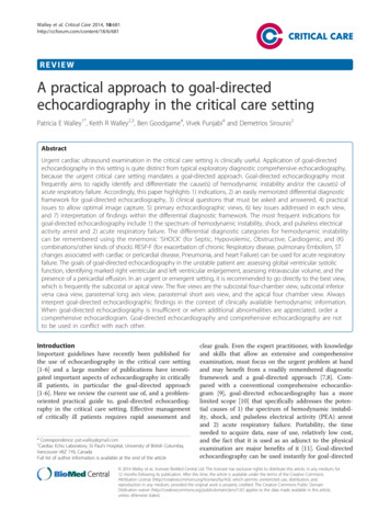
nTech ologylurna of VeteJo&ary SciencerinISSN: 2157-7579Journal of VeterinaryVeterinary Science &TechnologyGazi et al., J Veterinar Sci Technolo 2015, 6:3DOI: 10.4172/2157-7579.1000228Review ArticleOpen AccessAdvances of Echocardiography in Equines Practice - A ReviewGazi MA*, Makhdoomi DM, Abbas HY, Parrah JD, Ganai AM, Dar SH and BA MoulviDivision of Surgery and Radiology FVSc and A.H SKUAST-K, IndiaAbstractEchocardiography has become routinely procedure used in the diagnosis, management, and follow-up ofpatients with any suspected or known heart diseases. It is one of the most widely used diagnostic modality incardiology. It can provide a wealth of helpful information, including the size and shape of the heart, pumping capacity,and the location and extent of any tissue damage of heart. An Echocardiogram on other hand gives physician’sestimates of heart function such as a calculation of the cardiac output, ejection fraction, and diastolic function. It alsohelps to evaluate cardiac defects such as, atrial septal defects, AV valve stenoses, coronary artery defects, occultDCM, coronary artery disease, feline heartworm disease, persistent left cranial vena cava, canine hypertrophiccardiomyopathy and feline diastolic dysfunction may be identified. This Review details principle, uses, technique,types and limitations of Echocardiography in equines.Keywords: Heart; Horse; Ultrasound; EchoIntroductionEchocardiography is ultrasonography of heart. Equineechocardiography uses ultrasound technology to display images of ahorse’s heart. Echocardiography has emerged to become a standarddiagnostic procedure in equine cardiology. Development of M-modeechocardiography and introduction of 2D real-time echocardiographyallowed evaluation of internal cardiac structure, size and functionin horses. Development of Doppler echocardiography leads tothe ability to assess blood flow characteristics in the equine heart.Echocardiography is essential for diagnosing cardiac diseases in horsesas well as in other species [1]. The aim of this presentation is to givean overview on the general principles of equine echocardiography,indications for an echocardiographic examination, its clinicalrelevance, and current limitations of equine echocardiography [2].Echocardiography provides a substantial amount of structural andfunctional information about the heart. Still frames provide anatomicaldetail. Dynamic images tell us about physiological function. The qualityof an echo is highly operator dependent and proportional to experienceand skill, therefore the value of information derived depends heavilyupon who has performed it. Moreover, any deviation from the normalprocedure or protocol, leading to various complications, would attractlegal liability both at personal as well as institutional level.The idea of compiling this article was a typical case of Horse whichwas presented to our clinics with Unthriftyness, Jugular pulsation, fasterrespirations on work, Sudden falling while pulling Tonga, Crossed legs(Figure 1), dehydrated and loss of condition. The animal was put toECG at Clinical complex revealing heart ventricular enlargement.indices of cardiac function (e.g. fractional shortening and fractionalarea change) will be altered [4,5].Imaging modes used in echocardiographyReal time two-dimensional (2D) or B-mode (“brightness” mode),M-mode (motion mode), color flow Doppler, pulsed-wave (PW)Doppler, and continuous-wave (CW) Doppler are some of the possiblemodes that are employed for the procedure. M-mode and 2D equineechocardiography allow users to see the heart structure and function[6]. The structures nearest to the transducer are displayed at the topof the screen, the dorsal (in the long axis views) and cranial (in theshort-axis views) structures of the heart, respectively, are displayed toFigure 1: Animal presented to clinics with crossed leg appearance.Principles of Equine EchocardiographySedation may or may not be required prior to Echocardiographyin animals. The horse should not be sedated unless his behavior isuncooperative. Sedation can manipulate dimension and blood flowresults, leaving only the structural data to be legitimate. Second,the horse’s size, anatomical build of the thorax and the type ofechocardiography equipment used can affect image results [3]. Fourviews of the heart are taken-a long and short view from both the rightand left pasternal areas. While structural information is still valid in asedated horse, some cardiac dimensions (e.g. endsystolic left ventriculardiameter, interventricular septal thickness, and free wall thickness) andJ Veterinar Sci TechnoloISSN: 2157-7579 JVST, an open access journal*Corresponding author: Gazi MA, Division of Surgery and Radiology FVSc andA.H SKUAST-K, India, E-mail: mohsingazi9975@gmail.comReceived January 06, 2015; Accepted April 28, 2015; Published April 30, 2015Citation: Gazi MA, Makhdoomi DM, Abbas HY, Parrah JD, Ganai AM, et al. (2015)Advances of Echocardiography in Equines Practice - A Review. J Veterinar SciTechnol 6: 228. doi:10.4172/2157-7579.1000228Copyright: 2015 Gazi MA, et al. This is an open-access article distributed underthe terms of the Creative Commons Attribution License, which permits unrestricteduse, distribution, and reproduction in any medium, provided the original author andsource are credited.Volume 6 Issue 3 1000228
Citation: Gazi MA, Makhdoomi DM, Abbas HY, Parrah JD, Ganai AM, et al. (2015) Advances of Echocardiography in Equines Practice - A Review. JVeterinar Sci Technol 6: 228. doi:10.4172/2157-7579.1000228Page 2 of 5the right side of the screen Doppler ultrasonography works similarlyto Doppler weather radar by showing direction, volume, and speed offluid movement [7].There are 3 types of echocardiography used clinically: M-mode, twodimensional (2-D, B-mode or real time) and Doppler echocardiography.The different types of echocardiography are routinely used in eachechocardiographic examination and the findings from one type ofechocardiographic examination compliments those from the otherportions of the examination. Simultaneous electrocardiography shouldbe performed during the echocardiographic examination.M-mode echocardiographyThe M-mode echocardiogram yields a one-dimensional view of thecardiac structures moving over time. The echoes from various tissueinterfaces along the axis of the beam are moving during the cardiaccycle and are swept across time, providing the dimension of time.The lines on the recordings correspond to the position of the imagedstructures in relation to the transducer and other cardiac structures atany instance in time. More accurate placement of the M-mode cursorwithin the heart is performed by using the two-dimensional (2-D)real-time image as a guide. The M-mode echocardiogram uses a highsampling rate and can yield cleaner images of cardiac borders, allowingthe echocardiographer to obtain more accurate measurements ofcardiac dimensions and more critically evaluate cardiac motion [5].Careful placement of the M-mode beam at the appropriate locationswithin the heart and obtaining clean echoes of endocardial surfaces arecritical to obtain accurate measurements and to make the calculationsperformed from these measurements, meaningful. Standard M-modeviews are obtained from the right parasternal position. The M-modecursor should be positioned within the heart using the right parasternalshort axis view, to avoid inclusion of a papillary muscle within the leftventricular free wall thickness. The standard M-mode views utilizedin veterinary medicine include the left ventricle (at the level of thechordae tendineae), the mitral valve and the aortic root (aorta/ leftatrial appendage) view.Two-dimensional echocardiographyTwo-dimensional echocardiography allows a plane of tissue(both depth and width) to be imaged in real time. Thus, the anatomicrelationships between various structures are easier to appreciatethan with M-mode echocardiographic images. An infinite numberof imaging planes through the heart are possible; however, standardviews are used to evaluate the intra and extra cardiac structures [8].The standard views are obtained from either the right parasternalwindow in all species and from the left parasternal window in adultlarge animals or in other species when imaging the heart from the leftside is desirable. Occasionally, images are obtained from subxiphoid(subcostal) or thoracic inlet (suprasternal) positions. These views areusually only feasible to obtain in small animals or young large animals.The standard views include the right parasternal long axis viewsof the 4 chambers (4 chamber view), left ventricular outflow tract, andright ventricular outflow tract and the short axis views perpendicularto this plane (left ventricle at the chordal level, mitral valve, and aorta/left atrial appendage). In large animals left parasternal long axis viewsof the mitral valve, aorta and pulmonary artery are also obtained whenindicated.Doppler echocardiographyDoppler imaging allows evaluation of blood flow patterns,J Veterinar Sci TechnoloISSN: 2157-7579 JVST, an open access journaldirection, and velocity; thus, it permits documentation andquantification of valvular insufficiency or stenosis and cardiac shunts.Estimations of blood flow and cardiac output can also be made.Doppler echocardiography is based on detection of frequency changes(the Doppler shift) occurring as ultrasound waves reflect off individualblood cells moving either away from or toward the transducer [9].Calculation of blood flow velocity is possible when the flow is parallelto the angle of the ultrasound beam. Since calculations becomeincreasingly inaccurate as the angle of incidence of the ultrasoundbeam and the path of blood flow diverges from 0 degrees, measurementof maximal blood flow velocity requires that the ultrasound beam be asclose to parallel with the path of blood flow as possible.Doppler echocardiography uses color to map the blood flow inthe heart. Doppler echo is used to determine whether the blood flowis too fast in certain areas, such as with subaortic stenosis or pulmonicstenosis (congenital heart defects that cause narrowing of the openingsthat blood flows through) [7,10]. Doppler echo can determine theseverity of the defect by measuring the velocity of blood flowingthrough these narrowed areas. Doppler echo also can detect holes inthe wall of the heart, such as occur with ventricular and atrial septaldefects. Doppler echo can also detect leakage of the heart valves [11].Three-dimensional (3D) echocardiography is available on some echomachines. It gives an accurate image of the heart but currently is usedmainly for teaching purposes.Two types of Doppler echocardiography are used clinically:pulsed wave and continuous wave. Pulsed wave (PW) Doppler usesshort bursts of ultrasound transmitted to a point (designated the“sample volume”) distant from the transducer [8]. The advantage ofthis type of Doppler is that blood flow velocity, direction and spectralcharacteristics from a specified point in the heart or blood vessel canbe calculated. The main disadvantage is that the maximum velocitythat can be measured is limited because the pulse repetition frequencyis limited. Continuous wave (CW) Doppler uses dual crystals so thatultrasound waves can be simultaneously and continuously sent andreceived. There is no maximum measurable velocity with CW so highvelocity flows can be measured. The disadvantage with CW Doppler isthat sampling of blood flow velocity and direction occurs all along theultrasound beam, not in a specified area [9].Color flow Doppler echocardiography is a form of PW Dopplerultrasonography which combines the M-mode and 2-D modalities withblood flow imaging. With color flow Doppler, multiple sample volumesare analyzed along multiple scan lines. The mean frequency shiftobtained from these many sample volumes is color-coded for directionand velocity. Several types of mapping are usually available. Mostsystems code blood flow toward the transducer as red and flow away asblue. Differences in relative velocity of flow can be accentuated, and thepresence of multiple velocities and directions of flow (turbulence) canbe indicated by different maps which utilize variations in brightnessand color [12].Contrast echocardiographyContrast echocardiography, or Contrast-enhanced ultrasoundis the addition of ultrasound contrast medium, or imaging agent, totraditional ultrasonography. The ultrasound contrast is made up oftiny microbubbles filled with a gas core and protein shell. This allowsthe microbubbles to circulate through the cardiovascular systemand return the ultrasound waves creating a highly reflective image.The most commonly used types of ultrasound contrast are known asDefinity , Optison . Both have been approved by the FDA. There areVolume 6 Issue 3 1000228
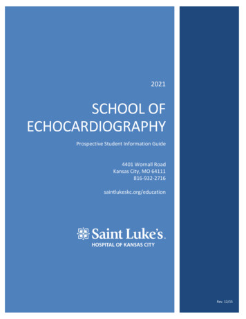
Citation: Gazi MA, Makhdoomi DM, Abbas HY, Parrah JD, Ganai AM, et al. (2015) Advances of Echocardiography in Equines Practice - A Review. JVeterinar Sci Technol 6: 228. doi:10.4172/2157-7579.1000228Page 3 of 5multiple applications in which contrast-enhanced ultrasound can byuseful. The most commonly used application is in the enhancement ofLV endocardial borders for assessment of global and regional systolicfunction. Contrast may also be used to enhance visualization of wallthickening during stress echocardiography, for the assessment of LVthrombus, or for the assessment of other masses in the heart. Contrastechocardiography has also been used to assess blood perfusionthroughout myocardium in the case of coronary artery disease.Indications and clinical use of echocardiography in horsesEchocardiography is performed when heart disease is suspected orchest radiographs (x-rays) show that the heart is enlarged [13,14] . Theechocardiogram (echo) shows the size of the heart chambers and howwell the left side of the heart is functioning. It shows whether the heartvalves are normal or thickened.An echo can detect the presence of extra fluid in the pericardial sacaround the heart and sometimes the presence of tumors in the heartthat are causing the extra fluid. Although heart murmurs may indicatevalvular disease or congenital malformations, they are also found in alarge number of clinically normal horses and foals. Horses can sufferfrom congenital cardiac malformations, valvular disease, myocardialdisease, other aquired cardiac defects, and cardiac arrhythmias. Heartmurmurs are frequently detected on cardiac auscultation in horses.Differentiation of physiologic (functional) from pathologic heartmurmurs can be difficult or impossible based on physical examinationand auscultation alone. Cardiac arrhythmias occur frequently in horses,either as primary disorders of impulse generation and conduction,or secondary to underlying structural cardiac disease. Detection ofany underlying cardiac disease may have important implications onprognosis and treatment of arrhythmias. Occasionally, cardiac diseasecan lead to very unspecific clinical signs such as poor performance orfever.Echocardiography can be used to identify a cardiac disease, makethe correct anatomical diagnosis, assess hemodynamic consequences,provide important prognostic data, monitor progression of the disease,and identify complications of a known diagnosis [15]. Evaluation of heartmurmurs, assessment of clinical significance of pathologic murmurs,dysrhythmias, detection of underlying cardiac disease, suspectedcongenital defects, evaluation of heart murmurs, unexplained cyanosis,or signs of heart failure in neonates [16,17], exercise intolerance /poor performance, detection of cardiac disease, muffled heart sounds,detection of pericardial effusion , fever of unknown origin, detectionof endocarditis, unexplained collapse / episodic weakness, detectionof cardiac disease, clinical signs of congestive heart failure, causeof heart failure, assessment of severity, monitoring of progressionand response to treatment severe respiratory disease. Detection ofpulmonary hypertension, detection of patent foramen ovale in foalswith respiratory disease form the indications of echocardiography.The Two-dimensional examination reveals enlargement ofthe left ventricle and left atrium. It is still uncertain as to howfrequently the ductus arteriosus itself can be visualized. The M-modeEchocardiographic study does not increase our understanding ofthis disorder. On the M-mode examination the measured thicknessof the interventricular septum is increased as well as that of the rightventricular free wall. The motion of the interventricular septum isnoted to be abnormal demonstrating a flat motion in systole [18].The Doppler Echocardiographic study reveals the presence ofturbulence in the main pulmonary artery which occurs in both systoleJ Veterinar Sci TechnoloISSN: 2157-7579 JVST, an open access journaland diastole. This picture of turbulence in the main pulmonaryartery is similar to that seen with pulmonic stenosis, however withpulmonic stenosis this pattern of turbulence is noted only in systole.Furthermore, the Doppler Echocardiographic examination is ideal toidentify the co-existence of other congenital cardiac disorders [18]. TheDoppler Echocardiographic study yields data which usually definitivelyestablishes the diagnosis of aortic stenosis and provides evidence of theseverity of the disorder. Doppler Echocardiography determines thevelocity of blood flow as it exits the left ventricle. The normal maximalvelocity of blood flow exiting the left ventricle is approximately 1.5meters per second. Thus velocities detected in excess of 1.5 m/s suggeststenosis of the column of blood flow (especially velocities in excess of2.0 m/s) [19].Echocardiography is of outstanding value to confirm a presumptivediagnosis of aortic stenosis or subaortic stenosis. In this disorder,short of cardiac catheterization, there is no other method available toconfirm the existence of aortic stenosis [19]. Although routine Twodimensional Echocardiography may give us clues to the existence ofaortic stenosis, in many cases this test is inconclusive. In severe cases ofaortic stenosis, the Two-dimensional exam may reveal left ventricularconcentric hypertrophy and a discrete subvalvular lesion, in the case ofsubaortic stenosis. The M-mode Echocardiographic examination mayreveal a clue to the presence of co-existent aortic valve insufficiency(diastolic fluttering of the anterior leaflet of the mitral valve) prematureclosure of one cusp of the aortic valve. As well this modality shouldindicate evidence of left ventricular concentric hypertrophy [12].Preparation of AnimalLittle preparation is needed for an echo examination. A few animalsneed to be tranquilized, but most do not. Aggressive cats that cannot behandled easily when awake may require general anaesthesia or sedationto perform this procedure. It is best not to feed your animal on themorning of the procedure, just in case sedatives are needed. In orderto get the best possible contact between the echo transducer and theskin, the hair is usually shaved on both sides of the chest [20]. Animalswith thin hair coats may not be clipped, but if the hair is not clipped,the quality of the echo image may not be sufficient to make a diagnosis.Most animals are required to lie on their sides for this procedure. Moreexperienced ultrasonographers may perform the examination with theanimal standing or sitting up. The procedure is done in a quiet room,with a minimal amount of stress to the animal [21].Description of TechniqueEchocardiography is a type of ultrasound examination. All typesof ultrasounds bounce sound waves off an object and record thereturning sound waves [22]. Special probes are placed on the animal’schest. These probes send and receive the sound waves or echoes [23].The echo machine converts these sounds waves into images of theheart. It takes special training and months of experience to becomeproficient in performing echocardiograms (Figure 2). Several typesof echocardiography exist and may be performed in sequence. Twodimensional (2D) echocardiography shows the heart as it is moving,as well as the inner chambers and outer walls of the heart. Echo in 2Dallows gross (major) abnormalities to be detected and identifies areasof the heart to be examined more closely [3]. Tumors, extra fluid in thepericardial sac, and clots in the heart can be found with this technique.Abnormal heart rhythms (arrhythmia) can be identified, and mostecho have built-in electrocardiographic capability, so the arrhythmiacan be examined or recorded during the echo procedure [24].Volume 6 Issue 3 1000228
Citation: Gazi MA, Makhdoomi DM, Abbas HY, Parrah JD, Ganai AM, et al. (2015) Advances of Echocardiography in Equines Practice - A Review. JVeterinar Sci Technol 6: 228. doi:10.4172/2157-7579.1000228Page 4 of 5procedure with no long-term or short-term side effects. The onlypotential complications are those that may arise post administrationof tranquilizers, sedatives, or general anesthesia. Side effects of thesedrugs are uncommon. Any carelessness in any of these procedureswould give rise to various kinds of legal liabilities [27]. Such liabilitycreates both personal as well as institutional liability. In such cases,both the doctor as well as the institution (hospital) is held liable. Thisis a matter of grave concern particularly in relation to equines becausethey are a source of income to a large number of families in Jammuand Kashmir. Such negligence in post procedure treatment may alsoamount to cruelty of animals and would attract criminal prosecutionas well.ReferencesFigure 2: Preparing a horse for echocardiography.Once the heart has been examined with 2D echo, an area is selectedfor examination with M-mode echo. This form of echocardiographyis used to measure the chambers of the heart and to determinehow well the left heart is functioning. Because M mode freezes themotion of the heart, it makes measuring the different areas easier.M-mode measurements must be done properly, because in accuratemeasurements can underestimate or overestimate problems.Stress EchocardiographyA stress echocardiogram, also known as a stress echo or SE, utilizesultrasound imaging of the heart to assess the wall motion in response tophysical stress. First, images of the heart are taken “at rest” to acquire abaseline of the patient’s wall motion at a resting heart rate. The patientthen walks on a treadmill or utilizes another exercise modality toincrease the heart rate to their target heart rate. Finally, images of theheart are taken “at stress” to assess wall motion at the peak heart rate.A stress echo assesses wall motion of the heart; it does not, however,image the coronary arteries directly. Ischemia of one or more coronaryarteries could cause a wall motion abnormality which could indicatecoronary artery disease (CAD) [17]. The gold standard test to directlyimage the coronary arteries and directly access for stenosis or occlusionis a cardiac catheterization. A stress echo is a non-invasive test and isperformed in the presence of a licensed medical professional, such as acardiologist, and a cardiac sonographer.Limitations and Potential ComplicationsEchocardiography is a well-established diagnostic method inequine medicine, and allows for diagnosis of heart disease, assessmentof cardiac function, and better understanding of the normal physiologyof the heart. Animal size, anatomical characteristics of the equinethorax, and technical features of (human) echocardiography equipmentsometimes limit the ability to image the equine heart [25]. While thisis usually less problematic for standard 2D imaging, determinationof blood flow velocities using Doppler imaging techniques is oftendifficult, due to the inability to achieve adequate alignment withblood flow [21]. Alignment for Doppler echocardiographic studiesof left ventricular inflow and outflow can be improved by usingtransesophageal echocardiography [26]. Although this technique hasbeen described in horses, it is not routinely used in a clinical setting.As echocardiographic technology advances, it may be possible thatthis technique will be applied for the evaluation of cardiac functionin resting and exercising horses [7]. Echocardiography is a very safeJ Veterinar Sci TechnoloISSN: 2157-7579 JVST, an open access journal1. Blissitt KJ, Marr CM, Rossdale PD, Green (1995) RE: Equine CardiovascularMedicine. Equine Veterinary Journal, Suppl. 19.2. Boon JA: Manual of Veterinary Echocardiography. Williams &Wilkins,Baltimore, 1998.3. Schwarzwald CC Schober KE, Berli AS, Bonagura JD (2009) Left ventricularradial and circumferential wall motion analysis in horses using strain, strainrate, and displacement by 2D speckle tracking. J Vet Intern Med 23: 890-900.4. Wideman RF Jr, Wing T, Kirby YK, Forman MF, Marson N, et al. (1998)Evaluation of minimally invasive indices for predicting ascites susceptibility inthree successive hatches of broilers exposed to cool temperatures. Poult Sci77: 1565-1573.5. Wu Y, Tobias AH, Bell K, Barry W, Helmes M, et al. (2004) Cellular andmolecular mechanisms of systolic and diastolic dysfunction in an avian modelof dilated cardiomyopathy. J Mol Cell Cardiol 37: 111-119.6. Schwarzwald CC, Hamlin RL, Bonagura JD, Nishijima Y, Meadows C, et al.(2007) Atrial, SA nodal, and AV nodal electrophysiology in standing horses:normal findings and electrophysiologic effects of quinidine and diltiazem. J VetIntern Med 21: 166-175.7. Jawad IA (1996) A Practical Guide to Echocardiography and Cardiac DopplerUltrasound. 2nd Edn., Lippincott Williams and Wilkins, Boston, pp: 432.8. Ohmer JK, Seward JB, Tajik AJ (1994) The echo manual: Little, Brown andCompany pp 24.9. Roldan CA (2005) The ultimate echo guide: Lippincott Williams & Wilkins pp 22.10. Goldstein DS, Brush JE Jr, Eisenhofer G, Stull R, Esler M (1988) In vivomeasurement of neuronal uptake of norepinephrine in the human heart.Circulation 78: 41-48.11. Rebecca E. Gomp (2000) Paso Robles Veterinary Medical Clinic 805; 2384622.12. Tempkin BB (1999): Ultrasound scanning: principles and protocols. 2nd edn.USA: WB Saunders Company.13. Hunsaker WG (1971) Round heart disease in four commercial strains ofturkeys. Poult Sci 50: 1720-1724.14. Liao R, Nascimben L, Friedrich J, Gwathmey JK, Ingwall JS (1996) Decreasedenergy reserve in an animal model of dilated cardiomyopathy Relationship tocontractile performance. American Journal Circulation Research. 78: 893-902.15. Patteson MW (1996): Equine Cardiology. Blackwell Science, Oxford.16. Gwathmey JK, Kim CS, Hajjar RJ, Khan F, DiSalvo TG et al. (1999) Cellularand molecular remodeling in a heart failure model treated with the beta-blockercarteolol. American. Journal of Physiology. Heart Circulation. Physiology, 276:H1678-H1690.17. Mandinov L, Eberli FR, Seiler C, Hess OM (2000) Diastolic heart failure.Cardiovasc Res 45: 813-825.18. Sutton MG, Oldershaw PJ, Kotler MN (1996): Textbook of echocardiographyand Doppler in adults and children: Blackwell Science.19. Sokolow M, Mciiroy MB (1986): Clinical cardiology, 2nd edn: Appleton CenturyCrofts.20. Otto CM: (2000) Textbook of Clinical Echocardiography. W.B. Saunders,Philadelphia.Volume 6 Issue 3 1000228
Citation: Gazi MA, Makhdoomi DM, Abbas HY, Parrah JD, Ganai AM, et al. (2015) Advances of Echocardiography in Equines Practice - A Review. JVeterinar Sci Technol 6: 228. doi:10.4172/2157-7579.1000228Page 5 of 521. Bonagura JD, Herring DS, Welker F (1985) Echocardiography. Vet Clin NorthAm Equine Pract 1: 311-333.22. Reef VB: (1998) Equine Diagnostic Ultrasound. W.B. Saunders, Philadelphia.25. Liao R, Charles M, Gwathmey JK (1997) Animal models of cardiovasculardisease for pharmacologic drug development and testing: Appropriateness ofcomparison to the human disease state and pharmacotherapeutics. AmericanJournal of Therapeutics. 4: 149-158.23. Nesheim MC, Austic RE, Card LE (1990) Poultry Production. 12th Edn., Leaand Febiger, Philadelphia. pp 325.26. Vlahović A, Popović AD (1999) [Evaluation of left ventricular diastolic functionusing Doppler echocardiography]. Med Pregl 52: 13-18.24. Marr C (1999) Cardiology of the Horse. W.B. Saunders Company, London.27. Bangia RK (2010) Law of Torts, Universal Law Publication, New Delhi. p. 185.J Veterinar Sci TechnoloISSN: 2157-7579 JVST, an open access journalVolume 6 Issue 3 1000228
echocardiography uses ultrasound technology to display images of a horse’s heart. Echocardiography has emerged to become a standard diagnostic procedure in equine cardiology. Development of M-mode echocardiography and introduction of 2D real-time echocardiography allowed evaluation of