Transcription
LASIK:BASIC PRINCIPLES INSURGICAL TECHNIQUEGregory J. Pamel, M.D.Pamel Vision and Laser Group Clinical Assistant ProfessorNew York University School of MedicinePamel Vision and Laser GroupESCRS Course 2014London
Lasik over the last 14 years:US Experience 2001-2003 Wavefront guided lasik2003-2004 Wavefront optimized lasik2004-2005 Intralase- “bladeless lasik”2004- Phakic IOLs receive FDA approval2005-present Resurgence of Surface Ablation2005-present Ectasia and Presbyopic IOLs.2007-present Crosslinking for post lasik ectasia2008-present further decrease in indications forlasik.
Lasik over the last 14 years:U.S. Experience cont’d 2010 No. of people undergoing lasik in U.S.levels off at 600-700K (down from peak of1.5M) 2011-2014 New technologies to be used incombination with lasik: prebyopic inlays,and LasikXtra Number of websites reporting the negativeeffects of lasik has increased.
Trends in Refractive Surgery PRK is accounting for at least 20-25% of laservision correction procedures as surgeons havebecome more vigilant about avoiding lasik incertain types of patients. Lasik market has been declining as Presbyopicmarket (baby boomers) is increasing-more andmore patients are having presbyopic correctingIOLS instead of Lasik. Phakic IOLs are being used for lower levels ofmyopia (-6D) as an alternative to Lasik. Identifying risk factors for ectasia
LASIK and the Media The lasik procedure has been unfairlyportrayed negatively in the U.S. mediaincluding on October 3, 2013 on the Dr. OZshow. Nationally syndicated talk show with over 3million viewers per day. Embed Link here
LASIK and the Media Falsely claimed that ophthalmologists donot undergo lasik surgery themselves. Guy Kezerian, M.D. took an informalsurvey of lasik surgeons in the U.S. and61% responded that they had undergone theprocedure themselves. Eye Care specialists are among the highestnumber of healthcare professionals who’veundergone lasik.
Media Distortions Also claimed based on the FDA reporteddata from the Wavelight Clinical trial that1/5 (20%) people saw worse after theprocedure than before. The data reported was that 81% had UCVAbetter or equal to BCVA with no nomogramadjustments or enhancements.
Trends in 2014 Improvement in technology has madelasik a safer procedure with betteroutcomes. Lasik volume has decreased because ofdemographic and economic reasons. Negative reporting of lasik hasincreased.
Pre-Operative PreparationPatient Selection is one of the mostimportant factors in insuring success ofthe lasik procedure.
Patient Selection Turning down a potential candidate couldbe the best decision you ever made. Most importantly, the surgeon mustdiscuss with the patient his expectationsand goals for the surgery as part of thescreening process and informed consent.This is especially important for presbyopes!
Patient Selection Unrealistic expectations is an absolutecontraindication to undergoing lasik. As the surgeon you may develop your ownparameters that may differ from publishedindications and guidelines.
Patient SelectionGeneral guidelines: Patient Age- 18yrs of age or older (FDA); 21 yrsof age or older (AAO guidelines). Children may benefit from lasik surgery-highametropes or anisometropia Refractive stability defined as 0.50D or lesschange in sphere or cylinder for at least 1 yearprior to the procedure.
Patient SelectionGeneral guidelines: Degree of refractive error- approved up to -14D ofmyoia; 6D of hyperopia; and up to -6D ofastigmatism. Most surgeons will not treat up to theselevels because the quality of vision isreduced, complication rates increase and becausethere are better procedures available.
Patient SelectionGeneral Guidelines: Lasik surgeons should be familiar with allthe available technologies to treat refractiveerrors. The indications for lasik havenarrowed as our experience has increased. Patients with a predisposition to ocularinjury: athletes, boxers etc. will often bebetter PRK/LASEK candidates.
Patient Selection:2014 Refractive surgery involves different technologies fordifferent candidates. LASIK: -1D to -9D depending on the CT for myopia andup to 4.50for hyperopia Advanced Surface Ablation: For thin corneas and irregularcorneas with the use of mitomycin C Phakic IOLs: For myopia -6D –reversible technology butusually for those pts / -9.0D Premium IOLs (multifocals/accomodating): For thoseolder hyperopes ( 50 yrs and / 3.00D) and those whodesire good uncorrected distance and near vision.
PatientSelection Poor EpithelialAdhesion/EBMD Corneal Disorders:Contraindications Lattice- GranularDystrophy Fuchs Dystrophy Steep ( / 46D) or Flat ( / 40D) Corneas-finalpostop Ks should bebetween 34D and 49D
Patient Selection Vascularized CorneasExposure KeratopathyHerpetic Eye DiseaseGlaucomaCataractStrabismusDry Eyes andAllergies
Patient Selection Autoimmune diseasesincluding RA, SLE,Wegener’s, Sjogrens. Uncontrolled CVD absolutecontraindication which canlead to thinning, melts andirregular healing. A number of publicationshave reported no seriouscomplications in pts. withwell controlled CVD.Collagen Vascular Disease-RA
Patient Selection Collagen Vascular Diseases: Patients with RA predisposed to dry eye andcan develop severe dry eye after lasik. Lasik should not be performed in thesepatients.
Patient SelectionAmiodaroneVericillata HIVDiabetesHx of Keloid formationPregnancy and NursingCertain medications:Isoretinoin (Accutane);Amiodarone (Codarone);Sumatriptan (Imetrex)
Patient Selection:Relative Contraindications to LASIKLarge Pupils: Some studies have found no correlation of pupilsize with subjective postop glare after 6 months(Schallhorn et al; Ophthalmology 2003; Pop, M etal Ophthalmology 2004). Others (Trattler et al) have reported a definitecorrelation between increased glare usingobjective glare testing and pupil size. Wavelight laser offers 6.5 and 7.0 mm OZ. Informed Consent
Preoperatively Screen Patients:Relative Contraindications Exposure Keratopathy Ocular SurfaceDisease K Sicca
Dry Eye Disease and Lasik 10-20% of post-lasik patients may sufferfrom chronic dry eye lasting greater than 6months. Dry eye symptoms peak at 1 week to 3months after lasik. FS flaps result in less dry eye disease thanmicrokeratome flaps. Greater loss of corneal sensitivity seen withlasik vs advanced surface ablation.
Ocular Surface Disease-Dry EyeDiseaseRisk Factors Preop Schirmer scores below 10mm Contact lens wear Older Age Females Atopic disease/Ocular Allergies
Ocular Surface Disease-Dry EyeDiseaseCauses of Tear Dysfunction Post Lasik: Loss of Goblet cells Neurotropic Epitheliopathy Changes in the corneal contour lead to abnormaltear distribution. Ablation depth Transection of corneal nerves with loss of cornealinnervation.
Ocular Surface Disease-Dry EyeDisease Characteristics of flap hinge includingwidth and location. Flap hinge located horizontally wouldpreserve the long posterior ciliary nervesand should preserve postop cornealsensation. Studies report conflicting results. Flap thickness and flap hinge angle notassociated with dryness.
Pre-op Evaluation of the OcularSurface SchirmersTBUTOcular surface stain Lissamine greenAssessing Meibomian glandsTear OsmolarityMMP-9 testing
Pre-op Management of theOcular Surface Artificial tearsTopical CyclosporineTopical SteroidsNutritional Supplementation- Omega 3sPunctal plugsToda et al. AJO 2014: 616-622
Pre-op/Post-op Management ofthe Ocular Surface Topical Cyclosporine 0.05% emulsion(Restasis ) may increase recovery ofcorneal sensitivity. Peyman et al JRS 2008 May also promote faster visual recoveryand refractive stability after lasik and PRK.Salib et al JCRS 2006
Other Treatment Modalitiesfor Dry Eye Autologous Serum Tears* Lipid Flow device Intense Pulsed Light Therapy* Noda-Tsuruya T et al JRS 2006
Patient SelectionAbsolute Contraindication to LASIK: Keratoconus or Form Fruste KC Pellucid Marginal Degeneration Thin cornea 490 micron
Form Fruste Keratoconus Has been reported as high as 33% of eyesscreened for refractive surgery Corneal topography units have variousindices and detection programs toaid the topographic diagnosis of KC. Rabinowitz-McDonnell Indices-using ISvalues 1.4 D suggestive of KC and centralcorneal power 47.2D
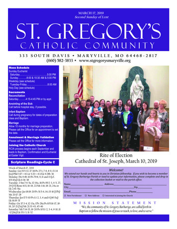
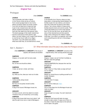
Are These Topographies Normal?(Courtesy Michael Belin, MD)
Are These Topographies Normal?(Courtesy Michael Belin, MD)
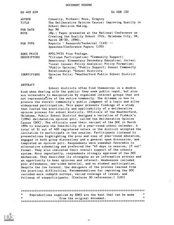
Would you perform lasik on thisindividual
Pentacam necessary to r/o high riskpt
Ectasia Lasik alters the biomechanics of the cornea whichin some instances can lead to ectasia. Ectasia is defined as surgically inducedkeratoconus following excimer laser surgery. More commonly seen in eyes that have undergonemyopic lasik surgery. Also reported eyes that have undergone hyperopiclasik and less commonly PRK.
Ectasia Incidence of keratoconus in the generalpopulation is approximately 1:2000. There are those patients who will undergolaser vision correction who do have KCat the time of surgery but will subsequentlydevelop KC and therefore will fall under thecategory of ectasia.
Risk Factors for Ectasia-identifiedbut not proven Pachymetry 500 micronsCalculated Residual bed 250 micronsSteep corneas ( 47D)High attempted correctionFF Keratoconus or asymmetrical topoEyes that have undergone enhancementsPregnancy
Pregnancy and Ectasia Hafezi et al (JRS April 2012) reported a series of 5cases of late onset ectasia that occurred inpregnant women. One cases occurred as late as 9 years after lasik. High levels of estradiol can result in a reduction ofthe biomechanical stiffness of the cornea (Spoerl). Patients should be counseled about the risk ofectasia developing during pregnancy.
Randleman et al: Ectasia Risk ScoreSystem – 5 Parameters Preoperative Topography: rule out formefruste KC Residual Stromal Bed Age Preoperative corneal thickness Manifest Refraction (Sph Eq)
Randleman et al: Ectasia Risk ScoreSystem The main criticism of this data is thatintraoperative pachymetry was performed in only11.8% of the cases. Intraoperative pachymetry is used to determine the“true” flap thickness and the residual stromal bed. Study estimated flap thickness based on themicrokeratome plate that was used. Flap thickness determines residual stromal bedthickness.
Factors determining Flap Thicknesswith a Microkeratome Preoperative corneal thicknessTranslational speed of microkeratomeBlade qualityFirst eye vs second eye useIOPAll of these factors can influence the trueflap thickness vs the microkeratome plate
Flap Thickness and Residual StromalBed Example:Preop MRx: -6.00DPreop CT: 540 micronsIntraoperative pachymetry: 410 micronsCalculated Microkeratome Flapthickness 130 microns Residual stromal bed (assuming15microns/D) 410-90 320 microns
Flap Thickness and Residual StromalBed Example:Preop MRx: -6.00DPreop CT: 540 micronsMicrokeratome Manufacturer indicates a160 micron flap Assumed Flap thickness 160 microns Residual stromal bed (assuming15microns/D) 380-90 290 microns
Flap Thickness and Residual StromalBed From the last example, if themicrokeratome manufacturer indicates a160 micron flap but the true flapthickness turns out to be 200 microns, Then the true residual stromal bed isnow 250 microns.
Ectasia Risk Scoring System Randleman study may have missed those eyesthat had no risk factors and developed ectasiabecause the true flap thickness was thicker thanindicated on the microkeratome plate, the residualstroma bed was thinner centrally and peripherally. Microkeratome error and not preoperative riskfactors could lead to the development of ectasia. to the de
Other Preoperative Ectasia RiskFactors Family history of KCHx of Eye RubbingAsymmetry in refraction ( 1D)Asymmetry in topographyAsymmetry in corneal apex and thinnestpoint in the cornea
Microkeratomes Microkeratomes make meniscus shapedflaps which are thicker in the entrance andexit sites. If a flap thickness centrally measures 130microns, it may measure 180 microns in theperiphery meaning more lamellar fibershave been cut than predicted.
Ectasia Can Still Develop in EyesWith No Risk Factors Never assume that Depth of Cutcorresponds to MicrokeratomeManufacturerʼs label Newer generation microkeratomes andFemtosecond lasers make more predictableflaps Corneas with the same thickness may havedifferent biomechanical strengths.
Avoiding Ectasia Preoperative topography and tomography(Pentacam) is the most important diagnostictool for screening lasik candidates. Always attempt to perform intraoperativepachymetry to determine the true flapthickness and residual stromal bed.
Ectasia Binder reported in 2007 a retrospectivereview of 10,000 eyes on which heperformed lasik for myopia. He found no cases of ectasia in eyes with 1 year follow up who had: residual stromalbed 250mcn; K readings 47D; eyesyounger than 25 yrs; or eyes withpreoperative CT 500 microns
Preop Keratometry Values Preop Keratometry Values will influenceflap size and potential risk for flapcomplications esp. using microkeratomes Flap complications rates range from0.3%-14% in the literature. Excessively flat ( 40D ) Ks and steep( 46D) increase the risk of flapcomplications.
Keratometry Values Final postop Ks should be between 34D and49D. Quality of vision including contrastsensitivity are more likely to decrease whenKs end up outside this range. Patients who may end up with excessivelyflat or steep K values would be better offhaving phakic or pseudophakic implants.
Preop Keratometry Values Mechanical microkeratomes flatten aconstant surface of the cornea. With steeper corneas, more tissue entersinto the suction head to be cut thereforesteeper corneas produce larger flaps. With flatter corneas, less tissue enters intothe suction head producing smaller flaps.
Mechanism by which variation in flap size isdependent upon preop K valuesAlbelda-Valles, JC et al JRS 2007 Vol 23:
Steep Preop Keratometry Values Steeper Ks leads to more buttonholes-excess tissuebeing compressed beyond applanation by thekeratome footplate causing apical cornea to buckleinward. Epithelial abrasions higher with steeper Ks Thin, irregular flaps more common with steeperKs. Published complication rates vary withMicrokeratome manufacturers
Flat Preop Keratometry Values Higher incidence of free caps Higher incidence of incomplete flaps
Microkeratome Factors thatdetermine Flap architecture Quality of the blades cutting edgeSpeed of microkeratome passSpeed of blade oscillationEase of pass on the corneaAdvancement of the microkeratome alongthe track of the suction ring
Femtoflap vs Microkeratome Planar flaps vs. meniscus shaped flaps. More predictable flap thickness and lower stddeviation. Less chance of ectasia Less chance of epithelial defects Less chance of button holes, free caps Able to adjust the hinge width and side cut angle. Surgically induced refractive change is less withfemto (0.05 0.11D) vs microkeratome (Moria)(0.42 0.28D)
Femtoflap vs Microkeratome Higher reported incidence of DLK in earlygeneration femto lasers. Transient Light Sensitivity reported with earliergeneration of Intralase which had higher pulseenergy. Opaque bubble layer. These complications have not been seen with thenewer generation FS lasers-Ziemer LDV.
Advantages to Surface Ablation/PRK PRK should be considered as an alternativeto lasik: Thinner corneas and therefore includepatients who are not good lasik candidates. Corneas which are slightly irregular (e.g.forme fruste KC) corneas. Steep or flat corneas BM dystrophy
Advantages to Surface Ablation/PRK Eyes that have undergone prior surgery maybe better suited for ASA:Scleral bucklePKPRKIncomplete LasikRetreatments on thin corneas
PRK in KC Suspects JCRS 2013* Guedj et al published 5 yearresults of patients who had forme fruste KCand underwent PRK. In 42 patients/62 eyes no eyes developedectasia during this period. Topo-guided PRK with CXL may be thebest treatment for these patients. *JCRS 2013 39; 66-73.
Preoperative Evaluation Ophthalmic History- Does the patient havepoor night vision?, glare?, halos? Corneal Anatomy- corneal topography andpentacam examining posterior curvature andpachymetry. Quality of vision- contrast sentivity,wavefront aberrometry
Preoperative Evaluation Contact lens history is very important. Patients should be out of soft contact lensesfor at least 1 week. Toric contact lenses should be removed forat least two weeks. Rigid gas permeable lenses for at least threemonths.
Preoperative Measurements Manifest/Cycloplegic refractions-look for stableprescriptions: 0.5D change within a 6 monthperiod is considered unstable. Pupillometry with infrared device (e.g. Covard) Corneal Pachymetry Corneal Topography-measure both the anterior andposterior surface-Pentacam becoming the standard ofcare Wavefront Aberrometry
Preoperative Measurements Slit lamp exam looking at the health of thecorneal surface-dry eyes, BM dystrophy,blepharitis. Retinal Evaluation-Myopes are at a higherrisk for RD throughout their lifetime
Treat Dry Eyes Aggressively BeforeSurgery Dry Eyes:1. Document with Clinical history2. Perform Appropriate tests-Lissamine Green, RoseBengal, Schirmers test.3. Treat with ATS, Restasis and Punctal plugs preop.4. Inform the patient that dry eyes will worsen after lasik.5. Nasal hinge lasik flaps tend to induce less drynessbecause nasal corneal nerves spared from resection.6. Consider surface ablation in pts with severe dry eyes.
Treat Blepharitis AggressivelyBefore Surgery Lid ScrubsWarm CompressesDoxycycline 75 mg BIDAzasiteHealthcare workers should receiveprophylaxis against MRSA
Patient Selection-Informed ConsentInformed consent is a process that begins when thepatient comes in for the initial evaluation.It may include:1)having the patient watch a video on the procedure;2)discussing the risks and benefits with the patient andreviewing the patientʼs expectations;3)presenting the patient with information to read(brochures, website) which may stimulate additionalquestions at a later time.4)Having the patient read and sign a detailed consentform.
Thank you
LASIK-Presurgical Preparation
Pre-surgical Preparation As a beginning lasik surgeon you should beinvolved in every step of the procedure eventhose steps performed by ancillary staff
Patient Preparation Treat blepharitis and ksicca preop with lidhygiene, lubricants and punctal occlusion.Restasis not readily available in Europe but youcan have local pharmacy prepare topicalcyclosporin. Begin 4th generation fluoroquinolone 1 day beforeto insure adequate levels in the cornea Review operative procedure and postoperativecare with patient. Instill 1 gtt proparacaine prior to betadine prep
LASIK Pre-surgical Preparation Review patient data prior to entering into laser.Minimum of two refractions plus auto refractionand topography. Record room temp. (18-24 C) and humidity(30-50%) Calibrate laser Enter treatment Rx into laser computer Verify correct entry including axis on computerscreen.
Microkeratome vs. Femto Laser2014 Some studies favor Femto laser while othersfavor microkeratomes while others show nodifference between rthe two. Over 80% of cases are being performed by afemtosecond laser in US. Cost benefit analysis needs to beconsidered. Marketing of “bladeless surgery”.
Presurgical PreparationInspection of Equipment: BladeMicrokeratomePower ConnectionsAlways test the microkeratomeReview Patient Data
Patient Preparation Position patient correctly on table:head and chin alignment Visually inspect head position from the side Have several speculums available Achieve adequate exposure by isolating lidand lashes with tape
Patient Preparation Maintain sterile conditions by using sterilepowder free gloves and mask Use copious irrigation to eliminate debrisand tear lipids-minimize chances for DLK Mark the cornea with marking pen
Laser technology-Improvement inOutcomes Wavefront optimized and Wavefront guidedlasers have improved outcomessignificantly. Debate continues which results in betteroutcomes
When Lasik is not Indicated-High Myopes
Verisyse Phakic IOL ImplantationVersus LASIKInvestigators conducted a prospective, randomizedcomparison of the Verisyse Phakic IOLimplantation with Excimer LASIK in myopic patientscorrecting between -9.00 and -19.50 diopters.(El Danasoury MA, et al. Ophthalmology. 2002;109:955-964) n 61 patients (90 eyes) mean SE refraction –13.5 D 29.5% of patients received a Verisyse lens inone eye and LASIK in the other eyeThe Verisyse Phakic IOL is sold under the Artisan name outside the US.
Author Conclusions The Verisyse Phakic IOL and LASIK werefound to be similarly effective, stable andreasonably safe for the correction ofmoderate to severe myopia. The Verisyse Phakic IOL offeredsignificantly better UCVA and BSCVA,better contrast sensitivity, and a lowerenhancement rate.
Author Conclusions (Cont.) Patient preference was significantly in favor ofthe Verisyse Phakic IOL, mainly because ofthe higher quality vision; 72.2% of patientswho received both surgeries preferred theVerisyse Phakic IOL.
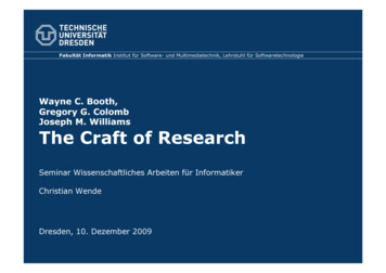
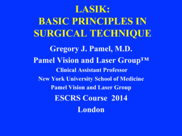
Contrast Sensitivity with the Verisyse IOLMesopic With GlareContrast Sensitivity300n 31*10030PreopPostop103.0p 0.031.53612Cycles per Degree*One eye analysis18
Indications For Lasik Upper Limit of Myopia Treated is now under--9.00 diopters in my practice. Upper Limit of Hyperopia is 4.00 DTreating presbyopic patients with multifocalIOLs (ReZoom/Restore IOLS) and RLE Any Irregular cornea is a contraindication forwavefront optimized lasik and a considerationfor topography guided lasik or surface ablation
Bioptics: Lasik After VerisysePhakic Lens Implantation For myopic patients who have residualmyopia and/or astigmatism after phakiclens implantation. For patients whose myopia hasprogressed after phakic lens implantation
Postoperative Care Provide patient with written instructions andexpectations to read prior to and aftersurgery. Advise patient to go home and sleep for afew hours which will facilitate healing andrecovery Advise patient to avoid reading and TV theday of the procedure
THANKYOU
Gregory J. Pamel, M.D. . Clinical Assistant Professor New York University School of Medicine Pamel Vision and Laser Group ESCRS Course 2014 London . Lasik over the last 14 years: US Experience 2001-2003 Wavefront guided lasik . 2013 on the Dr. OZ show. Nationally syndicated talk show with over 3