Transcription
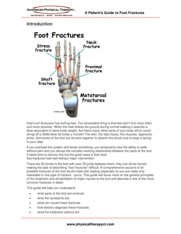
A Patient's Guide to Foot FracturesIntroductionFeet hurt! Everyone has aching feet. The remarkable thing is that feet don’t hurt more oftenand more severely. When the heel strikes the ground during normal walking it absorbs ablow equivalent to twice body weight. Are there many other parts of your body which couldshrug off a 300lb blow 30 times a minute? The skin, the fatty tissue, the muscles, ligaments,joints, and bones of the foot are all work together to absorb this shock and to keep a springin your step.If you overload the system and break something, you temporarily lose the ability to walkwithout pain and you disrupt the complex working relationship between the parts of the foot.It takes time to recover this but the good news is that mostfoot fractures heal well without major intervention.There are 26 bones in the foot with over 30 joints between them; they can all be injured,making the task of describing "foot fractures" difficult. A comprehensive account of allpossible fractures of the foot would make dull reading especially as you are really onlyinterested in one type of fracture - yours. This guide will focus more on the general principlesof the treatment and rehabilitation of major injuries to the foot and describe a few of the morecommon fractures in detail.This guide will help you understand what parts of the foot are involved what the symptoms are what can cause these fractures how doctors diagnose these fractures what the treatment options arewww.physicaltherapyct.com
A Patient's Guide to Foot FracturesAnatomyWhat structures are most commonly injured?The foot has 26 bones, but it also has four layers of muscles and tendons, joints, veins,arteries, nerves and fatty tissue. It is very important to remember that a force severe enoughto break a bone will damage some of these other tissues as well. Injury causes swelling andbleeding which eventually forms scar tissue. During the healing process, this scar tissue canbind together the muscles and tendons which normally glide over one another. How well thefoot functions after a fracture depends on how well both bone and soft tissue heal.The names of the bones going from the heel forwards are:Calcaneus – Heel boneTalus – Ankle BoneNavicularMedial, Intermediate (Middle) and Lateral CuneiformCuboidMetatarsals (Lesser Toes)1st Metatarsal (Big Toe, Hallux)1st Proximal Phalanx (Big Toe, Hallux)1st Distal Phalanx (Big Toe, Hallux)Proximal, Middle & Distal Phalanges (Lesser Toes)The big toe is officially called the hallux. Many parts of the foot that are associated with thehallux include the word hallucis in the name. For example, the long muscle that flexes(bends) the big toe downwards is called flexor hallucis longus.www.physicaltherapyct.com
A Patient's Guide to Foot FracturesThere are a large number of joints between the bones in the foot. The important ones are:The ankle joint between the end of the shin and the talus (ankle bone). It allows up and downmovement of the foot.The subtalar joint between the talus and the calcaneus (heel bone). It allows side to sidemovement so that you can stand on the side of a sloping surface.The transverse tarsal joint (also called Chopart’s joint) is formed by the talus and calcaneuson one side and the navicular and cuboid on the other. It allows some pivoting movements ofthe forefoot on the hindfoot and flattening of the arch.The tarsometatarsal joint (also called Lisfranc’s joint) is between the distal tarsal bones, (thethree cuneiforms and the cuboid) and the metatarsals. Its complex anatomy allows for a littlemovement of the longitudinal arch of the foot and a bit more movement at the outer border ofthe foot Dislocation of this joint is a serious injury often associated with fracture of themetatarsals and/or cuneiform bones.Metatarsophalangeal (MTP) joints. There are five of these. These joints are between theheads of the metatarsals and the proximal phalanges. They allow flexion and extension ofthe toes. When you stand on tiptoe the MTP joints are extended and you are bearing weighton the toes and the joints.Interphalangeal joints. These nine joints are located between the phalanges (toe bones) andallow the toes to curl. Loss of this function is not usually a severe handicap.The big muscles that power walking by moving the ankle, foot and toes, are located in theleg. If you wiggle your toes and put your fingers on the muscle on the outer side of the frontof the leg just below the knee, you can feel the muscles contracting. Their pull is transferredto the bones of the foot by long thin tendons that extend from the muscle in the leg, downwww.physicaltherapyct.com
A Patient's Guide to Foot Fracturesacross the ankle and into the foot to end in the toes. These tendons glide inside specialtendon sheaths. These tendon sheaths may be damaged when the foot is injured.Other intrinsic muscles of the foot form the flesh of the sole of the foot. There are actuallyfour layers of muscles on the sole of the foot. They are important in maintaining the arch ofthe foot, a critical part of the shock absorption system. These muscles are protected fromweight bearing by the plantar fascia, a tough layer of fibrous tissue next to the skin on thesole of the foot. Bleeding into the sole of the foot from a fracture may result in scar tissuewhich may bind all these layers together and interfere with function.Viewed from the side there are other interesting aspects of foot anatomy. The heel pad is aspecialized region situated between the heel bone and the skin. It has an elastic honeycomblike structure with the "cells" filled with fatty tissue. These cells work like a shock absorber.When the heel comes down the cells stretch and flatten, absorbing some of the shock. Aninjury severe enough to break the calcaneus (heel bone) is also often severe enough todisrupt the heel pad. It may heal with solid scar replacing the specialized heel pad structure.This reduces its ability to absorb shock.Further forward is the arch of the foot. This is a dynamic feature of the foot only partly due tothe shape of the bones. When you are standing, the muscles that maintain the arch relaxand it partially flattens. When you rise on the ball of the foot to take a step the arch isrestored. The ball of the foot itself is at the level of the metatarsophalangeal (MTP) jointsbetween the metatarsals and the toes.You bear quite a lot of weight on the heads of the metatarsals as you spring off the foot atthe end of a pace. The smaller toes do not contribute much to walking but a good deal offorce goes through the big toe. In a normal pace the weight of the body first meets theground at the heel then is gradually transferred forward along the outer margin of the foot,across the ball of the foot ending on the big toe. Fractures of bones along this path havemore serious consequences for foot function.CausesHow do fractures of the foot commonly happen?Bones break when the force applied to them exceeds their strength. In the foot this type ofoverload happens with falls from a height, from crushing injuries when something falls on thefoot, from kicking something, or from catching the foot in something and twisting. Catchingthe foot in machinery may cause devastating crush injuries. A rare but well recognized typeof foot fracture results from fatigue failure. This stress fracture is seen when repetitive loadsare excessive.TypesCalcaneusThe calcaneus, or heel bone, is the most frequently fractured tarsal bone, accounting for 2%of all fractures seen in adults and more than 60% of tarsal fractures.www.physicaltherapyct.com
A Patient's Guide to Foot FracturesAvulsion fractures (3%) can occur by over-pull of the heelcord (achilles tendon) usuallyduring a major effort in sports or jumping. It can be the result of fatigue failure withmultiple repetitive effort.A fracture occurring into the joint between the talus and the calcaneus(70%) is one of themost serious foot fractures. It is caused by axial over-loading, driving the talus (ankle bone)down into the heel bone. Motor vehicle accidents and falls from ladders are common causes.There is quite a high risk of other injuries with fractures of the other heel, fractures of thespine and injuries to the kidney all being quite common. Disruption of the heel pad is alsocommon and compartment syndrome of the foot can occur. Because it makes a major weightbearing joint surface irregular, this injury may be treated by surgery to restore the joint tosmoothness.www.physicaltherapyct.com
A Patient's Guide to Foot Fractureswww.physicaltherapyct.com
A Patient's Guide to Foot FracturesA fracture may also that does not involve the joint. This fracture is caused in a different way.A direct blow to the side of the heel or trapping and twisting the heel may cause a fracturewhich does not go into the joint. Most often these can be treated without surgery.A fracture of the anterior calcaneus process is a rare fracture that can be caused either byavulsion of the attachment of a ligament when the foot is twisted inwards or by compressionwhen the foot is twisted outwards.www.physicaltherapyct.com
A Patient's Guide to Foot FracturesA portion of the calcaneus that juts out to support the talus is called the sustentaculum. Itmay be broken in an accident in which the foot is twisted inwards while bearing a significantamount of weight.www.physicaltherapyct.com
A Patient's Guide to Foot FracturesTalusA fracture of the head of the talus accounts for 5 to 10 percent of all fractures of the talus.This fracture is caused by a compressive force when the foot is pointed downwards. Thefracture enters the joint between the talus and the navicular. If it is displaced it is oftentreated by surgery (ORIF), becasue it involves a joint surface.www.physicaltherapyct.com
A Patient's Guide to Foot FracturesA fracture of the neck of the talus is a serious injury. This fracture passes transversely acrossthe talus and isolates the part of the bone that forms the ankle joint. Forces that bend thefoot up beyond its limit are responsible for this pattern of fracture. Open fractures arecommon. There is a high incidence of avascular necrosis of the dome of the talus. Avascularnecrosis is a condition where the blood supply to a bone is damaged. This results in death ofthe area of bone that is normally supplied by the blood vessels that are injured. Over time,this bone may collapse causing severe damage to the joint and post traumatic arthritis.www.physicaltherapyct.com
A Patient's Guide to Foot FracturesThe lateral process is a small bump of bone on the outside of the talus which is theattachment of ligaments of the ankle joint and the subtalar joint. Severe twisting forces tothese joints may tear the ligament (ankle sprain) or pull off the piece of bone (lateral processfracture). The treatment and outcome of this fracture depends on how much of the nearbyjoint surfaces are damaged.The posterior process of the talus may be fractured. Either the inner (medial) or outer(lateral) side of the back of the talus may be broken off when the ankle is severely twisted.Ligaments of the ankle and subtalar joints attach here. The surface of the joint with the heelbone may be damaged.www.physicaltherapyct.com
A Patient's Guide to Foot FracturesA fracture of the joint surface of the talus is called an osteochondral fracture. In some anklesprains a portion of the joint surface of the talus can become separated from the rest. This isdifficult to see on x-ray. In some cases the fragment will heal if the joint is immobilized. Inothers it may need to be removed.www.physicaltherapyct.com
A Patient's Guide to Foot FracturesNavicularA cortical avulsion fracture may be caused by over-pull of muscle or ligament attachments.These injuries occur when the foot is twisted. Normally, they heal without intervention exceptfor resting the foot. However, if the fragment is displaced 1 cm or more it is advisable tooperate to replace it and fix it back in position.www.physicaltherapyct.com
A Patient's Guide to Foot FracturesA navicular tuberosity fracture is also an avulsion fracture caused by over-pull of the tibiallisposterior muscle. This is a powerful muscle that inverts (turns the foot inward) the foots andmaintains the arch. Surgery is often required to replace and fix the fragment.www.physicaltherapyct.com
A Patient's Guide to Foot FracturesFractures of the navicular body often pass through the joints with the talus, the cuneiform orboth. If there is any displacement they need to be treated surgically with ORIF.www.physicaltherapyct.com
A Patient's Guide to Foot FracturesA stress fracture of the navicular is a relatively common fatigue failure fracture in athletesand military recruits who undertake repetitive training exercises that load the foot. Diagnosisis often delayed because these fractures do not initially show up on X-ray. Simply resting thefoot for several weeks is usually successful treatment.CuboidA isolated fracture of the Cuboid is relatively uncommon. The bone is more often brokenwhen a major force injures the midfoot and fractures the cuneiforms or the talus and/or thenavicular as well. Treatment depends on the overall situation.The nutcracker injury results from a force pushing the foot outwards and crushing the cuboidbetween the calcaneus and the 4th & 5th Metatarsals. If the cuboid is badly crushed theouter border of the foot will be distorted even after the bone heals. In some cases the lengthof the cuboid can be restored by an open operation to reduce and fix the fracture thensupplement the crushed bone with bone graft.www.physicaltherapyct.com
A Patient's Guide to Foot FracturesMedial Cuneiform, Intermediate Cuneiform, Lateral CuneiformIsolated fractures of these bone are quite rare but may occur from direct impact – droppingsomething on the foot. More commonly, fractures to these bones occurs in association withfractures of other bones of the midfoot and hindfoot. An injury that disrupts thetarsometatarsal joint often also causes fracture of one or more cuneiform bones.One relatively common injury occur at the tarsometatarsal (Lisfranc’s) joint. Partial orcomplete dislocation with or without fractures of one or more of the metatarsals is called aLisfranc Dislocation. It is caused by axial loading the foot when standing on tiptoe or byimpact or twisting the mid-foot. It is sometimes difficult to diagnose on standard x-rays.Stress views of the mid-foot show that the joint between the 2nd metatarsal and thecuneiforms is disrupted. This is an unstable injury which often requires operative treatment.1st – 4th MetatarsalsInjuries of these metatarsals usually occur as fractures of the neck region, shaft fractures orstress fractures. Neck fractures often affect more than one metatarsal bone and are causedby dropping a weight on the instep, kicking or loading when on tiptoe. Even though thefractures may be quite displaced they usually heal without intervention and cause nosignificant long term problems.www.physicaltherapyct.com
A Patient's Guide to Foot FracturesMetatarsal shaft fractures are also usually caused by impact or crushing. Treatment isindividualized but many such fractures can be treated non-operatively.Stress fractures occur in the proximal region of the metatarsal shaft. They are caused byrepetitive activity and were first described in army recruits who were expected to undertakelong marches and drill activities. These injuries are fatigue failures of the bone and usuallyheal if the activity which causes them is stopped and the foot is allowed to rest for severalweeks.Insufficiency fractures occur in people with osteoporosis and the metatarsal can break withvery little force. These are fragility fractures which are a warning that there is a greater risk ofspinal and hip fractures in the patient. Treatment of the fracture is often quite simple with restand non-weight bearing resulting in healing. However, the patient should be assessed forosteoporosis and may need treatment for that condition.Related Document: A Patient's Guide to Osteoporosis5th MetatarsalA very common foot fracture that sometimes occurs during an ankle sprain is the avulsionfracture of the tuberosity, or base, of the fifth metatarsal. The injury is caused by inverting orturning the ankle over - the same movement that occurs when an ankle sprain occurs. Oneof the muscles that attempts to prevent this is the peroneus brevis. Over-action by thismuscle causes an avulsion fracture of the tuberosity of the 5th metatarsal where themuscle’s tendon attaches. This injury will heal without intervention and should be treatedquite simply with rest and protection.www.physicaltherapyct.com
A Patient's Guide to Foot FracturesA more serious type of 5th metatarsal fracture is known as the Jones fracture. This injury isalso caused by an inversion twist of the foot. It is a fracture of the upper end of the 5thmetatarsal as well but slightly further out, or distal, than the avuslion fracture. This fracturealso enters the joint with the 4th metatarsal. This fracture is more serious primarily because itmay not heal and may require surgery.Stress fractures can also occur in the 5th metatarsal. Patients may present with a painfulouter border of the foot without a clear history of an accident. Although it seems very similarto the avulsion fracture, the stress fracture behaves very differently with a high rate of nonunion. Some surgeons prefer to treat this fracture by surgery even if it is an acute injury;chronic injuries or stress fractures are very often treated by surgery (ORIF).Phalanges (Toes)The little bones that make up the toes are collectively called phalanges. Fractures to thesebones are common and can be caused by kicking, stubbing the toe or when something isdropped onto the toe. The fractures can be roughly divided into those that affect the joint atone end or the other (intraarticular fractures) and those which do not (shaft fractures).Restoring the smoothness of the joint surface may sometimes require surgery (ORIF) but it isactually rare for either type of fracture to require operation. Most people do not put a lot ofweight through their toes so they don’t have to heal straight.www.physicaltherapyct.com
A Patient's Guide to Foot FracturesSymptomsWhat symptoms do foot fractures cause?Pain and tenderness are the most frequent symptoms that arise from a foot fracture. Boththe pain and the tenderness are usually localized to the broken bone. Moving the foot orhanging it down tend to increase the pain while elevation, ice, and immobilization make itbetter. Bruising and swelling are also very common and arise within a few minutes of theinjury. Wounds in the foot need to be carefully evaluated in case there is an underlying openfracture.Foot fractures are quite common in multiple injuries – such as motor vehicle accidents.Where there are other major symptoms from fractures of the long bones, head injuries, orabdominal injuries, the symptoms from foot fractures may not be very prominent. It is notunusual for foot fractures to be unrecognized for a few days in these circumstances.EvaluationHow will my fracture be evaluated?At the scene of the injury, first aid for foot fractures consists of dressing any wounds, treatingshock, taking weight off the foot, and transferring the patient to hospital. Sometimes it isadvisable to splint the foot.In the Emergency Room the focus of the treating team is on treating pain and shock,diagnosing the foot injury accurately, making sure that there are no other significant injuries,and arranging for referral to the appropriate specialist. Many foot fractures can be treated bythe Emergency Room Physician and followed by your Family Doctor, but others requirereferral to an orthopaedic surgeon or podiatrist.The evaluation will include taking an account of the accident and the type of force that hurtyour foot. The foot will be examined for wounds, tenderness, swelling, bruising, or anyabnormality of the shape of the foot or toes. It may be helpful to compare it with the uninjuredside. Many of the accidents that cause a fracture on one side also cause a less prominentbut still significant injury on the other. Footwear and socks must be removed for theexamination and this may require cutting off the shoe or boot to prevent further pain.X-rays of the foot, toes, or ankle will be taken if there is a suspicion of fracture. The standardviews are often sufficient but special x-rays, including stress views, can be ordered forfractures of the talus and injuries of Lisfanc’s joint.The specialist’s assessment will repeat the history and physical examination. The patient’sgeneral medical status, occupation, sports activity, and expectations are all importantaspects which may affect the treatment plan. The surgeon will evaluate the x-rays and mayorder more specific views to make sure the full extent of the injury is known.For fractures of the calcaneus, ankle bone and mid-foot a Computerized Tomography (CT)Scan may be useful. Once the anatomy of the fracture is completely understood the surgeonwill discuss the treatment options with you. This discussion may take into account the exactnature of the fracture and its prognosis as well as your expectations, preferences, andgeneral medical status.www.physicaltherapyct.com
A Patient's Guide to Foot FracturesTreatmentWhat treatments should I consider?Nonsurgical TreatmentMost foot fractures recover well without surgery. The common fractures of the toes andmetatarsals do not need 100% accurate reduction for your foot to heal and perform normally.In these cases the goals of treatment are to relieve pain, protect the foot against further harmand allow the bones to heal.Pain medication, rest, elevation, and avoidance of weight bearing are the principle treatmentmethods. It is not always necessary to apply a cast or splint to the foot as this does not affectthe healing process. A splint may be applied to help with pain relief or to protect the foot.These splints may be discontinued after a few days. Crutches are used to allow you to getaround without bearing weight on the fracture, until there are signs of healing on x-ray –usually six weeks.Toe fractures which are in an unacceptable position may need to be reduced beforesplinting. This can usually be done under local anesthetic. The toe can then be pulledstraight and splinted to its neighbor. This is commonly referred to as buddy taping.SurgeryUnstable foot fractures are likely to lose the correct position and fail to heal (nonunion) orheal with deformity (malunion). The Jones fracture of the 5th Metatarsal is a commonexample. These fractures may be treated by reducing the fracture fragment back intoposition by manipulation and holding it there with pins passed through the skin and acrossthe fracture. The foot would be protected by a splint after this procedure and the pin(s) wouldstay in place until there was evidence of healing on x-ray. This generally takes about sixwww.physicaltherapyct.com
A Patient's Guide to Foot Fracturesweeks. Pin removal is a minor procedure which is usually done in the office or clinic anddoes not require an anesthetic.Surgery is needed for open fractures. The bone and surrounding muscles and tendons arecontaminated by bacteria which have entered the wound. All dirt and dead tissue must beremoved from the wound and this may mean quite an extensive opening up of the wound tomake sure that the tendons that glide over the bone are cleaned up.Fractures that disrupt important joints such as calcaneus fractures, fractures of the talus andinjuries of Lisfranc’s joint are often treated by open reduction and internal fixation (ORIF).The aim is to restore the joint surface to smoothness and minimize future post traumaticarthritis of the joint. Calcaneus fractures can be treated by applying a plate along the outerside of the bone. Many other situations can be treated by fixing the fragments in place withscrews.Where ORIF has been performed, the implants (screws and plates) used to hold the fracturefragments in position are usually left in place. Sometimes protruding screw heads or tipscause symptoms and this prompts hardware removal. Many patients worry about theoperation to remove hardware and expect it to be as painful as the original fracturetreatment. This is rarely the case because the bone is healed and there is much lesscollateral damage in the way of swollen tissue and torn muscle. Recovery after hardwareremoval is usually quite rapid and most people are very pleased to have it done.Regardless of the amount of intervention, the management plan usually involves close followup. It is important to know that the fracture is behaving as predicted and the fragments areremaining in an acceptable position. You should make sure that you are able to return forfollow up as recommended.RehabilitationWhat happens as I recover?After treatment the foot stays symptomatic for several weeks. It hurts and swells when ithangs down; bruising may be evident for some time also. Rather mysteriously, it is commonfor fractures of the hind-foot, ankle & mid-foot to result in bruising between the toes. Becausethe bones are deep inside the muscle layers the bleeding may track to distant areas ratherthan coming to the skin right under the broken bone.Most foot bones show evidence of healing by six weeks post fracture. Until then it is oftenmore comfortable to avoid bearing weight on the affect foot, so you should use crutches andkeep the foot off the ground. Once the pain and swelling has settled you may be able to bearsome weight, depending on the location and nature of the fracture. Take your doctor’s adviceabout this. If you put too much weight on the foot too soon, you risk displacing the fracture orbreaking the pins or screws if you have had surgery.Overall it takes about 18 months for fractures to heal fully. The first six weeks is the fastest;during that time a scar is formed between the fracture fragments and stiffened with calciumdeposits. This achieves about 50% of the eventual strength of the healed bone. After thatthere is a slower process of maturation and consolidation of the healing area of the boneuntil it has the strength and appearance of normal bone. One can normally use your healedfoot for all normal activities including sports by about three months. However, there will stillbe some aching and swelling continuing with use but getting better for another year or more.Although some foot fractures are treated in a cast, brace, or special shoe, many need nosupport or protection. Toe fractures are often treated with buddy taping and weight bearingon the ball of the foot. You can get a shoe with a build up under the ball of the foot to keepwww.physicaltherapyct.com
A Patient's Guide to Foot Fracturesweight off the toe(s). If some form of support is needed it may be discontinued at six weeks ifthe fracture shows evidence of healing.Physical therapy may be needed to start you off walking safely on crutches. However, thereis little need for it until after the bone has healed. Once you can put weight through thehealing area it may be helpful to have an exercise program to recover movement, strengthand endurance.Fractures that damage weight bearing joints in the foot may cause post traumatic arthritis.The intent of treatment is to limit this possibility but if the accident crushes or scrapes off thejoint surface it does not recover or re-grow. In time the joint will become painful and stiff.Much more often the results of healing of a foot fracture are very good with complete returnto normal function, work and sports being the norm.ComplicationsWhat are the potential complications of these fractures?Complications are rare problems that make recovery from the injury longer or more"complicated". The intent of treatment is to keep these complications as rare as possible;that is why orthopaedic surgeons pay so much attention to them.Compartment SyndromeBleeding from broken bones or torn muscles deep inside the foot may cause swelling insidethe muscle compartment. This compartment cannot enlarge much so the pressure may risehigh enough to cut off circulation to the living muscle that occupies this region. Now thismuscle too is at risk of dying and swelling more in turn. This dangerous sequence causessevere pain when the muscles are moved actively or passively. If the condition is diagnosedearly, before any muscle death occurs then surgery to open up the muscle compartments willsave the situation and allow the muscle to heal normally. The treating staff are very much onthe look out for this problem, even though it is rare. The observations getting you to moveyour toes right after surgery make sure that a compartment syndrome is not developing orthat it is detected right away.MalunionWhen a bone heals with a deformity and looks different from the normal shape it is said to bemal-united. Malunion of a joint surface leaves it rough and irregular. Movement of the jointmay then be limited or painful and there is a higher risk that the joint would wear out. Theassessment of the injury and the choice of treatment including operation looks at thelikelihood and consequences of malunion. Malunion in areas of the bone outside the jointmay be better tolerated even if the bone heals with a "kink" in it.If poor alignment is identified before the bone has healed (mal-alignment) it may be possibleand desirable to improve the position either by manipulation or by surgery. If the bone hastruly healed (malunion) the position can only be improved by cutting the bone and re-aligningit. It may not be worth doing something as elaborate as that.Post Traumatic Arthritis (PTA)PTA means that the joint has become painful and stiff because it has worn out. The jointwears out because it was injured or because it healed with deformity (malunion). Thisarthritis is the same process
bleeding which eventually forms scar tissue. During the healing process, this scar tissue can bind together the muscles and tendons which normally glide over one another. How well the foot functions after a fracture depends on how well both bone and soft tissue heal. The names of the bone


