Transcription
Investigation of and Responseto 2 Plague Cases, YosemiteNational Park, California, USA, 2015Mary Danforth, Mark Novak, Jeannine Petersen, Paul Mead, Luke Kingry, Matthew Weinburke,Danielle Buttke, Gregory Hacker, James Tucker, Michael Niemela, Bryan Jackson,Kerry Padgett, Kelly Liebman, Duc Vugia, Vicki KramerThis activity has been planned and implemented through the joint providership of Medscape, LLC andEmerging Infectious Diseases. Medscape, LLC is accredited by the American Nurses Credentialing Center (ANCC),the Accreditation Council for Pharmacy Education (ACPE), and the Accreditation Council for Continuing MedicalEducation (ACCME), to provide continuing education for the healthcare team.Medscape, LLC designates this Journal-based CME activity for a maximum of 1.00 AMA PRA Category 1Credit(s) . Physicians should claim only the credit commensurate with the extent of their participation in the activity.All other clinicians completing this activity will be issued a certificate of participation. To participate in thisjournal CME activity: (1) review the learning objectives and author disclosures; (2) study the education content; (3)take the post-test with a 75% minimum passing score and complete the evaluation athttp://www.medscape.org/journal/eid; and (4) view/print certificate. For CME questions, see page 2241.Release date: November 16, 2016; Expiration date: November 16, 2017Learning ObjectivesUpon completion of this activity, participants will be able to: Interpret laboratory and epidemiologic findings regarding 2 human cases of plague in 2015 in patientswith recent travel history to Yosemite National Park Assess environmental findings regarding 2 human cases of plague in 2015 in patients with recenttravel history to Yosemite National Park Identify critical risk reduction measures used to help prevent plague transmission to Yosemite visitorsand staff.CME EditorP. Lynne Stockton Taylor, VMD, MS, ELS(D), Technical Writer/Editor, Emerging Infectious Diseases. Disclosure: P.Lynne Stockton Taylor, VMD, MS, ELS(D), has disclosed no relevant financial relationships.CME AuthorLaurie Barclay, MD, freelance writer and reviewer, Medscape, LLC. Disclosure: Laurie Barclay, MD, has disclosedthe following relevant financial relationships: owns stock, stock options, or bonds from Pfizer.AuthorsDisclosures: Mary E. Danforth, PhD; Mark Novak, PhD; Jeannine Petersen, PhD; Paul Mead, PhD, MPH;Luke Kingry, PhD; Matthew Weinburke, MPH; Danielle Buttke, DVM, PhD, MPH; Gregory Hacker, MSc;James R. Tucker, MS; Michael Niemela, MS; Bryan T. Jackson, PhD; Kerry Padgett, PhD; Kelly Liebman, PhD,MPH; Duc Vugia, MD, MPH; and Vicki Kramer, PhD, have disclosed no relevant financial relationships.Author affiliations: California Department of Public Health,Sacramento, California, USA (M. Danforth, M. Novak, G. Hacker,J. Tucker, M. Niemela, B. Jackson, K. Padgett, K. Liebman,D. Vugia, V. Kramer); Centers for Disease Control and Prevention,Fort Collins, Colorado, USA (J. Petersen, P. Mead, L. Kingry);National Park Service, El Portal, California, and Fort Collins,Colorado, USA (M. Weinburke, D. Buttke)DOI: http://dx.doi.org/10.3201/eid2212.160560In August 2015, plague was diagnosed for 2 persons whohad visited Yosemite National Park in California, USA. Onecase was septicemic and the other bubonic. Subsequentenvironmental investigation identified probable locations ofexposure for each patient and evidence of epizootic plaguein other areas of the park. Transmission of Yersinia pestis was detected by testing rodent serum, fleas, and rodentcarcasses. The environmental investigation and whole-genome multilocus sequence typing of Y. pestis isolates fromEmerging Infectious Diseases www.cdc.gov/eid Vol. 22, No. 12, December 20162045
SYNOPSISthe patients and environmental samples indicated that thepatients had been exposed in different locations and that atleast 2 distinct strains of Y. pestis were circulating amongvector–host populations in the area. Public education effortsand insecticide applications in select areas to control rodentfleas probably reduced the risk for plague transmission topark visitors and staff.Plague is a zoonotic disease caused by the gram-negativebacterium Yersinia pestis; the organism’s reservoir isrodents and the vectors are fleas (1,2). Transmission to humans can occur through bites by infected fleas or throughhandling Y. pestis–infected rodents (1,2). Epidemics ofplague still occur on the continents of Africa, Asia, andNorth and South America (3). Plague was introduced toCalifornia in 1900 (1,4–6), where over the next 25 years itcaused occasional outbreaks in rats commensally residingwith humans in urban areas (2,4,6). Shortly after its introduction, Y. pestis moved into wild rodent populations, establishing a sylvatic transmission cycle (7,8). In subsequentdecades, plague spread across California and other westernstates (9) periodically affecting humans (4–6,10–13).The human risk of contracting plague is higher duringepizootic transmission when Y. pestis is amplified amongsusceptible rodent hosts (2), such as the California groundsquirrel (Otospermophilus beecheyi), the golden-mantledground squirrel (Callospermophilus lateralis), and certainchipmunk species (Tamias spp.) (2,3,14,15). Higher mortality rates among these animals lead to the release of infectious fleas into the environment (2). The California groundsquirrel plays a major role in human exposure in Californiabecause its predominant flea species, Oropsylla montana,is a competent Y. pestis vector (1,2) that is often abundant on this rodent and in its burrows (16) and will readilybite humans (1,11). Since the 1980s, evidence of Y. pestistransmission in rodents in the Sierra Nevada mountains hasbeen generally restricted to locations at elevations 1,200meters (California Department of Public Health, unpub.data, 1983–2015). Despite ongoing sylvatic transmission,human plague remains rare in the western United States(17–19), including in California, where no cases have beenconfirmed since 2006 (20,21).During the summer of 2015, the Los Angeles CountyDepartment of Public Health (LACDPH) and the GeorgiaDepartment of Public Health reported 2 cases of plague inpersons who had recently travelled to Yosemite NationalPark (Yosemite). The California Department of PublicHealth (CDPH), in collaboration with the US Centers forDisease Control and Prevention (CDC) and the NationalPark Service (NPS), investigated the increased Y. pestistransmission in Yosemite. We summarize the epidemiologic, laboratory, and environmental findings and the public health response.2046MethodsEpidemiologic and Laboratory InvestigationWe defined a case of plague as clinically compatible illness and isolation of Y. pestis from a person with a historyof travel to Yosemite during the 7 days before illness onset. Clinically compatible illness included fever, headache,chills, and malaise in conjunction with regional lymphadenitis, septicemia, or pneumonia (22). Patients were identified by their county or state health department and reportedto CDPH or CDC.Diagnosis of plague was made after PCR testing ofclinical specimens, including blood and bubo aspirates;Laboratory Response Network assays and culture wereused. Recovered isolates were confirmed as Y. pestis bybacteriophage lysis (23). For whole-genome multilocussequence typing (MLST), DNA extracted from Y. pestisisolates was sequenced by using the PacBio RS II platformand sequence reads were assembled by using a hierarchalgenome assembly process (Pacific Biosciences, MenloPark, CA, USA). Allele calls for 3,979 Y. pestis open reading frames (ORFs) (4,046,060 bp) and cluster analyseswere performed as described (24).Local and state public health officials interviewed patients with confirmed cases and their family members whohad traveled with them. Respondents were asked abouttheir illness history, travel, activities, and interactions withrodents in and around the Yosemite area during the weekbefore illness onset.Environmental InvestigationThe environmental investigation was prioritized by patient travel itineraries and historical evidence of Y. pestis transmission at these locations or in similar habitats.To assess the scope of Y. pestis transmission and thepotential exposure risk for visitors and park personnel,the investigation was expanded to include additional locations in Yosemite. At prioritized locations, visual riskassessments were conducted to evaluate the presenceand abundance of rodents, the type of human activitiesin the area, and the potential for human exposure to infective fleas (25). In areas with suspected Y. pestis transmission, a 30 30 cm flannel cloth was used to samplefleas from rodent burrow entrances. Rodents were livetrapped for plague serologic testing and flea collection(25). For rodent trapping, Sherman (H.B. Sherman Traps,Tallahassee, FL, USA) and Tomahawk (TomahawkLive Trap, Hazelhurst, WI, USA) live traps were baitedonce with a mixture of grains and opened either fromovernight through the following midday or from earlymorning through noon. Relative rodent abundance wasestimated by calculating the ratio of captured rodents tothe total number of traps set and is referred to as the trapEmerging Infectious Diseases www.cdc.gov/eid Vol. 22, No. 12, December 2016
Plague, Yosemite National Park, 2015success rate. Captured rodents were anesthetized withisoflurane, identified to species, brushed to collect fleas,and subjected to collection of 0.1 mL of blood for Y.pestis antibody testing. Deer mice (Peromyscus maniculatus) collected near structures were euthanized; all otherrodents were marked with a numbered ear tag (National Band & Tag Company, Newport, KY, USA) and released near the point of capture. Small mammal handlingtechniques were reviewed and approved by the CDPHInstitutional Animal Care and Use Committee, protocol2015-14. In addition to live rodent trapping, rodent carcasses reported by Yosemite staff or visitors were collectedfor testing.Flea and rodent specimens were tested for Y. pestis byCDPH, CDC, and NPS. Blood samples from trapped rodents were sent to CDPH for concurrent testing by passivehemagglutination and passive hemagglutination inhibitionto detect antibodies against Y. pestis F1 antigen (23). Allpositive passive hemagglutination titers 1:32, the lowestdilution tested, that were negative by passive hemagglutination inhibition were considered positive (23). Fleas collected by burrow swabbing, from live-captured rodents, orfrom rodent carcasses were sent to CDPH or CDC to beidentified to species according to standard taxonomic keys(26) and to be tested for Y. pestis. Fleas of the same speciesfrom the same burrow or rodent host were sorted into poolsof up to 10 fleas and then homogenized in brain–heart infusion broth by using glass beads and Mixer Mill MM301(Retsch, Haan, Germany).Rodent carcasses were tested at CDPH, CDC, andNPS. Spleen and liver tissues were removed; for direct fluorescent antibody testing, slide touch tissue preparationswere incubated with fluorescein isothiocyanate–labeledrabbit anti-F1 antibodies, washed, and then viewed byfluorescence microscopy (23). DNA was extracted fromflea homogenates and carcass tissues by using the MagNA Pure Compact Nucleic Acid Isolation Kit (RocheDiagnostics, Basel, Switzerland) and amplified by usingTaqMan primers and probe targeting the caf1 gene (27).Animal and flea specimens positive by PCR were inoculated onto sheep blood agar plates or onto cefsulodinirgasan-novobiocin agar plates to enable isolation of Y.pestis from contaminated environmental samples (23).Isolates were confirmed as Y. pestis by bacteriophage lysis and typed by whole-genome MLST (24) with the exception that genome sequencing was performed with theMiSeq platform (Nextera XT library preparation, MiSeqReagent Kit v2, 300 cycle; Illumina, San Diego, CA,USA). Read corrections and assemblies were generatedby using SPAdes 3.6 (28). All diagnostic tests were performed by using standard negative and positive controls.ResultsLaboratory and Epidemiologic FindingsOn August 2, 2015, LACDPH presumptively diagnosedsepticemic plague for a 14-year-old male resident of LosAngeles County (patient 1) and reported the suspected caseto CDPH and CDC (L. Tovar Padua, David Geffen UCLASchool of Medicine, pers. comm., 2016 Jan 7). LACDPHlater confirmed the diagnosis. The patient became symptomatic on July 18, after camping at Crane Flat Campground in Yosemite July 12–17 and visiting Yosemite Valley and Rainbow Pool Day Use Area (Stanislaus NationalTable 1. Yersinia pestis transmission risk assessments in and around Yosemite National Park, California, USA, August–October 2015*Elevation, AssessmentY. ection1Patient 1 visitedRainbow Pool Day Use Area Stanislaus National Forest850V, BNone2Patient 1 visitedCrane Flat CGYosemite National Park1,890V, B, T, CSerology ,flea pool 3Patients 1 and 2 visitedYosemite ValleyYosemite National Park1,220H, V, T, CNone4Patient 2 visitedGlacier PointYosemite National Park2,190V, B, TSerology 5Patient 2 visitedSentinel DomeYosemite National Park2,470V, BNone6Patient 2 visitedVernal FallsYosemite National Park1,510V, BNone7Patient 2 visitedBass LakeSierra National Forest1,040HNone8Patient 2 visitedLewis CreekSierra National Forest1,280V, BNone9Patient 2 visitedNelder GroveSierra National Forest1,640V, BNone10Expanded investigationWhite Wolf CGYosemite National Park2,400VNone11Expanded investigationPorcupine Flat CGYosemite National Park2,480VNone12Expanded investigationTamarack Flat CGYosemite National Park1,940V, B, TSerology 13Expanded investigationHodgdon Meadows CGYosemite National Park1,450VNone14Expanded investigationTuolumne MeadowsYosemite National Park2,620V, B, T, CSerology ,flea pool ,carcass 15Expanded investigationCrane Flat–NatureBridgeYosemite National Park1,890VNoneCampus16Expanded investigationWawonaYosemite National Park1,220CNone17Expanded investigationBridalveil Creek CGYosemite National Park2,130VNone*B, burrow swabbing; C, carcass collection; CG, campground; H, historical review; T, rodent trapping; V, visual assessment; , positive for Y. pestis.Emerging Infectious Diseases www.cdc.gov/eid Vol. 22, No. 12, December 20162047
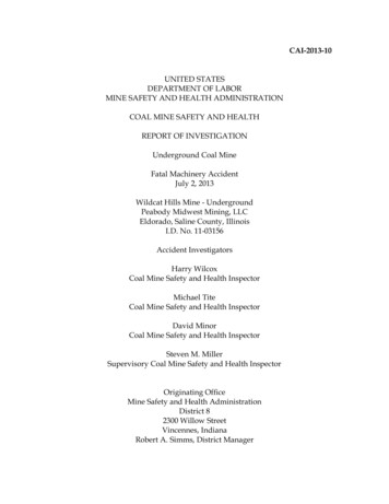
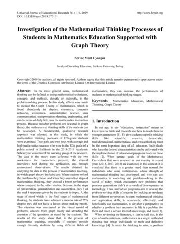
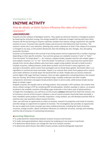
SYNOPSISEnvironmental FindingsPlague risk assessments were conducted for 9 locations inYosemite and the surrounding national forests visited bythe patients (Table 1; Figure 1). Within the park, 8 moresites were also evaluated for Y. pestis transmission and potential risk areas for transmission to humans.Sites Visited by Patient 1Figure 1. Locations of plague transmission risk assessments inand around Yosemite National Park, California, USA, August–October 2015.Forest) during this period (Table 1; Figure 1). He reportedthat he fed squirrels but did not touch them (L. Tovar Padua, pers. comm., 2016 Jan 7).On August 14, the Georgia Department of Public Healthpresumptively diagnosed bubonic plague for an 18-year-oldfemale resident of Georgia (patient 2) after she had becomesymptomatic on August 11. On the same day, CDC was notified of the suspected case and later confirmed the diagnosis.During the prior week, this patient had stayed in a rental homein Oakhurst, California, and visited Yosemite Valley, VernalFalls, Glacier Point, and Sentinel Dome in Yosemite, as well asNelder Grove, Lewis Creek, and Bass Lake in the adjacent SierraNational Forest (Table 1; Figure 1). Patient 2 reported havingobserved numerous squirrels in her vicinity at Vernal Fallsand Glacier Point but did not report having had any contactwith them.Neither patient reported seeing dead rodents. Wholegenome MLST showed that the genome sequence of Y.pestis isolates from each patient (blood culture from patient 1, bubo aspirate from patient 2) differed at 21 ORFs(Table 2; Figure 2), including 18 single-nucleotide polymorphisms (SNPs). Each patient was accompanied on thetrip by family members who did not become ill.2048On August 4, the Rainbow Pool Day Use Area (Table1; Figure 1) was evaluated and deemed to be an area oflow risk because of the lack of historical documentationof plague at this habitat and elevation, low abundance ofCalifornia ground squirrels observed in the day use area,and lack of other diurnal rodent species that are known Y.pestis reservoirs in this region. Limited burrow swabbingcollected no fleas. A visual evaluation of Yosemite Valley (Table 1; Figure 1) was postponed because it similarlylacked historical documentation of local Y. pestis transmission and because no reports of sick or dying rodents fromthis heavily visited area had been received. The next week,numerous healthy California ground squirrels were notedin Yosemite Valley. At Crane Flat Campground (Table 1;Figure 1), visual assessment and subsequent burrow swabbing suggested recent epizootic activity; California groundsquirrel abundance seemed to be very low relative to thenumber of burrows in the campground, abandoned burrowswere noted, and 134 Y. pestis–negative fleas were collectedfrom the entrances of 29 (31.5%) of 94 burrows sampled.Carrion flies (Calliphora latifrons) were also collected orobserved at several burrows. Few chipmunks were observed in the campground, but several Douglas squirrels(Tamiasciurus douglasii) were noted. Rodent trappingconducted the following day corroborated the low abundance of rodents (trap success rate 6.9%; Table 3). Y. pestis antibodies were detected in 1 (12.5%) of 8 Californiaground squirrels (Table 4), and a pool of 8 O. montana fleascollected from a seronegative California ground squirreltested positive by PCR for Y. pestis (Table 3). Subsequentwhole-genome MLST of Y. pestis recovered from this fleapool demonstrated 100% sequence identity across all ORFswhen compared with the isolate from patient 1 (Figure 2).Sites Visited by Patient 2The 7 locations visited by patient 2 were evaluated on August 18 and 19 (Table 1; Figure 1). Bass Lake was not visually assessed because of the historic lack of plague activity in this area. Visual assessments and burrow swabbingat Sentinel Dome, Vernal Falls, Nelder Grove, and LewisCreek found no obvious indications of Y. pestis transmission or increased human risk. Initial evaluation at GlacierPoint revealed several abandoned California ground squirrel burrows in close proximity to pathways and picnic areas. From the entrances of the 2 burrows swabbed, 21 fleasEmerging Infectious Diseases www.cdc.gov/eid Vol. 22, No. 12, December 2016
Plague, Yosemite National Park, 2015Table 2. MLST alleles in whole-genome sequences of Yersinia pestis isolates recovered from humans, animals, and fleas, YosemiteNational Park, California, USA, August 2015*†Y. pestis strain CO92MLST allele, ORFgenome positionMutation type‡Group 1 isolates§Group 2 1932114466-bp VNTR–LossYPO04454675491-bp INDEL–DeletionYPO0776Multiple9-bp VNTRLossGainYPO089498008915-bp PO13321498571SNPTCYPO1422161772518-bp PO22532531428SNPTAYPO255628718526-bp 13886839SNPCTYPO34903898668SNPTCYPO382842966998-bp INDEL–InsertionYPO40684587603SNPGT*INDEL, insertion/deletion; MLST, multilocus sequence typing; ORF, open reading frame; SNP, single-nucleotide polymorphism; VNTR, variable numberof tandem repeats; –, none.†PacBio (Pacific Biosciences, Menlo Park, CA, USA) sequencing of patient isolates yielded an average read length of 15,541 bp with 83,608 averagefiltered subreads and an average quality and coverage of 84.5% and 202 , respectively. Illumina MiSeq sequencing of flea and animal isolates yielded anaverage ContigN50 of 40,440 for the 6 assemblies and an average read coverage depth of 84.35 .‡Gain/loss and
SYNOPSIS the patients and environmental samples indicated that the patients had been exposed in different locations and that at least 2 distinct strains of Y. pestis we