Transcription
Welcome! This is the freePDF version of this book.Feel free to share and e-mailit to your friends.If you find this book useful,please support this projectby buying the printedversion at Amazon.com.Here is the Timothy Root, M.D.Root Eye DictionaryA "Layman's Explanation" of the eyeand common eye problemsWritten and Illustrated byTimothy Root, M.D.www.RootEyeDictionary.com1
Contents:IntroductionThe Dictionary, A-ZExtra Stuff- Abbreviations- Other Books by Dr. Root2
Intro 3
INTRODUCTIONGreetings and welcome to the Root Eye Dictionary. Inside these pagesyou will find an alphabetical listing of common eye diseases and visualproblems I treat on a day-to-day basis.Ophthalmology is a field riddled with confusing concepts andnomenclature, so I figured a layman's dictionary might help you "decode"the medical jargon. Hopefully, this explanatory approach helps removesome of the mystery behind eye disease.With this book, you should be able to:1. Look up any eye "diagnosis" you or your family has been given2. Know why you are getting eye "tests"3. Look up the ingredients of your eye drops.As you read any particular topic, you will see that some words areunderlined. An underlined word means that I've written another entryfor that particular topic. You can flip to that section if you'd like furtherexplanation, though I've attempted to make each entry understandableon its own merit. I'm hoping this approach allows you to learn moreabout the eye without getting bogged down with minutia . but if you areinterested in a topic, you can dive in as deep as you like!Even though this book is full of information, I discourage you fromtrying to diagnose your own eye problems . or opening your own eyeclinic in your garage. Many eye diseases present with similar symptomsand can only be diagnosed by examination under a microscope. When indoubt, it is best to consult with an actual eye doctor. You should lookupon this book as supplemental education and NOT as medical advice.With that said, I hope you find this information useful.Timothy Root, M.D.4
Root's DisclaimersHere are a couple of things to keep in mind as you read this book:Accuracy: With many of the topics here, I faced a dilemma betweenfactual accuracy and maintaining "understandability." When in doubt, I'vedecided to err on the side of understandability. You may occasionallyfind information that is not 100% technically accurate. This is a bookdesigned to improve comprehension through intuitive examples andmetaphor. If you need more exact definitions, I can point you towardmedical textbooks and journal articles that I've written for this purpose.Cartoons: A "dictionary" can be a rather boring textbook to read. Tokeep this book interesting, I've added many cartoons and comic strips.These silly jokes are not meant to downplay eye disease or disrespectpatients. Rather, this lighthearted approach is meant to keep this bookpalatable and easy to read. No offense is intended, and I hope youapproach these cartoons with the same good-natured outlook that I hadwhen I illustrated them.Opinion: If you see another doctor, you may find information in thisbook that conflicts with what you've been told about the eye. When indoubt, listen to the doctor who has actually examined your eyes. Thisbook reflects my own personal beliefs, medical experiences, and isspecific to the greater Daytona Beach area.5
6
A 7
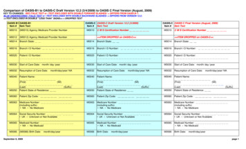
accommodation.Accommodation is the process by which the eyefocuses to see near objects. A normal eye, that is to say, an eye that isneither nearsighted nor farsighted, is naturally focused to see distantobjects clearly. To see close-up objects, such as when reading, theflexible lens inside the eye changes shape and becomes "rounder." Thisprocess is called accommodation and is quite versatile when young.After the age of 40, however, our lens becomes stiff and accommodationbecomes more challenging. We lose our ability to accommodate and webecome more dependent on glasses and bifocals with time. This loss ofaccommodation is called presbyopia.With accommodation, the lens becomes rounder.acetazolamide. An oral water pill that is used to treat glaucoma. Thispill is a diuretic and will dehydrate the body, but it will also dehydrate theeye and decrease eye pressure. This medication is also known as Diamoxand is normally used in cases of extremely high eye pressure where we'veexhausted our topical eye drop options, or used immediately after othereye surgery to avoid pressure spikes. Don't confuse this withacetomenophine (Tylenol). If you switch these, you might be up all nightpeeing instead of curing your headache. This pill is also used for peoplegoing to high altitudes to avoid mountain sickness. Side effects are minorbut can include tingling sensations in the fingers/toes and carbonatedsoft drinks may taste odd.8
Acular. This is an eye drop in the NSAID class of medicines. It is ananti-inflammatory drop with a mechanism similar to Advil or Motrin. Itis occasionally used to help with ocular discomfort but mainly used aftereye surgery. This class of medicines is good at decreasing the chance ofmacular edema (retinal swelling) after cataract surgery. It can sting a littlegoing in, however.acute glaucoma.Acute glaucoma is when the pressure inside theeye goes up suddenly. This usually occurs because of a sudden closure ofthe drainage "angle" inside the eye. With no drainage, the aqueoushumor fluid builds up and causes a spike in eye pressure that can lead torapid vision loss. Symptoms include extreme eye pain along with nauseaand halos seen around lights. Treatment is geared toward lowering thepressure and "breaking the attack," often with a laser, eye drops, anddiuretic pills like Diamox. Acute glaucoma is less common in the USA asmost people with glaucoma have chronic "open-angle" glaucoma. If aneye appears to be at risk for having an attack, then we will sometimesperform a prophylactic laser procedure called a laser peripheral iridotomy(LPI) to decrease the likelihood of this problem.acyclovir. An antiviral pill used for viral infections such as shingles(chicken pox) and herpetic eye disease. This medication is cheap andeffective, but requires a lot of pills to get the correct dosing. A similarmedicine we use is called Valtrex (valacyclovir). Some people with theserecurring infections will take a maintenance dose of acyclovir to decreasethe chance of a new outbreak.Adie's pupil.This is a neurologic disorder in which one eye becomesdilated. Most patients have no symptoms or visual complaints, but afriend points out that one of their pupils is now much larger than theother. Also, the pupil does not seem to constrict normally with light. AnAdie's pupil usually occurs from damage to one of the nerve clustersbehind the eye (inside the eye socket) that controls pupil constriction.This damage can occur from an otherwise harmless viral infection suchas a common cold. The problem is usually temporary and goes away aftera few months. The opposite condition is called Horner's syndrome.Horner's causes pupil constriction and is potentially dangerous.9
after-cataract.This is a cloudy membrane that forms on the backsurface of an implant lens inside the eye after cataract surgery. Thisopacity can form months or years after a successful cataract operationand can cause blur and glare symptoms (similar to the original cataract).These "after cataracts" are not a complication from cataract surgery, butrather a continued proliferation of tissue inside the eye (similar to scartissue). After-cataracts are easy to treat with a laser. A YAGcapsulotomy can be performed to create a hole through the opaquemembrane. This is a simple, painless procedure, and once performed the"after cataract" does not typically reoccur.Alaway. An effective over-the-counter allergy drop. Alaway containsthe medicine ketotifen and is usually used twice a day. Allergy drops aregood for itching and swelling, and can make the eye feel less sensitive.Alaway and Zaditor (which contains the same active ingredient) are twoof my favorite over-the-counter allergy drops.allergic conjunctivitis.Theeyes are particularly sensitive toenvironmental allergens. Symptoms areusually bilateral, with both eyes beingitchy and puffy. Eyelid swelling can beso bad that you look like you've been ina fight . we call these "allergic shiners."Treatment for allergic conjunctivitisinvolves cool compresses, antihistamineallergy drops, and occasionally mildsteroid drops.allergy drops.Allergy drops are commonly used to treat ocularitching and swelling. There are several types of allergy drops on themarket. The first generation antihistamine drops like Opcon-A areeffective but tend to give short-lived relief. Second generationantihistamines like Alaway/Zaditor are more effective and what Irecommend for most of my patients. Prescription strength allergy dropsare also available such as Bepreve, Pataday, and Lastacaft.10
Alphagan. A glaucoma drop used to lower eye pressure. This dropwent generic so many people have switched to generic brimonidine ormoved up to Alphagan P.Alphagan P.This is a glaucoma eye drop designed to lower eyepressure. It is actually a "new" formulation of brimonidine that has alower concentration of active drug and a less harsh preservative in it.Despite the decreased concentration, the drop appears to have the sameefficacy as the old Alphagan but with less irritating side effects.Alrex.A mild steroid eye drop. Useful in cases of ocular inflammationand irritation. This drop has the same ingredient (loteprednol) asLotemax but with a third of the steroid concentration. By reducing thesteroid concentration, this decreases the chance of untoward reactionssuch as premature cataract formation and glaucoma pressure spikes.ALT.This stands for Argon Laser Trabeculoplasty and is a laserprocedure designed to lower the eye pressure in people with glaucoma.This procedure involves using a "hot" laser to burn spots into thetrabecular meshwork (the drainage filter of the eye). By doing this, scartissue forms that opens up the meshwork and creates better flow. Whileeffective and well tolerated, the pressure improvements of ALT tend towear off in a couple of years. The procedure can only be done oncebecause of the scar formation. ALT is largely being replaced by a similarprocedure called SLT. With SLT a "cold" YAG laser is used to createsimilar spots on the trabecular meshwork but instead of creating heatinduced scars, the drainage cells are merely stimulated. This promotesbetter flow through the drain without creating permanent tissue damage.This means that SLT can be repeated if it wears off. SLT is slowlybecoming first-line therapy for many doctors treating glaucoma.11
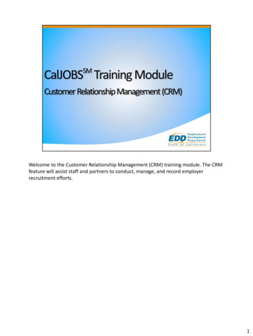
amblyopia.Also known as"lazy eye." Amblyopia occurs at ayoung age from disuse when an eyedoesn't see well. A child's visualnervous system is still developinguntil age seven. If during thisdevelopmental period, one eye haspoorer vision, the "brain wiring"for that eye does not form asstrongly as the better eye. This htedness or from other visualproblems such as congenitalcataract. This imbalance can alsooccur if the eyes are in pooralignment (like being cross-eyed).If detected early, amblyopia can bereversed.Thisistypicallyaccomplished with glasses andpatching therapy - by patching the"good eye" closed, this forces thelazy eye to "work" and reform itswiring. There is no way to fix alazy eye in adulthood as the brainwiring has already formed and theamblyopic eye will never see quiteas well.amiodarone.This is a commonly used oral medication that is usedto help with abnormal rhythms of the heart (arrhythmias). Whileeffective, amiodarone can occasionally cause changes in the eye. One ofthese changes is "corneal verticillata," which are pigment deposits in theclear cornea that can be seen with the slit lamp microscope. Thesecorneal changes rarely cause any appreciable vision problems, but ifsevere may prompt a change in medication.12
amphotericin B.An antifungal medicine that can be compounded(see fortified antibiotics) and used to treat fungal eye infections. Thereare not many antifungal eye medications out there - the only other eyedrop easily available is Natamycin.Amsler grid.A checkered pattern used at home for detecting retinaldistortions, such as from macular degeneration or an epiretinalmembrane. To use, a patient is instructed to look at the central dot whilecovering an eye. If the surrounding lines are missing or look distorted,then the surface of the retina (which acts like "film in a camera") may bedistorted as well. This prompts further evaluation and retinal scans likean OCT to find these problems. My patients are often sent home with acopy of this grid to monitor their vision at home. Stop by my office for afree copy if you don't already have one.anesthetic drops.There are several drops we use to anesthetizethe surface of the eye. The most common one is called proparacaine,though we occasionally use tetracaine. These drops are very similar tothe "novacaine" that a dentist uses . but fortunately we don't have to usea needle to apply it! Numbing drops make it easier to check eyespressure using applanation tonometry. We also use these drops prior tocataract surgery to minimize discomfort. Unfortunately, anesthetic eyedrops are not safe for home use. The medications are toxic to thecorneal surface when used repeatedly and will keep surface wounds fromhealing. For pain, we prescribe ointments, bandage contact lenses, andcan even patch an eye shut if needed (see patching).angle. In regards to the eye, the "angle" usually refers to the drainageangle inside the eye where excess ocular fluid (aqueous) is reabsorbedback into the blood stream. This angle is located at the intersection ofthe iris and the white sclera of the eye . in other words, in a 360-degreering where the "white" of the eye meets the "colored part" of the eye. Ifthis angle closes down, then you can have an angle-closure glaucoma,also known as acute glaucoma.13
anisocoria. This is when the pupils are of unequal size. Manypeople have slightly different sized pupils and this is considered normal.Large differences between the eye is not normal, however. See dilatedpupil for more information.anterior chamber.This is the fluid-filled space in the front partof the eye, located immediately behind the cornea but in front of the iris.This "chamber" is filled with clear aqueous fluid and easy to examine bythe doctor using the slit lamp microscope in the office. In cases oftrauma or iritis, the anterior chamber may be filled with inflammatorycells that can be detected in the office. In more severe cases of trauma,blood can fill this space . this is called a hyphema. If you have acuteglaucoma, the anterior chamber can be shallow as the aqueous fluidcannot drain out properly.14
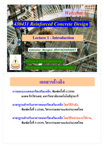
The "anterior chamber" is the fluid-filled front portion of the eye.This fluid is called "aqueous."antibiotic.This usually refers to a drop or pill that is designed to killor decrease the proliferation of bacteria. The eye is well protected frominfection by the conjunctiva and the corneal epithelium. In addition, thetear film contains antimicrobials and the tear flow itself tends to washaway pathogens. The eye also harbors a host of non-pathogenic bacteriathat competitively prohibit new bacteria growth. However, these eyedefenses can be breached by trauma, improper tearing, or contact lenswear and lead to an infection. Topical antibiotics work best for the eyegiven the avascular nature of the cornea.anti-VEGF.This is a class of medicines that are designed to combatneovascularization inside the eye and decrease blood vessel leakage.They are usually used for treating problems like macular edema, causedby wet macular degeneration, though occasionally they are used fortreating swelling from other sources such as diabetic retinopathy orcentral retinal vein occlusion. These medicines work by decreasingleakage of fluid across abnormal blood vessels in the retina. The originalanti-VEGF medication used was the injection medicine Avastin whichwas originally formulated for combating colon cancer. Lucentis and Eyleaare newer anti-VEGF medications that may be more effective with lesssystemic side effects, but are quite costly when compared to Avastin.Refer to the entries on VEGF and neovascularization to betterunderstand how these medications work.15
Anti-VEGF medications target leaky capillaries in the retina.aphakia. This is when the natural lens has been removed from the eye(such as after cataract surgery) but has not been replaced with a new lensimplant. In the early days of eye surgery, cataracts were removed but notreplaced with anything. The vision was better but "aphakic people"required thick coke-bottle glasses to see well. Today, most people receivea new implant so aphakia is rare. Most people with aphakic eyes havehad some kind of trauma or complicated cataract surgery that precludesthe placement of a modern lens implant.applanation tonometry.This is a method for checking thepressure inside the eye. The eyeball is a closed ball of fluid so that there isno good way to measure the internal pressure of the eyeball directly.However, we can estimate the eye pressure by pushing on the surface ofthe eye and feeling "how hard" is seems. This is similar to kicking a cartire to estimate the air pressure inside. With applanation tonometry, aflat probe is pushed onto the surface of the cornea (the clear window thatmakes up the front of the eye). The probe is pushed hard enough toflatten a small round area of the cornea. By looking at the size of the16
circle flattened, and examining the amount of pressure used, the internaleye pressure can be calculated. The machine we use for this is called aGoldmann Applanation Tonometer. It is attached to the slit lampmicroscope and it looks like a blue light when in use. You don't feel thismeasurement as we use anesthetic drops ahead of time.aqueous. The aqueous "humor" is the fluid that fills the front part ofthe eye (the anterior chamber). This clear fluid maintains the shape ofthe eye and affects the eye pressure. Glaucoma can occur if the pressuregets too high, and most glaucoma treatments are geared towardregulating the production and drainage of the aqueous fluid. Severalstructures in the eye, such as the cornea and lens, contain no bloodvessels and rely on the aqueous humor to provide them with nutritionalsupport.arcus senilis.This is a white haze or ring on the cornea that occurswith age. The cornea is the clear window that makes up the front of theeye. This living tissue has no blood vessels running through it because itneeds to be perfectly clear. To get its nutrition, the cornea depends uponthe tear film on the outside, the aqueous fluid on the inside, and bloodvessels on the sclera (the white of the eye) that run right up to the edgeof cornea before stopping. Lipid and cholesterol fats travel in our bloodstream, and over a lifetime, can leach out of the blood vessels anddeposit themselves in a ring in the cornea. This white ring is called arcussenilis and is a normal aging change that has no effect on vision. When Isee this in only one eye (or in a young person) I start looking into otherproblems such as circulation or cholesterol abnormalities.AREDS Study.This stands for the Age-Related Eye Disease Study.This large study was conducted to study the effects of vitaminsupplements in slowing the progression of macular degeneration. Thestudy showed that certain antioxidants were more helpful, such asVitamin A, Vitamin C, and Vitamin E and the metals zinc and copper(cupric acid). While these vitamins are found in a healthy diet, the highdoses used in the AREDS trial were much higher than normalmultivitamin tablets and are difficult to obtain through normal foodintake.
Greetings and welcome to the Root Eye Dictionary. Inside these pages you will find an alphabetical listing of common eye diseases and visual problems I treat on a day-to-day basis. Ophthalmology is a field riddled with confusing concepts and nomenclature, so I figured a layman