Transcription
Optics, Photonics and Lasers:Proceedings of the 3rd International Conferenceon Optics, Photonics and Lasers (OPAL' 2020)21-22 October 2020Tenerife (Canary Islands), SpainEdited by Sergey Y. Yurish
Sergey Y. Yurish, EditorOptics, Photonics and LasersOPAL’ 2020 Conference ProceedingsCopyright 2020by International Frequency Sensor Association (IFSA) Publishing, S. L.E-mail (for orders and customer service enquires): ifsa.books@sensorsportal.comVisit our Home Page on http://www.sensorsportal.comAll rights reserved. This work may not be translated or copied in whole or in part without the written permissionof the publisher (IFSA Publishing, S. L., Barcelona, Spain).Neither the authors nor International Frequency Sensor Association Publishing accept any responsibility orliability for loss or damage occasioned to any person or property through using the material, instructions, methodsor ideas contained herein, or acting or refraining from acting as a result of such use.The use in this publication of trade names, trademarks, service marks, and similar terms, even if they are notidentifies as such, is not to be taken as an expression of opinion as to whether or not they are subject to proprietaryrights.ISBN: 978-84-09-24421-8BN-20201015-XXBIC: TTB
3d International Conference on Optics, Photonics and Lasers (OPAL' 2020)21-22 October 2020, Tenerife, SpainContentsContents . 3Foreword . 5Increasing Microcirculation in the Surface Layer of Lower Limb Tissue with Diabetic FoodUncler by Laser Stimulation . 6K. S. Beloshenko, I. A. Riabenko, K. S. GalichevaCan the Near-field Optics be used for Antiviral Therapy? . 10V. Lozovski,, N. Rusinchuk and V. LysenkoOptical Sensing of Biological Liquids by Means of Gold Nanoparticles with Biospecific Surfaces. 12V. Z. Lozovski and M. A. RazumovaSpirally Polarized Sources with Cosine-circular Coherence. 14J. C. G. de Sande, G. Piquero, R. Martínez-Herrero , M. Santarsiero and F. GoriSpectroscopy of Stationary Ultracold Ca-40 Plasma Prepared by Continuous Wave Lasers . 16B. B. Zelener, S. A. Saakyan, K. P. Galstyan, E. V. Vilshanskaya and V. A. SautenkovSimulation of Mueller Matrix Polarimetry with Full Poincaré Beams and a CCD Camera . 18J. C. Suárez-Bermejo, J. C. González de Sande, M. Santarsiero and G. PiqueroTwo-wavelengths Interferometry as Confocal Microscope Extension to Provide 3D ImagingCapabilities . 20M. Stašík, F. Kaván, M. Mach, K. Sedláčková, J. Kredba and M. ŠpínaFresnel-Kirchoff Integral Method Comparison with Scattering Theory for the Studyof Volume Dielectric Bodies . 26I. Taleb, C. Neipp, F. J. Martínez, M. L. Álvarez, E. Calzado, A. Márquez, J. Francés, S. Gallegoand A. BeléndezNew Ultrashort OPCPA Petawatt Class Beamline for Vulcan Laser Facility . 30N. Baktash, M. Galimberti, P. Oliveira, I. Musgrave, and C. Hernandez-GomezOptical Phase Readout Instrument for Picometer-level Precision Heterodyne Interferometers. 32J.J. Esteban Delgado, M. Dovale Alvarez, T. Schwarze, C. Vorndamme, I. Bykov, P. Martínez Cano,G. Fernández Barranco, K. Yamamoto, A. Pizzella, Fahri Öztürk, D. Penkert, Jens Reiche and G. HeinzelRecent Progress on High Harmonic Generation Source at ELI Beamlines. 34O. Hort, M. Albrecht,, O. Finke,, M. Jurkovič,, R. Lera, D. D. Mai, E. Klimešová, S. Espinoza,S. Richter, J. Gautier, S. Sebban, R. Antipenkov, F. Batysta, J. Novák, J. T. Green, M. Krikunova,,J. Andreasson, and J. NejdlGhost Modalities with Classical Correlated Photons Emitted by Semiconductor Devices: QuantumMetrology 64 Years after Hanbury-Brown & Twiss . 37Wolfgang ElsäßerSpectroscopic Analysis of SWCNT Dimer: An Ab Initio Approach . 40D. SharmaGeneral Framework to Derive Casimir Force of Electromagnetic Field in Non-inertial ReferenceFrames . 42B. Markowicz, K. Dębski, M. Kolanowski, W. Kamiński, A. DraganOptical Development Steps of a Fully Integrated Miniature Spectrometer Designed for InjectionMolding Fabrication . 44M. Haupt, U. H. P. Fischer-HirchertIn Vitro Study of Ti Implants with Laser-functionalized Surface . 49Yu. Karlagina, G. Odintsova, E. Egorova, N. Shchedrina, C. Zernitskaia, K. Doll, B. Chichkovand V. VeikoParticle Size and Density of Dispersion Phase Measurement by Light Scattering Methods . 51Sh. M. Ismailov, Yu. D. Arapov, V. G. Kamenev and A. A. Tavleev3
3d International Conference on Optics, Photonics and Lasers (OPAL' 2020)21-22 October 2020, Tenerife, SpainDetermination of Sound Speed in Optical Fibers Based on the Stimulated Mandelstam-BrillionScattering Effect . 54A. A. Tavleev, Yu. D. Arapov, P. V. Kubasov, Sh. M. Ismailov4
3d International Conference on Optics, Photonics and Lasers (OPAL' 2020)21-22 October 2020, Tenerife, SpainForewordOn behalf of the OPAL’ 2020 Organizing Committee, I introduce with pleasure these proceedingsdevoted to contributions from the 3rd International Conference on Optics, Photonics and Lasers(OPAL’ 2020). The conference is organized by the International Frequency Sensor Association (IFSA)in technical cooperation with our media partners: Institute of Physics (IOP), UK and MDPI Photonicsjournal (ISSN 2304-6732), Switzerland.In keeping with tradition begun in 2018 in Barcelona, Spain and in 2019 in Amsterdam, The Netherlandsthe Conference attracts researchers and practitioners in the related fields, from around the worldincluding 4 keynote speakers from Germany and India.The conference program provides an opportunity for researchers interested in optics, photonics andlasers to discuss their latest results and exchange ideas on the new trends and challenges. The mainobjective of the Conference is to encourage discussion on a broad range of optic related topics and tostimulate new collaborations among the participants.The conference proceedings contains all papers of regular and keynote presentations. We hope thatthese proceedings will give readers an excellent overview of important and diversity topics discussedat the conference. Based on the proceeding’s contributions, selected and extended papers will besubmitted by the authors to the special issue of ‘Sensors & Transducers’ journal (ISSN: 2306-8515,e-ISSN 1726-5479) or Photonics journal (ISSN 2304-6732). The limited number of articles, publishedin ‘Sensors & Transducers’ journal will be invited to be extended for ‘Advances in Optics: Reviews’,Vol. 5, Book Series, which will be published in 2021.We thank all authors for submitting their latest work, thus contributing to the excellent technicalcontents of the Conference. Especially, we would like to thank the individuals and organizations thatworked together diligently to make this Conference a success, and to the members of the InternationalProgram Committee for the thorough and careful review of the papers. It is important to point out thatthe great majority of the efforts in organizing the technical program of the Conference came fromvolunteers.Prof., Dr. Sergey Y. YurishOPAL’ 2020 Chairman5
3d International Conference on Optics, Photonics and Lasers (OPAL' 2020)21-22 October 2020, Tenerife, Spain(006)Increasing Microcirculation in the Surface Layer of Lower Limb Tissuewith Diabetic Food Uncler by Laser StimulationK. S. Beloshenko 1, I. A. Riabenko 1, K. S. Galicheva 11V. N. Karazin Kharkiv National University, 4 Svobody Sq., Kharkiv, 61022, UkraineTel.: 380989824245, fax: 380577050241E-mail: kbeloshenko@karazin.uaSummary. This work is devoted to the study of the mechanisms of laser radiation effect on tissues of patients with diabeticfoot ulcer (DFU). When exposed to a laser beam (radiation power of 50 mW, λ 635 nm), patients experience increasedmicrocirculation due to the increase in the mass flow of blood in the capillaries. To confirm the working hypothesis, sugarconcentration in the blood, temperature distribution over the lower limbs before and after laser irradiation, systolic and diastolicblood pressure are measured. Wound healing is much faster, taking into account the daily procedures on the Barva apparatus.Skin condition, color and turgor had a healthier appearance after the 3 procedures. The approach in treatment and rehabilitationwith non-invasive methods available is studied. The building of a physical-mathematical model of the process based on thepolyelastic model of the sample is planned. The data obtained fully confirm the hypothesis.Keywords: Barva apparatus, Diabetic foot ulcer, Laser irradiation, Thermography.1. IntroductionAccording to the International Diabetes Federation,at the end of 2013, more than 382 million patients withdiabetes mellitus (DM) were registered in the world,expected to rise to 592 million by 2035 [1]. It is alsoknown that every 15 years the number of patients withDM doubles [2]. One of the most dangerouscomplications of DM is diabetic food uncler (DFU).This complication is observed in 15-25 % of patients[3-5]. To date, the treatment of DFU in most cases endswith lower-limb amputations (LLAs). In the world,every 40 seconds one such operation is performed [6].The UN calls on all countries of the world to take thenecessary measures to reduce the number ofamputations at least twice. Moreover, more than atrillion US dollars are spent annually on the fightagainst DM and its complications worldwide [7].DFU is associated with a significant deteriorationin blood microcirculation in the lower extremities inpatients with DM [8]. Therefore, the aim of the workis to study the laws of action of low molecular weightelectromagnetic radiation of the red spectrum on themicrocirculation of blood in the lower extremities ofpatients with DM.2. Materials and Methods2.1. Physical Model of Fluid RedistributionThe methods of laser medicine and the mainmechanisms of interaction of laser radiation withtissues have been known for a long time [9, 10]. Photothermal and photochemical transformations ofcontinuous laser radiation can lead to significantresults both in the prevention of diseases [10] and intheir treatment [11]. One of the methods of analyzingthe effect of laser radiation on the surface layer is givenby the thermodynamic approach. With the approach,we assume that one-photon absorption of laserradiation occurs in the surface layer of body tissues inaccordance with the Bugrea-Lambert-Behr law. Wecan estimate the penetration depth of laser radiationh 1 lnII0,(1)at which the radiation intensity is decreased by factore, if we take the extinction coefficient for λ 532 nmequal to α 3.33 cm-1 [12], then the penetration depthwill be 0.3 cm. Accordingly, the most of radiation isabsorbed by the skin, and we can simulate the skin asa black body. Photothermal conversion consists in thefact that the short-wave laser radiation is converted tolong-wave radiation, which was recorded using aFluke Ti400 thermal imager. From the Wien'sdisplacement law [13], we can calculate the maximumof the long-wavelength radiation band within the rangeof 8-14 μm, provided that the skin surfacetemperatures is within the range of 35-37 С. It is notedthat the integral power of the secondary radiation canbe estimated on the base of Stefan-Boltzmannlaw [13]:I T 4 ,(2)where I 0.05 W/cm2 at temperature T 300 K, ε 1,σ 5.67 10-8 W/(m2K4) is the Stefan-Boltzmannconstant.2.2. Anatomical Model of the DiseaseThe main causes of the development of DFU areperipheral arterial disease (PAD) [14]. Other factors6
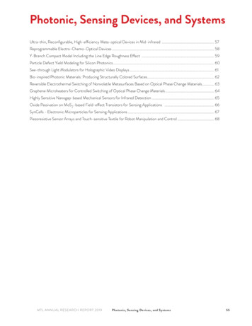
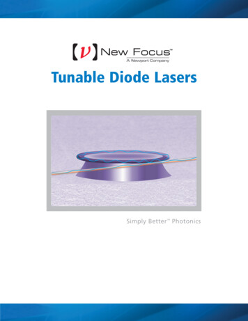
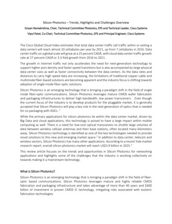
3d International Conference on Optics, Photonics and Lasers (OPAL' 2020)21-22 October 2020, Tenerife, Spainthat are responsible for diabetic foot infections arerepetitive trauma and super-imposing foot infections[15, 16].In the foot, the own plantar finger arteries(aa. Digitdles plantdres propriae) which are shown inFig. 1, the dorsal digital arteries (aa. Digitdlesdorsdles) and the calcaneal rete which are shown inFig. 2 (rete calcalcum) are primarily and potentiallydestroyed.Fig. 1. The dorsal arteries and nerves of the foot [17].known reactions of the conversion of ATP to ADP areexothermic, and therefore they maintain a specialtemperature inside tissue cells in the region of 37 C.Moreover, these reactions require oxygen supply in anamount of 7 ppm/min [18] to maintain thermodynamicequilibrium and chemical potential in the tissues of thesurface layer [19]. Otherwise, these reactionsattenuate, which leads to slowdown in metabolism,lowering the temperature inside the tissues andimbalance of the chemical potential [19], and this inturn leads to disturbance of microcirculationin the tissues.The experiment may serve as evidence ofmicrocirculation disturbance. In a random sample of30 people, the patient’s legs were irradiated with theBarva apparatus (radiation power of 50 mW,λ 635 nm) in the morning on an empty stomach for30 minutes. Before irradiation, a blood sampling wastaken to conduct an express blood test for sugarconcentration using an Accu-Chek Active glucosemeter. Blood pressure measurements were alsoperformed with Omron M300. The averaged data areshown in Tables 1, 2 and 3.Table 1. Change in blood glucose.Left armLeft legRight legBefore irradiation113.33.5After irradiation11.56.3 (toe)9.1 (toe)Table 2. Change in blood pressure.Fig. 2. Right leg, posterior view. Neurovascular structuresin the deep posterior compartment after partial removalof the triceps surae and the deep layer of the fasciaof the leg [17].Every vessel in the leg has tissues surrounding it,the parameters of which will be taken into accountwhen constructing the polyelastic model.To assess the redistribution of temperature, it isimportant to make a thermogram taking into accountthe capture of the anterior and posterior regions of theknee joint, the proximal and distal parts of the anteriorand posterior regions of the lower leg.3. ResultsThe study observed a temperature increase of0.5-2 C after phototherapy with a red laser. Whenassessing the ratio of power radiated by the skin ofpatients before and after the procedure, the change is12-13 %, but in absolute terms it is about 4 mW/cm2.From the above estimates, it can be concluded that70 % of the laser radiation is used for heating, and30 % is spent on photochemical transformations. Whentaking into account the photon energy of 3.73 10-27 J,it can be assumed that covalent bonds cannot be brokenonly by radiation, and laser radiation changes only thethermodynamic conditions for reactions in the surfacelayer of the patient's skin. It should be noted that theLeft armRight armBefore irradiation104/65103/74After irradiation123/81114/74Table 3. Pulse change.Left armRight armBefore irradiation7576After irradiation7173Thermodynamic control of the surface temperaturewas carried out using a Fluke Ti400 thermal imagerwith precompensation for the background temperature(T 296.15 K) and emissivity (ε 0.86). Theemissivity level was selected by comparison with theOmron Gentle Temp 720 thermal resistance. Thebackground temperature was measured on a foil fixednext to the patients. Thermograms frontal anterior andposterior planes of the feet of patients are shown inFig. 3a,b and Fig. 4a,b.Fig. 3. A thermoregulatory response of the front surfaceof lower legs (a-before irradiation, b-after irradiation).7
3d International Conference on Optics, Photonics and Lasers (OPAL' 2020)21-22 October 2020, Tenerife, SpainFig. 4. A thermoregulatory response of the back surfaceof lower legs (a-before irradiation, b-after irradiation).The data evident that the temperature distributionis equalized over the entire surface of the lower limb,which indicates changes in thermodynamicequilibrium. Zonal temperature distributions areshown in Fig. 5 a, b and Fig. 6 a, b.Fig. 5 а. Zonal temperature distributions of the front surfaceof lower legs before irradiation.Fig. 5 b. Zonal temperature distributions of the frontsurface of lower legs after irradiation.The maximum temperature in the front and rearareas of the knee joint is reduced by 0.9-1.2 C. In theproximal sections of the anterior and posterior regionsof the lower leg the maximum temperature decreasesby 0.8-1.4 C. In the proximal sections of the anteriorand posterior regions of the lower leg the maximumtemperature decreases by 0.8-1.4 C. In the distal partsof the anterior and posterior regions of the lower legthe maximum temperature decreases on average by1 C. In the surface layer of the foot the maximumtemperature increases by 0.5-1.4 C.Fig. 6 a. Zonal temperature distributions of the back surfaceof lower legs before irradiation.Fig. 6 b. Zonal temperature distributions of the backsurface of lower legs after irradiationThis suggests that in the own plantar finger arteries,dorsal finger arteries and the calcaneal rete, the amountof blood increases, so the inner plantar nerves and theheel of the tibial nerve in the affected leg receiveenough nutrients to improve the innervation of thesurface layer of the foot after irradiation.The increase in sugar level in the lower limbsindicates improvement in microcirculation. Thevenous capillaries, venules and veins, as well as thevenous part of the arterio-venous anastomoses,expanded in diameter, thereby increasing thedifference in osmotic potentials between the arterialand venous channels. With this change in the osmoticpotential of a large circle of blood circulation in thelower limbs, blood outflow will increase, which meansthat blood inflow will also be increased according toBernoulli’s law [20].All these qualitative changes inhibit thedevelopment of DFU and make it possible toavoid LLAs.However, we must bear in mind that one tion under conventional heating of thepatient. Since when heated, the heat transfer method isconvection. With convection heating in the humanbody, processes will start in which water willaccumulate in the surface layer of the skin (since water8
3d International Conference on Optics, Photonics and Lasers (OPAL' 2020)21-22 October 2020, Tenerife, Spainhas a heat capacity of 4200 J/(kg C), which is10 orders of magnitude higher than the heat capacity ofdry skin, therefore, the water layer in the transitionprocess is a good storage of thermal energy), therebyforming a protective layer on the skin surface, whichwill prevent heat transfer to deeper layers. In order toachieve energy transfer through radiation, it would benecessary to increase the emitter temperature over600 C, which is dangerous for biological processes.4. ConclusionsBased on the obtained data on the improvement ofmicrocirculation after irradiation with a red laser, wecan assume that the absorption of this wavelength wasdue to those tissues that do not reflect it, and these areall the tissues other than red. If we take into account alarge circle of blood circulation, then the absorption ofthis electromagnetic radiation was due to veins,venules and capillaries of the venous channel, whilethe rest of the tissues are considered «transparent» forthis spectrum and scattering is neglected. With thisabsorption, the energy that acted on the venous vessels,and specifically on their walls, increased the potentialenergy of the elements of the connective tissue. Themeasurement of the pulse and blood pressure ofpatients will be taken into account when constructing apolyelastic model of the system for further descriptionof the experiment.AcknowledgementsGood memory of Korobov Anatoly Mikhailovich,PhD, the head of the laboratory of quantum Biologyand quantum Medicine in V. N. Karazin KharkivNational University.References[1]. International Diabetes Federation, 6th Edition, Brussels,2013, IDF Diabetes Atlas, http://www.idf.org/diabetesatlas[2]. DIAMOND Project Group, Incidence and trends ofchildhood Type 1 diabetes worldwide 1990-1999,Diabetic Medicine, Vol. 23, Issue 8, 2006,pp. 857-866.[3]. P. Zhang, et al., Global epidemiology of diabetic footulceration: A systematic review and meta-analysis,Annals of Medicine, Vol. 49, Issue 2, 2017,pp. 106-116.[4]. N. C. Schaper, J. Apelqvist, K. Bakker, Theinternational consensus and practical guidelines on themanagement and prevention of the diabetic foot,Current Diabetes Reports, Vol. 3, Issue 6, 2003,pp. 475-479.[5]. J. A. Mayfield, et al., Preventive foot care in diabetes,Diabetes Care, Vol. 27, 2004, 63.[6]. S. H. Won, et al., Risk factors associated withamputation-free survival in patient with diabetic footulcers, Yonsei Medical Journal, Vol. 5, Issue 5, 2014,pp. 1373-1378.[7]. A. Raghav, et al., Financial burden of diabetic footulcers to world: a progressive topic to discuss always,Therapeutic Advances in Endocrinology andMetabolism, Vol. 9, Issue 1, 2018, pp. 29-31.[8]. N. Singh, D. G. Armstrong, B. A. Lipsky, Preventingfoot ulcers in patients with diabetes, Jama, Vol. 293,Issue 2, 2005, pp. 217-228.[9]. J.-L. Boulnois, Photophysical processes in recentmedical laser developments: A review, Lasers inMedical Science, Vol. 1, Issue 1, 1986, pp. 47-66.[10]. L. Goldman (Ed.), The Biomedical Laser: Technologyand Clinical Applications, Springer Science & BusinessMedia, 2013.[11]. P. Schaaf (Ed.), Laser Processing of Materials:Fundamentals, Applications and Developments,Vol. 139. Springer Science & Business Media, 2010.[12]. H.-J. Wei, et al., Determination of optical properties ofnormal and adenomatous human colon tissues in vitrousing integrating sphere techniques, World Journal ofGastroenterology, Vol. 11, Issue 6, 2005, 2413.[13].E. Hecht, 4th Edition, Optics, Addison Wesley Longman,Inc., 1998.[14]. M. H. Criqui, Peripheral arterial diseaseepidemiological aspects. Vascular Medicine, Vol. 6,Issue 1, 2001 pp. 3-7.[15]. A. J. M. Boulton, The pathway to foot ulceration indiabetes, Medical Clinics, Vol. 97, Issue 5, 2013,pp. 775-790.[16]. L. A. Lavery, et al., Diabetic foot prevention: Aneglected opportunity in high-risk patients, DiabetesCare, Vol. 33, Issue 7, 2010, pp. 1460-1462.[17]. M. Schuenke, E. Schulte, et al., General Anatomy andthe Musculoskeletal System (Thieme Atlas ofAnatomy), New York, Thieme, 2005.[18]. S. S. Simakov, A. S. Kholodov, Computational studyof oxygen concentration in human blood under lowfrequency disturbances, Mathematical Models andComputer Simulations, Vol. 1, Issue 2, 2009,pp. 283-295.[19]. M. Marcotte, M. Le Maguer, Mass transfer in cellulartissues. Part II: Computer simulations vs experimentaldata, Journal of Food Engineering, Vol. 17, Issue 3,1992, pp. 177-199.[20]. D. Chapelle, P. Moireau, General coupling of porousflows and hyperelastic formulations – Fromthermodynamics principles to energy balance andcompatible time schemes, European Journal ofMechanics-B, Vol. 46, 2014, pp. 82-96.9
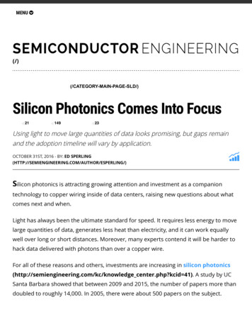
3d International Conference on Optics, Photonics and Lasers (OPAL' 2020)21-22 October 2020, Tenerife, Spain(008)Can the Near-field Optics be used for Antiviral Therapy?V. Lozovski 1,2, N. Rusinchuk 1 and V. Lysenko 21Institute of High Technologies Taras Shevchenko National University of Kyiv,56 Volodymyrska str., 01602 Kyiv, Ukraine2V. Lashkariov Institute of Semiconductor Physics, National Academy of Sciences of Ukraine,45 Nauki ave., 01000 Kyiv, UkraineTel.: 380445213566E-mail: v.lozovsk@gmail.comSummary: When nanoparticles solution is adding to viruses preparation the nanoparticles may adsorb on the virus surfacedue to Van der Vaals forces. As a result of such interaction a stable nano-sized system is formed. In such system the localelectric field is redistributed by the nanoparticles and viruses, which in turn leads to the local field enhancement effect andappearance of ‘hot spots’ on the virus surface. Consequently, there are local field gradients on the virus surface, and this is thereason for ponderomotive forces acting on the virus. Results of calculations show that on the surface of influenza virus nearthe Au nanoparticles of 5 nm diameter the forces may lead up to 70 nN. Existed experimental studies of mechanical propertiesof viruses indicate that mainly the force higher than 30 nN leads to irreversible deformation of the virus particle. Hence, themain idea of the work is to show that ponderomotive forces are the reason for destruction of viruses mixing with nanoparticles,which was demonstrated by microscopy studies for many times. This is the novelty of the work as for now the exact mechanismof the virus destruction under the action of nanoparticles is not known.Keywords: Nanoparticle, Virus, Antiviral action, Near-field interaction, Local field enhancement, Ponderomotive forces.1. IntroductionThe interaction between nanoparticles and virusescan be the reason of a lost by the virus its infectivity’s[1-6]. However, the mechanism of this action is notknown. There are different suppositions for thismechanism. We would like to discuss, to extend and toimprove the model of influence of nanoparticle on thevirus infectivity based on the local-field enhancementeffect proposed earlier in [3, 5]. The main feature ofproposed model is the arising of the domains of highlocal field (hot spots) at the virus surface. It was shownthat the higher the local field gradient is, the higherantiviral action is without explanation of thisdependence. Here we would like to consider thisdependence and try to explain it via the action ofponderomotive forces.2. The ModelThe most human viruses have the quasi-sphericalshape with linear dimension about 100 nm. Thecharacteristic dimensions of the nanoparticles areabout few nanometers. Then, we can use the modelwhen the virus is spherically shelled solid nanoparticlewhere core is characterized by dielectric constant 𝜀and the virus shell is characterized by dielectricconstant 𝜀 . The dielectric constant of thenanoparticle is 𝜀 . As is was mentioned, in the systemthere are the high local field on the virus surface. Thedomains of high field mean the existence of gradientsof the local field at the surface of virus shell. Themolecule-receptors at the surface of virus contain thepolar sites [7, 8]. Then, the dipole moments of the polarsites are under action of the inhomogeneous field. Itmeans that the forces acting to the molecule-receptorsarises (ponderomotive forces):F Pi E j / xi .(1)The action of the forces leads to damage the virussurface molecules (up to destruction of virus shell)which can lead to the loose the infection ability of thevirus. Sketch of the main stages of the mechanism areshown in Fig. 1.Fig. 1. Sketch of the proposed mechanism of nanoparticles action on the virus.10
3d International Conference on Optics, Photonics and Lasers (OPAL' 2020)21-22 October 2020, Tenerife, SpainThe effect of the local-field enhancement requeststhe existence of external field. In the normal conditionsthe virus-nanoparticle system is situated under daylight action. When the additional field source acts tothe system one can obtain the influence of the externallight on the virus infection ability. The influence wasstudied in [9] where the addition lighting lead toincrease the antiviral properties of nanoparticles. Thismeans that additional illumination by themonochromatic light with resonant wavelength mayintensify the antiviral action much. Hence, if we wantto evaluate ponderomotive forces at the virus surfacewe need to consider system of two nanoparticles: smallone and big one with a shell and at least, calculate thelocal-field distribution in the system.3. Results of CalculationUsing approach described in [10] we calculated theadsorption potential in the system, defined the distancebetween nanoparticles. Then, for this system of twonanoparticles we calculated the local field distributionand the ponderomotive forces distribution. Themaximum force acts at nearly the 1 nm inside the virusfrom its surface. Direction of forces is schematicallyshown in Fig. 1 and maximum force value is presentedin Table 1.Table 1. System parameters and results of calculation.ParameterVirusOuter radius, nmInner radius, nmDielectric constant of thevirus coreDielectric constant of thevirus shellNanoparticle materialNanoparticle radius, nmNanoparticle dielectricconstantPonderomotive force maxvalue, nNSystem 1System 2Influenza virus H1N1605024Au52010.5 1.3i755It can be seen, that for smaller nanoparticle themaximum force value is rather high. If we compare thisvalue with experimental results of study of mechanicalproperties of viruses, we can see that the maximumponderomotive force is higher than the force neededfor irreversible virus deformation [11]. Consequently,these forces may lead to the virus particle destruction,which can be seen o
3d International Conference on Optics, Photonics and Lasers (OPAL' 2020) 21-22 October 2020, Tenerife, Spain 5 Foreword On behalf of the OPAL' 2020 Organizing Committee, I introduce with pleasure these proceedings