Transcription
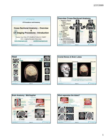
2/27/2009Overview Cross-Sectional Anatomy Head & Neck– Brain– VasculatureCT Procedures and Anatomy SpineCross Sectional Anatomy – Overview&CT imaging Procedures - IntroductionCarolyn Kaut Roth, RT (R)(MR)(CT)(M)(CV) FSMRTCEO, Imaging Education – Cervical– Thoracic– LumbarCTImages BodyB d– Chest– Abdomen– Pelvis– Vasculature Musculoskeletal– Upper extremities– Lower extremities2009Slide # 1HeadMRImagesSlide # 2Cranial Bones & Brain LobesCTImagesMRIImagesBrain LobesCranial BonesMidline Sagittal T1WIMidline Sagittal reformatParietalFrontalPara Para Sagittal reformatTemporalAxial PDWIAxialThe 5th lobe of the brain is known as the InsulaIt is located “inside” the Sylvian (Lateral) FissureCoronal T2WICoronal Direct or reformat20092009Slide # 3Brain Anatomy: Mid-SagittalWhat separates the lobes?Parietal lobeFrontal lobeFrontal boneTemporal lobeParietal lobeParietal boneOccipital lobeOccipital boneTentorium separatescerebrum fromcerebellumOccipital lobeFrontal lobeSylvian fissure(Insula located within)Sylvian Fissure or Lateral Fissure Separates the Frontal lobe from the parietal lobeInside is the 5th lobe of the brain, the “Insula” & the MCABrain SpecimenAxial CT ImageThe red line (box) indicates thepplocation of the midlineapproximatesagittal sliceParietal lobeFrontallobeParietal lobeOccipital lobeLongitudinal Fissure Right and Left lobes(inside is the Falx Cerebri)Longitudinal FissureOccipitallobeFrontallobeSlide # 4Sylvian FissureCerebellumCerebellumMidline SagittalMidline SagittalCT ImageMRI ImageTentoriumTentorium2009Slide # 52009Slide # 61
2/27/2009Brain Anatomy: Gray & White MatterSagittal PlaneMidline SagittalParasagittalBrain alMidline SagittalMidline e # 7Brain Anatomy: Corpus CallosumCorpus CorpusCorpusCallosum Callosum Callosum(genu) (body)(splenium)Midline SagittalMidline SagittalCT ImageMRI ImageSlide # 8Fractional AnisopetryCorpus CorpusCorpusCallosum Callosum Callosum(genu) (body)(splenium)Midline SagittalCT ImageMRI ImageHint‐ the Corpus callosum is the only white matter structure to cross midlineHint‐ white matter is made up of myelin20092009Slide # 9Brain Anatomy: Brain StemSlide # 10Brain Anatomy: Midline StructuresCerebralpedunclesPonsMedullaSpinal CordCerebralpedunclesPonsMedullaSpinal CordAnterior hornLateral ventricleThalamusThird ventricleOptic chiasmAnterior hornlateral ventricleThalamusThird ventricleSella turcicaPituitary stalkPituitary glandClivusClivusMidline SagittalMidline SagittalMidline SagittalCT ImageHint‐ we know that we are in the midline when we see the spinal cord2009Slide # 11Midline SagittalCT ImageMRI ImageMRI ImageHint‐ we know that we are in the midline when we see the pituitary and sella Turcica2009Slide # 122
2/27/2009Brain Anatomy: ellumCoronal PlaneMaxillary SinusTeethSulcigyriParietal lobepara SagittalMRI Imagecoronal slicemore anteriorOccipital lobeSylvian (lateral) fissureMCA flow throughThe Insula is insidecoronal slicemore posteriorFrontal lobeCerebellumTemporal LobeSagittalAxialCoronalpara Sagittal2009CT Image Slide # 13Slide # 14High Resolution Brain ProtocolCoronal Facial Bone AnatomyChristi galliCribriform plateOrbital roofNasal conchae MaxillaMaxillaMandibleFractureFrontal boneNasal boneMaxilla Temporal boneZygomaCoronal CT reformatted image of TraumaCoronal Oblique CT2009coronal slicevery posterior2009Lateral & AP Scout120 kv, 80 ma, 50 fovAxial120 kv, 220 ma, 28cm fov512 x 512 matrix1-3 mm to frontal sinus-straight- floor of maxillary sinusperpendicular to table- gantrystraight9 mm to top of heador5mm / 5mm through fossa8 -9mm entire head125 W 55 L HeadCoronal120 kv, 220 ma, 16-20cm fov512 x 512 matrix3 mm to frontal sinusangled perpendicular to mandible250 W 30 L (soft tissue)2000 W 350 L (bone)2009Slide # 15Slide # 16Soft tissue windowsBone windowsBrain Anatomy Coronal PosteriorCoronal Through Pituitary GlandOptic chiasmPituitary stalkThis red line indicates the location ofthe coronal sliceDo you see the seagull?Parietal lobeLongitudinal fssurePituitary gland (w/tumor)Pit stalk (infundibulum)Optic chiasmLateral ventriclesSeptum pellucidumTemporal lobesCoronal CT2009Cisterna Ambiensw/Pineal glandCerebral aqueductTentorium4th ventricleCerebellumCoronal MRSlide # 17This red line indicates the location ofthe coronal sliceCoronal MR imageCoronal CT image2009Slide # 183
2/27/2009Imaging PlanesAxial Slice LocationsAxial Slice locations5 4 3 2 1Axial – SLICE # 1Axial – SLICE # 2Axial – SLICE # 4Axial – SLICE # 5Angledg with base of skullAxial – SLICE # 320092009Slide # 19Brain Anatomy: Axial SuperiorSlide # 20Brain Anatomy: Axial @ VentriclesThis red line indicates thelocation of the axial sliceThis red line indicates thelocation of the axial sliceWhite matterGray matter ribbonsSuperiorsagittal sinusAnteriorAnterior Horns of theLateral VentriclesFrontal lobeWhite matterSeptum pellucidumLongitudinalfissurefalx cerebrienhanced onCT imageSuperiorsagittal sinus2009Axial MR image Superior locationAxial CT imageBrain Anatomy: Axial Basil GangliaAxial T1 weighted MR imageJust because the structure is gray, does not automatically make it gray matter!2009Slide # 21Slide # 22White Matter TractsAxialT1 weightedMR imageCaudate nucleusCaudate NucleusPosterior Horns ofthe LateralVentriclesSubcutaneusfatPosteriorAxial CT image Superior locationChroriod PlexusSulcigyriCortical boneParietal lobeSulci & GyriAxialproton density weightedMR imageGray matterLentiform nucleusExternal CapsulePutamen & globus pallidusInternal capsuleLentiform NucleusThalamusInternal CapsuleThalamusExternal capsuleClaustrumExtreme capsuleAxial CT imageAxial PD weighted MR imageJust because the structure is white, does not automatically make it white matter!2009Slide # 232009Slide # 244
2/27/2009Brain Anatomy: Axial OrbitsBrain Anatomy: Axial OrbitsThis red line indicates thelocation of the axial sliceCerebellumAt the level of thevermisThis red line indicates thelocation of the axial sliceFrontal sinusOrbitNasal conchaeLensGlobe of the eyeOptic nerveLateral rectus muscleRt. Medial rectus muscleLt. Medial rectus muscleTemporal lobeShenoid sinusTemporal LobeBasilar ArteryPons4th ventricleMastiod air cellsCerebellumAxial MR image2009Axial CT imageAxial CT imageAxial MR image2009Slide # 25Brain Anatomy: IAC’sSlide # 26Brain Anatomy: IAC’sThis red line indicates thelocation of the axial sliceThis red line indicates thelocation of the axial sliceThis red line indicates thelocation of the axial sliceThis red line indicates thelocation of the axial sliceTemporal LobeTemporal boneIACMastoid Air CellsCocleaSemicircular canalsTemporal LobeTemporal boneIACMastoid Air Cells7th & 8th cranial nervesAxial CT imageAxial CT imageAxial MR imageAxial MR image20092009Slide # 27Axial Brain Anatomy: MidbrainOverview Cross-Sectional AnatomyThis red lineindicates theapproximatelocation of theaxial sliceThis red lineindicates theapproximatelocation of theaxial sliceCerebellumAt the level of the vermisFrontal LobeFalx Cerebri Head– Anterior Circulation– Posterior CirculationSylvian FissureMCAocclusionSlide # 28– COW (circle of Willis)Temporal lobe NeckCerebral pedunclesRed nucleusCerebral AcqueductVermisCerebellum– Carotid ArteriesOccipital LobeAxial CT image2009– Vertebral ArteriesAxial MR imageSlide # 292009Slide # 305
2/27/2009Cerebral VasculatureCircle of Willis - CTASagittal2009Coronal2009Slide # 31Slide # 32AxialCerebral Circulation – Coronal (COW)Left (Anterior & Middle) Artery – CoronalSuperiorLeft (ACA ) AnteriorCerebral ArteryRightLeft(ACOM )AnteriorCommunicatingArteryLeft (MCA )Middle CerebralArtery(ICA )InternalCarotidArteryCoronal CTA ImageAP angiogram ‐ Left Internal Carotid(Anterior and Middle Circulation)Coronal CTA20092009InferiorSlide # 33Right & Left (Anterior & Middle) Artery – CoronalSlide # 34Right & Left Vertebral Arteries & Basilar Artery –Coronal(ACA ) AnteriorCerebral Artery(MCA ) Middle(ACOM ) AnteriorCerebral ArteryCommunicating Artery(PCA ) PosteriorCerebral ArteryAP imageAP imagegBasilar Artery(ICA ) InternalCarotid ArteryCoronal image2009Vertebral ArteryCoronal imageAP imageAP imageSlide # 352009Slide # 366
2/27/2009Cerebral Circulation – Coronal (COW)Intra-Cerebral Circulation - COWSuperior(ACA ) AnteriorCerebral Artery(MCA ) Middle(ACOM ) AnteriorCerebral ArteryCommunicating Artery(ACA ) AnteriorCerebral Artery(PCA ) PosteriorCerebral Artery(PCA ) PosteriorCerebral ArteryAP image(MCA ) MiddleCerebral Artery(ACOM ) AnteriorCommunicating ArteryBasilar Artery(ICA ) InternalCarotid ArteryRight(ICA ) InternalCarotid ArteryLeftBasilarArteryVertebral ArteryCoronal CTA Image2009Vertebral ArteryAP imageCoronal CTA2009Slide # 37Cerebral Circulation – Sagittal (COW)SuperiorSlide # 38InferiorAnterior & Middle Cerebral Arteries – Sagittal(ACA ) AnteriorCerebral Artery(MCA ) MiddleCerebral Artery(ICA ) InternalCarotid ArteryAnteriorSagittal CTA2009PosteriorInferior2009Slide # 39Internal carotid (Anterior andMiddle circulation)Lateral angiogramAnterior andd Middleddl circulationlLateral DSARight (ICA )Internal CarotidArtery(PCOM ) PosteriorCommunicatingArteryLeft (ICA )Internal CarotidArterySagittal CTAPosterior Cerebral Arteries - Sagittal(PCA) PosteriorCerebral ArterySlideLateral# 40 RadiographCerebral Circulation – Sagittal(PCA) PosteriorCerebral Artery(ACA ) AnteriorCerebral Artery(MCA ) MiddleCerebral A ) InternalCarotid ArteryVertebralArterySagittal CTAAnterior and Middle circulationLateral DSALateral RadiographPosterior cerebralcirculationLateral DSA2009Lateral RadiographSlide # 41Posterior cerebral circulationLateral DSA(PCOM ) PosteriorCommunicatingArtery2009Sagittal CTASlide # 427
2/27/2009Cerebral Circulation – Sagittal (COW)Cerebral Circulation – Axial (COW)Superior(ACA ) AnteriorCerebral ArterySuperior(MCA ) MiddleCerebral Artery(PCOM )PosteriorCommunicatingArtery(PCA ) PosteriorCerebralb l ArteryAnteriorPosteriorRightLeftBasilar Artery(ICA ) InternalCarotid ArteryVertebral ArterySagittal CTA2009AxialCTA2009InferiorSlide # 43Cerebral Circulation - Axial (Circle of Willis)AnteriorSlide # 44Anterior(ACOM ) AnteriorCommunicating Artery(ACA ) AnteriorCerebral Artery(ACOM ) AnteriorCommunicatingArteryInferior(ACA ) AnteriorCerebral ArteryIntra-CerebralCirculation – Axial COW(MCA ) MiddleCerebral Artery(ICA ) InternalCarotid Artery(MCA ) MiddleCerebral ArteryRightBasilarArtery(PCA ) PosteriorCerebral Artery(PCOM ) PosteriorCommunicating ArteryLeft(ICA ) InternalCarotid Artery(PCOM ) PosteriorCommunicatingBasilar Artery ArteryRight(PCA ) PosteriorCerebral ArteryAnteriorLeftLeftLeftAxial2009 CTA2009PosteriorSlide # 45Anterior Cerebral Arteries (right & left)Anterior(ACOM ) AnteriorCommunicatingArterySlide # 46Anterior(ACOM ) AnteriorCommunicating ArteryAxial COW(ACA ) AnteriorCerebral ArteryAnterior cerebral arteries(ACA ) AnteriorCerebral Artery–(ICA ) InternalCarotid Artery(MCA ) MiddleCerebral Artery(MCA ) Middle LeftCerebral ArteryRight(ICA ) InternalCarotid ArteryPosteriorAxial CTAAnteriorRightLeftLeftLeftAxial2009 CTAPosteriorSlide # 472009Slide # 488
2/27/2009Posterior Cerebral Arteries (right & left)AnteriorAxial COWPosterior cerebral arteriesAnterior–(PCOM ) PosteriorCommunicating ArteryRight(PCOM ) PosteriorCommunicatingArtery(PCA ) PosteriorCerebral ArteryAxial CTABasilarArteryLeftPosteriorBasilar ArteryRightLeft(PCA ) PosteriorCerebral ArteryAxial2009 CTA2009PosteriorSlide # 49Cerebral Circulation - Axial (Circle of Willis)Anterior(ACA ) AnteriorCerebral Artery(ACOM ) AnteriorCommunicatingArtery(ICA ) InternalCarotid Artery(PCOM ) PosteriorCommunicatingBasilar Artery ArteryRightLeft(PCA ) PosteriorCerebral ArteryAxial2009 CTACircle of Willis - CTA2009– 120 kv, 80 ma, 50 fovAxial120 kv, 420 ma512 x 512 matrix, 24cm fov.675 mm through COWthen9mm through entire head-straight- floor of maxillarysinus perpendicular to tabletablegantry straight-Contrast if 100 -120cc’snon-ionic 350 mg/ml- 3 cc/sec for 20g angiocath-delay scan 15-20 sec postinjection-usually reconstructedto a field of 128mm fromabove the cow to c22009PosteriorSlide # 51SagittalCTA-Rapid Brain ProtocolCTA\ AP & Lateral Scout (MCA ) MiddleCerebral ArterySlide # 50Slide # 52Head VeinsCoronalSlide # 53Axial2009Slide # 549
2/27/2009Overview Cross-Sectional AnatomyNeck Vasculature Head– Anterior Circulation– Posterior Circulation– COW (circle of Willis) Neck– Carotid Arteries– Vertebral Arteries20092009Slide # 55Carotid Arteries – SagittalSlide # 56Vertebral Arteries – Sagittal(ICA ) InternalCarotid ArteryBasilarArtery(PCA )PosteriorCerebralArtery(ECA ) ExternalCarotid ArteryRight and LeftCCA - commoncarotid arteriesVertebralArterySagittal Neck2009Sagittal NeckSagittal Neck2009Slide # 57Carotid Arteries – SagittalSagittal NeckSlide # 58Carotid Arteries – Sagittal(ICA ) InternalCarotid ArteryBasilarArtery(PCA )PosteriorCerebralArtery(ECA ) ExternalCarotid ArteryRight and LeftCCA - commoncarotid arteriesVertebralArterySagittal Neck2009Slide # 59Sagittal NeckSagittal Neck2009Sagittal NeckSlide # 6010
2/27/2009Overview Cross-Sectional AnatomyProtocol for Neck AP & Lat scout120 kv, 80 ma, 50cm FOV Axial120 kv 280 ma5/5 mm, 23 cm FOV512 x 512 matrix Scan frontal sinus to carinainclude mandible Contrast100 cc’s non-ionic 240-300mg/ml-2 cc/sec for 20g cath-delay scan 70 secondpost injection-250 W 30 L (soft tissue)-2000 W 350 L (bone) Head– Anterior Circulation– Posterior Circulation– COW (circle of Willis) Neck– Carotid Arteries– Vertebral Arteries20092009Slide # 61CTA‐Rapid Neck ProtocolScan Planning 2009CT ImagesAP & Lateral Scout– 120 kv, 80 ma, 50 cm FOVAxial120 kv, 420 ma512 x 512 matrix, 24cm FOV1 mm from the arch to abovethe frontal sinuses-straight- floor of maxillary sinusperpendicular to tabletable gantrystraight-Contrast if 100 -120cc’snon-ionic 300-350 mg/ml- 3 cc/sec for 20g angiocath-delay scan 15-20 sec postinjection-usually reconstructedto a field of 128mm2009Slide # 63Imaging PlanesSlide # 62Slide # 64Image Comparison in the C-spineMedian LineMid-sagittal PlaneParasagittal PlanesTransverseOr AxialPlaneSlicelocationFrontal orCoronal PlaneMRI SagittalSagittalReformatLateral radiographSagittal2009AxialSlide # 65Sagittal CTSagittal T1 MRICoronal2009Slide # 6611
2/27/2009Spinal Anatomy Brachial Plexus & Lumbar Plexuspinal Anatomy Vertebral bodies & Intervertebral disksC1C2C3FractureBrachial plexusCervical vertebraeCORONAL MRISpinal cordIntervertebral DiskAnulus (dark)Nucleus pulposis (bright)Conus medularisL4L5S1Lumbar vertebraeLumbar plexusSacral plexusSacrumSAGITTAL MRICORONAL CTCORONAL MRI2009Slide # 672009SAGITTAL CTImaging Planes – C Spine & NeckSlide # 68SAGITTAL MRINeck CT - LaryngoceleAnterior arch c1The densLaryngealCartilagesLarynxCervical vertebraPedicleDorsal and ventralnerve rootsSpinal cordLaminaSpinous processInternal LaryngoceleCauses stridorSpinal musclesAxial MR imageAxial CT image20092009Slide # 69“C-spine”Slide # 70“Arnold Chiari Malformation”Vertebral bodyPedicleLaminaforaminaCerebellar TonsilsSyrinxMRI AXIALCT AXIALC1Dens (C2)C‐7LordosisVert bopdiesDiskCordesophagustracheaSpinal musclesSAGITTAL MRIEpidural fat (bright)CSF (Cerebro spinal fluid)(dark)2009SAGITTAL CTSlide # 712009SAGITTAL MRISlide # 7212
2/27/2009Apical Herniation: Ehlers-Danlos syndromeSagittal T-spine AnatomyApproximatelocation forsagittalApproximatelocation forsagittalreformatCT dSagittalCTSlide # 73MRI AXIALT1 (1st thoracic vertebrae)T 12 (12th thoracic vertebrae)Spinal CordVertebral bodies((curvature kyphosis)yp)Intervertebral DiskSpinus processConusCauda equinaCSF around the nervesT1T2T3T7T8T9T10T11T12L1Posterior longitudinal ligament(along the posterior vertebral bodies from C1 –sacrum)Anterior longitudinal ligament(along the anterior vertebral bodies from C1 –sacrum)Slide # 74L3L4L5S1SAGITTAL MRIVertebral bodyPedicleLaminaFacet jointsApproximatelocation foraxialCT AXIALSAGITTAL CTSAGITTAL MRIMRI AXIALL1L5Vert bodieslordosisAortaVertebral bodyLungCosto‐vertebral jointDiskPedicleLigamentum flavumPosterior longitudinal ligamentRibCT AXIALL2Lumbar SpineThoracic SpineApproximatelocation foraxial2009LigamentumflavumT4T5T6Spinal canalSpinal cordCSF (cerebrospinal fluid)Transverse processLaminaSpinus processLigamentum flavumErector spinae musclesMRI AXIAL2009Slide # 75SAGITTAL MRISlide # 76SAGITTAL CTImaging Planes - TMJAnatomy Musculoskeletal SystemMedian LineTMJMid-sagittal PlaneTMJMid FrontalCoronal PlaneParasagittal PlanesAxialTransversePlaneCoronalSagittalCT Images2009Slide # 77MRI Images2009SagittalSlide # 78AxialCoronal13
2/27/2009Overview musculoskeletal Anatomy - TMJLocalizer for sagittal oblique sectionsCorocoid vs coronoid “There’s a sea (C) between two Nations”Localizer for coronal oblique sectionsShoulder radiographTemporal boneFossa(meniscus within)MandibularcondyleMandibleShoulder CTSagittal reformatted CTSagittal Oblique TMJ2009Sagittal MR ElbowTMJ 3D CT reformatted imageCoronal Oblique TMJCoroNoid2009Slide # 79CoroCoidLateral radiography (Elbow)CoroNoidSlide # 80The lumps and bumps of the shoulderAnatomy Musculoskeletal SystemShoulderShoulder Shoulder– Scapula Spine of the scapulaprocess Coracoid p Acromion process– Clavicle– Humerus Head of the humerus Greater tubercle Lesster tubercle20092009Slide # 81Surgical FixationReformatted CT Image shoulderSlide # 82Overview musculoskeletal Anatomy - ShoulderMedian LineMidline Sagittal PlaneParasagittal PlanesAC JointFrontal orCoronal PlaneTransverseOr AxialPlaneClavicleAxialRibsThe sagittal oblique plane is acquired along thisdotted red line, parallel to the glenoid fossafSagittalThe coronal oblique plane is acquired along this dotted red lineon the axial shoulder image. Parallel to the supraspinatusmuscle & tendon or perpendicular to the glenoid fossa2009Slide # 832009Coronal obliqueSlide # 8414
2/27/2009Structures of the rotator cuff (SITS)Spine of the scapulaShoulder StructuresDeltoidSpine of the scapulaSupraspinatus tendon.Infraspinatus tendonTeres minor tendonSubscapularis tendonBicepts muscleReformatted CT ImageAxial Shoulder CTTrapeziusDeltoidRC (S.I.T.S.)SupraspinatusInfraspinatusTeres MinorSubscapularisAxial MR Image2009TrapeziusDeltoidSupraspinatus tendonRotator cuffGlenoid fossaGlenoid labrumCoronal oblique MR ImageAxial MR Image2009Slide # 85Bony Structures of the shoulderAcromionA/C joint(acromio‐clavicular joint)ClavicleHumeral HeadHumerusCorocoid processReformatted CT ImageSagittal Oblique ShoulderCoronal oblique MR ImageSlide # 86Anatomy Musculoskeletal SystemSpine of the scapulaElbowReformatted CT ImageSagittal Oblique ShoulderElbowAcromionRotator cuffHumeral HeadGlenoid fossaGlenoid rimScapulaHumerusCoronal oblique MR ImageAxial MR Image20092009Slide # 87Bones of the Elbow JointMuscles, Tendons & Ligaments of the Elbow JointHumerusOlecranon processJoint spaceTrochleaCapetellumCoronoid processUlnaSagittal Oblique ElbowHumerusOlecranon fossaCapetellumHumero‐radial jointCapetellumCoronal oblique MR Image“Cap onHead”2009(Capetellum on the head of the radius) Slide # 89Slide # 88Tricepts musclesBicepts MusclesExtensor tendonsBrachoiadialis muscleAxial MR ImageTrochleaHumero‐ulnar jointRadio‐ulnar jointAxial MR ImageSagittal Oblique ElbowTricepts musclesUlnar collateral ligamentBrachoiadialis muscleRadial collateral ligament2009Coronal oblique MR ImageSlide # 9015
2/27/2009Overview musculoskeletal Anatomy – WristAnatomy Musculoskeletal SystemNever Lower Tillie’s Pants Grandmother Might Come HomeNavicularLunateTriquetruimPisiformGreater MutlangularLesser MultangularCapatateHammateWristWristCoronal MRICoronal reformatted CT20092009Slide # 91Overview musculoskeletal Anatomy – WristSlide # 92Overview musculoskeletal Anatomy – WristSome Lovers Try Positions .That They Can’t ege complex)Coronal reformatted CT2009Carpal TunnellFlexor tendonsMedian pezoidCapetateHammateAneurysmRaidal arteruUlnar arteryTrapeziumTrapezoidCapetateHammateHook of theHammateTFCC (triangulo-Coronal MRIDistal RadiusDistal UlnaRadio‐ulnar jointCoronal MRIAxial MRI2009Slide # 93Wrist PathologySlide # 94HandshakeAneurysmFracture2009Slide # 952009Slide # 9616
2/27/2009Bony anatomy of the HIPAnatomy Musculoskeletal SystemAcetabulimIliumIshiumPubisFemoral HeadFemoral NeckGreater TrocanterLesser trocanterFemoral ShaftHipHipObturator foramenReformatted CT Image Hip20092009Slide # 97BONE METSSlide # 98Hips & PelvisIlumIliac arteriesPsoas MusclesObturator foramenCompressedvertebral bodyGleuteal MusclesCoronal reformatted CTMet to the pelvicboneReformatted SagittalCT Image SpineReformatted Coronal CT Image pelvis2009MassAxial pelvis CTObturator Internus musclesObturator Externus2009Slide # 99Femoral HeadFemoral NeckGreater TrocanterLesser trocanterFemoral ShaftStructures of the HIP(ASIS) IliumAcetabulimObturator foramenIshiumPubisCoronal CTSlide # 100NAVELNerve, Artery, Vein, Empty space, Lymph nodePsoas MuscleIliacus MuscleGleuteus MaximusGleuteus minimusQuadracepts MuscleHamstringsReformatted CT Image HipCoronal MRI of the HipsIliumAcetabulimGlenoid LabrumFemoral NeckFemurbladderObturator Internus musclesObturator ExternusCoronal MRI of the Hips2009Slide # 1012009Axial MRI of the HipsNerveArteryVeinEmpty spaceLymph nodesQuadracepts MuscleGleuteus MaximusSlide #Rectum10217
2/27/2009Anatomy Musculoskeletal SystemBones of the KneeCondylendyleeal plateauial spineCoronal knee MRIAxial kneeFemurPatellaPatello‐femoral jointKnee JointTibiaFibulaKneeKneeCoronal reformatted CT2009Sagittal knee MRI2009Slide # 103Slide # 104Muscles of the KneeLower Extremity AnatomyAnterior cruciate ligament (ACL)Axial pelvis CTPosterior cruciate ligament (PCL)Hamstring musclesQuadracepts TendonAttaches the quatracepts musclesGastrocnemius musclesQuadracepts musclesFemurHamstrings musclesQuadcepts tendonAxial femur CTMid sagittal slice (MRI)Patello- Femoral JointPatellar ligamentPara Sagittal knee MRILocations of the lateral collateral ligamentsQuatracepts musclesHamstring MusclesAxial femur CTFemoral coldylesMeniscus ([posterior horn, lateral meniscus)PatellaAxial knee CTPara2009sagittal slice (MRI)TibiaFibulaCoronalSlide # 105reformatted CTMeniscus and other structures of the kneeLateral aspect of thepatello‐femoral jointLateral retinaculumAxial Femur MRI2009Lower Leg FractureMedialretinaculumFractureAnterior horn of themeniscusFracturePosterior horn of themeniscusPara Sagittal knee MRIAxial CTSlide # 106AP ScoutAxial knee MRIMedial collateral ligamentMedial meniscusLateral collateral ligamentLateral meniscusLat Scout2009Slide knee# 107 MRICoronal2009Slide # 10818
2/27/2009Anatomy Musculoskeletal SystemFoot & ankle bones - Come To Cuba Next ChristmasTibiaFibulaCome (calcaneus)To (talus)Cuba (cuboid)Next NavicularCh i tChristmas (3 cuneiforms!)if!)Foot and ankle2009Foot and ankle2009Slide # 109Slide # 110Review, Peripheral Vascular AnatomyAnkle Ligaments and TendonsTibiaFibulaAbdominal Aorta(AAA- abdominal aortic aneurysm)Mortise jointMedial collateral ligamentsLateral collateral ligamentsIliac ArteriesFemoral ArteriesTibio‐talar jointPosterior longitudinal ligamenAchilles tendonPopliteal ArteriesTrifurcation (anterior tibial)(posterior Tibial)(Pernoeus Brevis)2009Slide # 111Coronal MRA2009Coronal reformatted CTASlide # 112Coronal reformatted CTWhat Do We Do With All The Images?Hand Vasculature View ImagesScroll ThroughWindow / LevelMagnificationPost ProcessingBasic Tasks2009Slide # 113Slide # 11419
2/27/2009Imaging PlanesCT ImagesMedian LineMid-sagittal PlaneFrontal orCoronal PlaneChest Coronal, Heart & LungsMRI ImagesParasagittal PlanesTransverseOr AxialPlaneMRAAxialAxialCTAHeartCoronalCoronal ReformatLungsSagittal Reformat2009SagittalEnhanced Coronal CTCoronal MRI2009SagittalSlide # 115Axial CoronalChest Coronal, VasculatureSlide # 116Chest Coronal, Lungs & AirwayUpper Lobe of the right lungUpper Lobe of the left lungMRACTABronchi ‐ carinaMiddle lobe of the right lung*remember there is nomiddle left because of the heart!Pulmonary ArteriesPulmonary VeinsLower lobe of the right lungLower lobe of the left lungDiaphragmCoronal MRICoronal CTEnhanced Coronal CT2009Coronal MRISlide # 117Chest Coronal Muscles of the Chest2009Slide # 118Sagittal Chest – “Candy Cane Shot”Pectoralis musclesHeart MuscleAortic ArchAxial MRIAxial CTAscending AortaDescending AortaTrapeziusLatissimusHeartIntercostal MusclesSagittal MRIDiaphragmCoronal MRICoronal CT2009Slide # 119Sagittal CT2009Slide # 12020
2/27/2009Axial chestAxial slice #1Axial slice #2Axial slice #3Axial slice #1Axial slice #2Axial slice #3Axial slice #1Axial slice #22009Axial slice #3Pectoralis MuscleAortic ArchLatissimus MuscleSpine (vertebral body)Ascending AortaPulmonary arteriesPulmonary veinsDescending AortaHeartSlide # 121Lung Nodule ‐ MDCTAxial slice #1Axial slice #2Axial slice #3Basic “Vanilla”Thorax Protocol AP Scout120 kv, 60 ma, 50 cm FOV Axial120 kvkv, 180 mama,36 cm FOV (fit anatomy)512 x 512 matrixgenerally 5mm1500 W & 500 L(lung windows)450 W & 30 L (mediastinal windows)30 sec (or less) breath-holdcan be modified2009Evaluation of Stent Placement3D rendering of normal lung2009Slide # 122Slide # 1232009Heart anatomyHeartMDCT evaluation of airway stentafter chemical injurySlide # 124CTA CORONALHeart flowIVC & SVC (Inferior & Superior vena cava)RA (right atrium)TriRvP valvePaLungsPvLaBiLvAortic valveAorta – coronaryCT AXIALarch2009Slide # 1252009MRI CORONALMRI AXIALSlide # 12621
2/27/2009Heart AnatomyHeart flowSVC (superior vena cava)RA (right atrium)Tri cuspid valveRvP valvePaLungsPvLaBiLvAortic valveAorta – coronaryarch2009Heart AnatomyHeart AnatomyCTA CORONALCT AXIALHeart anatomyCTA CORONALCTA CORONALSlide # 131Heart flowIVC & SVC (Inferior & Superior vena cava)RA (right atrium)Tri cuspid valveRV (right ventricle)P valvePaLungsP‐S vLaBiLvAortic valveAorta – coronaryCT AXIALarch2009MRI CORONALMRI AXIALHeart AnatomyMRI CORONALMRI AXIALHeart anatomyCTA CORONALMRI AXIALMRI CORONALMRI AXIALSlide # 130CTA CORONALHeart flowIVC & SVC (Inferior & Superior vena cava)RA (right atrium)Tri cuspid valveRV (right ventricle)Pulmonary valvePA (Pulmonary artery)LungsPV (pulmonary veins)LA (Left Atrium)Bi cuspid valveLVAortic valveAorta – coronaryCT AXIALArch2009MRI CORONALSlide # 128Heart flowIVC & SVC (Inferior & Superior vena cava)RA (right atrium)Tri cuspid valveRV (right ventricle)Pulmonary valvePA (Pulmonary artery)LungsPV (pulmonary veins)LABiLVAortic valveCT AXIALAorta – coronaryArch2009Slide # 129Heart flowIVC & SVC (Inferior & Superior vena cava)RA (right atrium)Tri cuspid valveRV (right ventricle)Pulmonary valvePA (Pulmonary artery)LungsPV (pulmonary veins)LA (Left Atrium)BiLVAortic valveAorta – coronaryCT AXIALArch2009MRI AXIALSlide # 127Heart flowIVC & SVC (Inferior & Superior vena cava)RA (right atrium)Tri cuspid valveRV (right ventricle)Pulmonary valvePA (Pulmonary artery)LungsPvLaBiLvAortic valveCT AXIALAorta – coronaryarch2009MRI CORONALCTA CORONALMRI CORONALMRI AXIALSlide # 13222
2/27/2009Heart anatomyCTA CORONALHeart flowIVC & SVC (Inferior & Superior vena cava)RA (right atrium)Tri cuspid valveRV (right ventricle)Pulmonary valvePA (Pulmonary artery)LungsPV (pulmonary veins)LA (Left Atrium)Bi cuspid valveLV (Left Ventricle)Aortic valveAorta – coronaryCT AXIALArch2009MRI CORONALMRI AXIALHeart anatomyHeart flowIVC & SVC (Inferior & Superior vena cava)RA (right atrium)Tri cuspid valveRV (right ventricle)Pulmonary valvePA (Pulmonary artery)LungsPV (pulmonary veins)LA (Left Atrium)Bi cuspid valveLV (Left Ventricle)Aortic valveAorta – coronaryCT AXIALArch2009Slide # 133CTA CORONALMRI CORONALMRI AXIALSlide # 134Aortic Arch – the “ A, B, C’s”Heart and Pulmonary ArteriesAscending aortaCoronariesBrachiocephalic (aka Innominate)right common carotidright subclavianright vertebralLeft common carotidLeft SubclavianLeft vertebralMRA CORONALCTA CORONAL20092009Slide # 135Aortic Arch – the “ A, B, C’s”Slide # 136Aortic Arch – the “ A, B, C’s”Ascending aortaCoronariesBrachiocephalic (aka Innominate)right common carotidright subclavianright vertebralLeft common carotidAscending aortaCoronariesBrachiocephalic (aka Innominate)right common carotidright subclavianright vertebralLeft common carotidLeft SubclavianLeft vertebralLeft SubclavianLeft vertebralMRA CORONALMRA CORONALCTA CORONAL2009Slide # 137CTA CORONAL2009Slide # 13823
2/27/2009Aortic Arch – the “ A, B, C’s”Coronary CalcificationsAscending aortaCoronariesBrachiocephalic (aka Innominate)right common carotidright subclavianright vertebralLeft common carotidLeft SubclavianLeft vertebralMRA CORONAL Calcium is an indicatorfor a disease artery(CAD) Calcification in thecoronary artery revealsthe condition ofcoronaryarteriosclerosis Leads to heart attacksGrossly calcified left coronaryarteryCTA CORONAL2009Slide # 139Situs Inversus20092009Slide # 140Cross Sectional AbdomenSlide # 1412009Slide # 142Frontal orCoronal PlaneMedian LineMid-sagittal PlaneImaging PlanesAbdomen ImagesParasagittal PlanesTransverseOr AxialPlaneSagittalAxialCoronalCT Images2009Slide # 143Axial2009CoronalreformatSagittalreformatSlide # 14424
2/27/2009Frontal orCoronal PlaneMedian LineMid-sagittal PlaneImaging PlanesCoronal AbdomenParasagittal PlanesApproximate SlicelocationApproximate SlicelocationTransverseOr alLiverCT ImagesPsoasMusclesGleuteusMusclesCoronal MR imageAxial2009CoronalCoronal CT imageSagittal2009Slide # 145Coronal AbdomenCoronal AbdomenApproximate SlicelocationApproximate SlicelocationApproximate SlicelocationSpleenLiverSlide # 146LiverSpleenSpleenStomachumorLiverMassCoronal CT imageCoronal
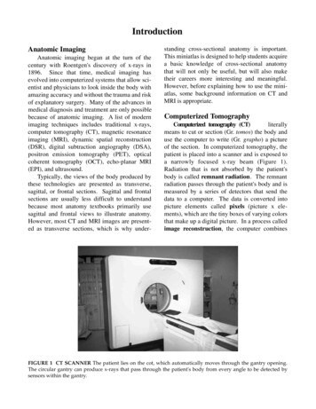
CT Procedures and Anatomy Cross Sectional Anatomy - Overview & 2009 Slide # 1 CT imaging Procedures - Introduction Carolyn Kaut Roth, RT (R)(MR)(CT)(M)(CV) FSMRT CEO, Imaging Education Associates www.imaginged.com candi@imaginged.com Overview Cross-Sectional Anatomy Head & Neck - Brain - Vasculature Spine - Cervical - Thoracic .
