Transcription
HindawiBioMed Research InternationalVolume 2022, Article ID 4302625, 12 pageshttps://doi.org/10.1155/2022/4302625Research ArticleStructural and Functional Annotation and Molecular DockingAnalysis of a Hypothetical Protein from Neisseria gonorrhoeae:An In-Silico ApproachLincon Mazumder , Md. Rakibul Hasan , Kanij Fatema , Md. Zahirul Islam ,and Sanjida Khanam TamannaDepartment of Microbiology, Jagannath University, Dhaka 1100, BangladeshCorrespondence should be addressed to Lincon Mazumder; linconjnu@gmail.comReceived 20 June 2022; Revised 17 August 2022; Accepted 23 August 2022; Published 5 September 2022Academic Editor: Paul HarrisonCopyright 2022 Lincon Mazumder et al. This is an open access article distributed under the Creative Commons AttributionLicense, which permits unrestricted use, distribution, and reproduction in any medium, provided the original work isproperly cited.Background. Worldwide, Neisseria gonorrhoeae-related sexually transmitted infections (STIs) continue to be of significant publichealth concern. This obligate-human pathogen has developed a number of defenses against both innate and adaptive immuneresponses during infection, some of which are mediated by the pathogen’s proteins. Hence, the uncharacterized proteins of N.gonorrhoeae can be annotated to get insight into the unique functions of this organism related to its pathogenicity and to finda more efficient therapeutic target. Methods. In this study, a hypothetical protein (HP) of N. gonorrhoeae was chosen foranalysis and an in-silico approach was used to explore various properties such as physicochemical characteristics, subcellularlocalization, secondary structure, 3D structures, and functional annotation of that HP. Finally, a molecular docking analysiswas performed to design an epitope-based vaccine against that HP. Results. This study has identified the potential role of thechosen HP of N. gonorrhoeae in plasmid transfer, cell cycle control, cell division, and chromosome partitioning. Acidic nature,thermal stability, cytoplasmic localization of the protein, and some of its other physicochemical properties have also beenidentified through this study. Molecular docking analysis has demonstrated that one of the T cell epitopes of the protein has asignificant binding affinity with the human leukocyte antigen HLA-B 15 : 01. Conclusions. The in-silico characterization of thisprotein will help us understand molecular mechanism of action of N. gonorrhoeae and get an insight into novel therapeuticidentification processes. This research will, therefore, enhance our knowledge to find new medications to tackle this potentialthreat to humankind.1. IntroductionN. gonorrhoeae, the etiological agent of Gonorrhea, first isolated in 1878, belonging to the Neisseriaceae family [1, 2], isa gram-negative, 0.6-1 micrometer in diameter [3], encapsulated bacterium [4]. It is fastidious [5], non-acid fast [6],oxidase-positive [7], and non-spore-forming in nature [8].In addition, it is a non-motile [9] and obligate-human pathogen [10] that can thrive aerobically or anaerobically in thepresence of nitrite [11]. These diplococci, kidney-shapedbacteria infecting both men and women can cause the sexually transmitted disease (STD) named gonorrhea [12, 13].Every year, 87 million new infections are being reportedfor this quick-spreading contagious disease. This STD hasalready emerged as a major problem in low- and middleincome countries in Africa, Asia, Latin America, and theCaribbean [1, 5, 12]. Gonorrhea can be asymptotic ordevelop with symptoms. It can manifest as urethritis inmen, with symptoms such as epididymitis, urethral stricture,and prostatitis. In women, it might manifest as urethritis orcervicitis, with symptoms including tubal infertility, chronicpelvic discomfort, severe pelvic inflammatory diseasesequelae, and ectopic pregnancy [3, 14]. Oropharyngealand anorectal gonococcal infections can be transmitted fromone person to another through kissing and during oral-analintercourse. Furthermore, gonorrhea can be caused by
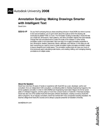
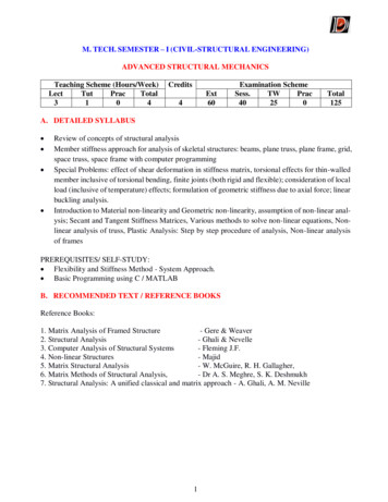
2contamination via cervical fluids [14, 15]. However, there isstill no effective treatment for gonococci and even no gonococcal vaccination is available yet. To make the situationworse, N. gonorrhoeae has been found resistant to severalantimicrobial drugs such as penicillins, tetracyclines, sulphonamides, fluoroquinolones, macrolides, azithromycin, andceftriaxone [12, 16, 17]. Hence, WHO recommends azithromycin and ceftriaxone as a dual therapy for the time beingagainst this disease [12]. All these things have now madethe discovery of novel antibacterial drugs and the development of alternative therapies a crying need for combatingthis disease [18].The genome size of N. gonorrhoeae varies from strain tostrain, about 2001 /-197 kbp [19]. For example, the genomeof N. gonorrhoeae NCCP11945 contains 2232.025 kbp in onecircular chromosome that encodes 2662 predicted openreading frames and 4153 bp that codes 12 predicted ORFs[20]. Additionally, N. gonorrhoeae is known to encode several proteins with unknown functions, known as hypothetical proteins (HPs). HPs are considered to be expressed in anorganism, but there is no experimental and chemical proofof their existence [21–23]. In most genomes, HPs coverapproximately half of the protein-coding regions, but theseproteins’ roles are yet to be discovered [21, 24, 25]. Althoughthere is no empirical evidence for the existence of these proteins, they can be predicted to be generated from an openreading frame (ORF) [23, 24]. As a result, the annotationof the functions of hypothetical proteins has becomeincreasingly popular [25]. The hypothetical proteins can becategorized as uncharacterized protein families (UPF) aswell as the domain of unknown functions (DUF) [23].Uncharacterized protein families (UPF) have been experimentally confirmed to exist, although they have yet to beidentified or connected to a known gene. On the other hand,DUFs are proteins that have been found experimentally buthave no known functional or structural domains [23]. Eventhough they have not been characterized, elucidating theirstructural and functional secrets can lead to the identification of new domains and motifs, pathways and cascades,structural conformations, protein networks, etc. [21, 22].These are crucial in understanding biochemical and physiological pathways, for example, in identifying pharmaceuticaltargets [21, 22, 25] and providing early detection and advantages for proteomic and genomic studies [21]. It is now easier to analyze hypothetical proteins utilizing a variety ofbioinformatics tools that provide benefits such as 3D structural conformation prediction, identification of newdomains and pathways, phylogenetic profiling, and functional annotation [22, 23].The purpose of this study is to characterize a hypothetical protein F0T10 13280 (plasmid) of N. gonorrhoeae withan integrated computational approach, with previously validated tools and databases, to get an insight into the HP’sphysical and structural information along with its potentialfunctions. Potential role of this HP in plasmid transfer, cellcycle control, cell division, and chromosome partitioningmay give insight into the pathogenic flexibility of N. gonorrhoeae. Analyzing the phylogenetic relationship between thisHP and other proteins, physicochemical properties analysis,BioMed Research Internationalprediction of the HP’s location in the cell, analysis of the secondary and tertiary structure, prediction of the potentialfunction of the HP, and evaluation of the active sites aresome of the main focuses of this research. Finally, thisresearch also aims to design an epitope-based peptide vaccine and validate it with a molecular docking study.Figure 1 illustrates the complete workflow and tools usedin this study. Table 1 depicts the entire framework, whichincludes all the tools used to annotate the structural andfunctional properties of HP of N. gonorrhoeae. A preprintof this research has previously been published by Mazumderet al. [26]2. Materials and Methods2.1. Sequence Retrieval and Phylogeny Analysis. The aminoacid sequence (accession No. QIH20856.1) was selected bysearching the NCBI protein database for HP of N. gonorrhoeae. The sequence was obtained in FASTA format. Toidentify sequence similarity, BlastP [27] was performed.MUSCLE v3.6 [28] was used to perform multiple sequencealignment. Phylogenetic analysis was carried out usingMEGA X [29].2.2. Physicochemical Properties Analysis. The physicochemical properties of the target protein sequence were investigated using ExPASy’s ProtParam program [30]. Themolecular weight, atomic composition, estimated half-life,theoretical isoelectric point (pl), extinction coefficient,amino acid composition, aliphatic index, stability index,the total number of positive and negative residues, andgrand average of hydropathicity (GRAVY) were all analyzedusing this tool.2.3. Subcellular Localization Prediction. It is crucial to knowthe subcellular localization of proteins in order to comprehend their functions [31] entirely. Computer analysis helpsin the discovery and localization of adhesion-like intercellular proteins [32]. In the last few decades, several computational tools have been developed that can efficientlydetermine and synthesize ORFs of various proteins (mitochondrial, cytoplasmic, nuclear, or extracellular) to convertthem into potential vaccine candidates. However, vaccinecandidates should be free of membrane or cytoplasmic localization [33]. CELLO v.2.5 [34] was first used to recognize thesubcellular localization of hypothetical protein F0T10 13280(plasmid) of N. gonorrhoeae. PSORTb v3.0.3 [35] was further used to anticipate subcellular location. To cross-checkthe results, we used PSLpred [36], a web server for predictingthe subcellular localization of gram-negative bacterialproteins.2.4. Secondary Structure Prediction. Secondary structure predictions of the hypothetical protein were performed usingthe SOPMA server [37]. The PSIPRED server [38] was alsoused to ensure the accuracy of the SOPMA results.2.5. 3D Structure Prediction and Quality Assessment. HHpredserver [39] provided a 3D model of the protein. The YASARAserver [40] (http://www.yasara.org/minimizationserver.htm)
BioMed Research International3Workflow of the structural and functional annotation of a hypothetical proteinRetrieval of hypothetical protein sequence from NCBIAnalysis ofphysicochemicalpropertiesExpasy protparamSubcellularlocalizationpredictionCELLO v.2.5Secondarystructureprediction3D structureprediction, energyminimization CKFunctionalannotationCD searchActive sitedetectionCASTp v3.0PfamPSORTb v3.0.3YASARAERRATPyMOL v2Verify3DPSIPREDINTERPROPSLpredMOTIFWorkflow of the molecular docking studyPrediction ofcytotoxic TlymphocytesNetCTLPrediction ofMHCI bindingallelesEpitopeprioritizationIEDBVaxiJen 2.0ToxinpredPeptide designingMoleculardocking analysisAPPTESTAutoDockVinaDiscovery studioAllerTop 2.0Figure 1: Complete flowchart of the hypothetical proteins (HPs) annotation process used in this study.was used to accomplish energy minimization. To visualizethe final model and perform structural analysis, PyMOLv2 [41] was employed. The SAVES server’s (https://services.mbi.ucla.edu) quality assessment tools were usedto assess the predictability of the hypothetical protein’sprojected 3D structural model. The Ramachandran plotwas built using the PROCHECK [42] tool to visualizethe backbone dihedral angles of amino acid residues. Withthe help of the ERRAT server [43], the quality of the protein 3D structure was evaluated. The Verify 3D server [44]was used to check whether an atomic model (3D) wascompatible with its amino acid sequence and comparethe results to standard structures.2.6. Functional Annotation. In order to make exact and reliable functional predictions of the HP, we used a variety oftools. INTERPRO [45], MOTIF [46], Pfam [47], and theconserved domain database of NCBI [48] are the databasesand tools being used for this requirement.2.7. Active Site Detection. For active site assessment andstructure-based ligand design, the shape and size of proteinpockets and cavities are crucial. The computed atlas of surface topography of proteins (CASTp) was utilized in thisexperiment to detect possible binding sites, pockets, and cavities from the 3D structure of the target protein [49].2.8. Prediction of CTL Epitope and MHC I Binding AlleleAnalysis. In order to design an epitope-based vaccine againstthe hypothetical protein, cytotoxic T lymphocytes (CTL)prediction was performed using the NetCTL server [50].The threshold parameter was set to 0.4 with 0.89 sensitivityand 0.94 specificity. To analyze the MHC I binding alleles,all CTL was evaluated with the immune epitope database(IEDB) utilizing the SMM method [51]. The MHC I allelesfor which the epitopes showed higher affinity(IC50 500 nM) were selected for further analysis.2.9. Epitope Selection for Docking and Epitope Prioritization.Among all the CTL epitopes, one epitope was selected basedon its interaction with the maximum number of MHC Ibinding alleles. The suitability of this epitope for vaccineconstruction was cross-checked with VaxiJen 2.0 [52], Toxinpred [53], and AllerTOP 2.0 [54] servers to investigate theantigenic, allergenic, and toxicity properties, respectively.The threshold parameter of the VaxiJen 2.0 server was setto 0.4, and all the parameters of the Toxinpred and AllerTop2.0 server were set to default.2.10. Peptide Designing and Docking Analysis. The threedimensional structure of the epitope was constructed withthe APPTEST server [55]. APPTEST server is a peptide tertiary structure prediction tool that predicts peptide structureusing a neural network architecture and simulated annealing
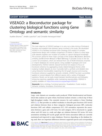
4BioMed Research InternationalTable 1: List of bioinformatics tools and databases used in this study for structural and functional analysis of the HP.S.N.Tools/serverURL(A) Sequence similarity SCLE3.MEGA X(B) Physiochemical characterizationExPASy –4.http://web.expasy.org/protparam/ProtParam(C) Subcellular localization identification5.PSORT B o.life.nctu.edu.tw/(D) Secondary structure predictionhttps://npsa-prabi.ibcp.fr/cgi-bin/npsa automat8.SOPMA.pl?page /NPSA/npsa red/(E) 3D structure prediction and quality Find similar sequences in protein databasesMultiple sequence alignment predictionPhylogenetic tree analysis272829Used for predicting physicochemical properties30Predict subcellular localizationPredict subcellular localizationPredict subcellular localization353634Predict the secondary structure of the protein37Predict secondary structure38Detect protein homologyUtilized to increase the stability ofthe 3D model structureUsed for Ramachandran plot analysisStructure verificationUsed to analyze the statistics of nonbondedinteractions between different atoms andverify protein structures39(F) Functional ov/Structure/cdd/wrpsb15.Used to search functional domains in a sequencedomain database.cgi16.Pfamhttp://pfam.xfam.org/Family relationship erpro/Used to search InterPro for motif Motif discovery(G) Active site identificationUsed to find, outline, and estimate e regions on protein 3D structuremethods. A molecular docking experiment was performed toscrutinize the binding interaction between the epitope andreceptor molecule. The crystal structure of HLA-B 15 : 01(PDB ID – 1xr8) was retrieved from the RCSB database[56] to perform docking analysis. The docking analysisbetween the peptide (ligand) and human receptor HLA-B 15 : 01 was performed using the AutoDockVina tool [57].The grid box size of the AutoDockVina tool was kept at12.702, 31.843, and 18.307, respectively, for X, Y, and Z.The binding interactions and residues in the interacting surface between the peptide and receptor were investigated withDiscovery Studio 2021 [58].3. Results and Discussion3.1. Sequence and Similarity Information. We selected ahypothetical protein (accession no. QIH20856.1) from theorganism N. gonorrhoeae. This hypothetical protein contains478 amino acids. The amino acid sequence for this protein404244434847454649was selected from the NCBI database and obtained inFASTA format. BlastP was performed to verify sequencesimilarity. The non-redundant protein sequences (nr) database (Table 2) and the UniProt/Swiss-Prot (SwissProt) database (Table 3) were examined to identify sequence similaritywith other known proteins by utilizing BlastP. The HPexhibits similarities with other MobA/MobL family proteins,according to the non-redundant protein sequence database.A phylogenetic tree showing the phylogenetic relatednessamong the sequences obtained from the non-redundantdatabase was constructed using the MEGA X program byneighbor-joining method with a bootstrap replication of1000, shown in Figure 2.3.2. Physicochemical Properties. According to the ExPASyProtParam server, the protein’s physical properties(Table 4) revealed that it includes 478 amino acids. The mostprevalent amino acids in the composition were Ala (37), Arg(30), Asn (23), Asp (26), Cys (3), Gln (47), Glu (55), Gly
BioMed Research International5Table 2: Similar protein obtained from non-redundant protein sequences (nr) database.DescriptionMobA/MobL familyMobA/MobL familyparainfluenzae]MobA/MobL familyhaemolyticus]MobA/MobL familyMobA/MobL familyparainfluenzae]Max Totalscore scoreScientific nameprotein [Neisseria gonorrhoeae]protein Haemophilusparainfluenza9849840100WP 032490546.1978978099.37WP 197561055.1Haemophilus haemolyticus977977099.16WP 140450219.1Neisseria P 127514845.1907907099.11MBS6191364.1protein [Proteobacteria]protein [Haemophilusprotein [HaemophilusEvalueTable 3: Similar protein obtained from UniProt/Swiss-Prot (SwissProt) database.Scientific nameMax scoreTotal scoreE valuePer. IdentAccessionEscherichia coliSalmonella enterica subsp. entericaserovar TyphimuriumAcidithiobacillus ferriduransBifidobacterium longum NCC2705Agrobacterium .1Description[Escherichia coli][Salmonella enterica subsp. entericaserovar Typhimurium][Acidithiobacillus ferridurans][Bifidobacterium longum NCC2705][Agrobacterium tumefaciens]79WP 032490546.1 MULTISPECIES: MobA/MobL family protein ProteobacteriaWP 127514845.1 MobA/MobL family protein Neisseria gonorrhoeaeQIH20856.1 hypothetical protein F0T10 13280 (plasmid) Neisseria gonorrhoeaeWP 140450219.1 MobA/MobL family protein Haemophilus haemolyticusWP 197561055.1 MobA/MobL family protein Haemophilus parainfluenzaeMBS6191364 .1 MobA/MobL family protein Haemophilus parainfluenzae8897Figure 2: Phylogenetic relationship among the hypothetical protein and other similar proteins obtained from the non-redundant databaseby BlastP search. The evolutionary distances were computed using the Poisson correction method and are in the units of the number ofamino acid substitutions per site.Table 4: ProtParam tool analysis result for the HP of Neisseriagonorrhoeae F0T10 13280.Number of amino acidsMolecular weightTheoretical pITotal number of negativelycharged residues (Asp Glu)Total number of positivelycharged residues (Arg Lys)FormulaInstability index (II)Aliphatic indexGrand average ofhydropathicity (GRAVY)The estimated half-life 37-1.179Thirty hours (mammalianreticulocytes, in vitro). 20 hours (yeast, in vivo). 10 hours (Escherichia coli,in vivo).(20), His (10), Ile (26), Leu (34), Lys (53), Met (7), Phe (17),Pro (11), Ser (28), Thr (15), Tyr (20), Trp (5), and Val (11).Its molecular weight is 56206.84 Dalton. The hypotheticalprotein has an instability index of 45.45, indicating that itis a stable protein. The numbers of negatively charged(Asp Glu) and positively charged (Arg Lys) residueswere calculated to be 81 and 83, respectively. The aliphaticindex was found to be 63.37, indicating that the protein isstable across an extensive temperature range. The protein’sGRAVY score of 1.179 suggested that it is water-soluble(hydrophilic). The protein’s pI was calculated to be 8.07,indicating that it is acidic (pH 7) in nature. The molecularformula of the HP was C2461H3884N716O774S10. Inmammalian reticulocytes (in vitro), yeast (in vivo), and E.coli, the putative protein’s half-life was calculated to be 30hours in mammalian reticulocytes (in vitro), 20 hours inyeast (in vivo), and 10 hours in E. coli (in vivo).3.3. Subcellular Localization Prediction. The environments inwhich proteins operate are determined by their subcellularlocalization. Protein subcellular localization is crucial forunderstanding protein function. Predicting an unknownprotein’s subcellular localization also provides valuable
6BioMed Research d, protein bindingExtracellularHelixPutative domain boundaryRe-entrant helix4050CoilMembrane interaction cytoplasmicDisorderedTransmembrane helixSignal peptideFigure 3: Secondary structure model predicted by the SOPMA 400Figure 4: Secondary structure model by PSIPRED server.Before energy minimizationAfter energy minimizationFigure 5: Predicted 3D structure of the hypothetical protein visualized by PyMOL (before and after energy minimization).information about genomic annotation and drug design[31]. The prediction of an unknown protein’s subcellularlocalization can be used to understand disease mechanismsas well as to develop drug or vaccine targets in a given pathogen genome [59]. The cytoplasmic proteins may serve asless suitable potential therapeutic targets, whereas surface
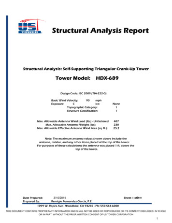
BioMed Research International7Saves180135Psi 45045Phi (degrees)90135180Error value⁎(a)99%95%20406080100120140160180Residue # (window :Y113:S119:Q11:A17:V–2.5Averaged scoreRaw score(c)Figure 6: (a) The PROCHECK program validated the Ramachandran plot of the predicted structure. (b) Quality factor 95.556 for ERRAToutput. Two lines on the error axis represent the level of confidence required to reject areas that exceed the error value. (c) Verify3Dprediction outcome showing 96.30% of the residues have averaged 3D-1D score 0.2.
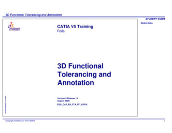
8BioMed Research InternationalTable 5: Ramachandran plot statistics of the predicted 3D modelfor studied protein.Ramachandran plot analysisNo. (%)Residues in the most favored regions [A, B, L]Residues in the additional allowed regions[a, b, l, p]Residues in the generously allowed regions[-a, -b, -l, -p]Residues in the disallowed regionsNo. of non-glycine and non-proline residuesNo. of end-residues (excl. Gly and Pro)No. of glycine residues (shown in triangles)No. of proline residuesTotal no. of residues159 (91.9%)13 (7.5%)1 (0.6%)0 (0.0)173 (100.0%)286189Table 6: Quality assessment score before and after energyminimization.CriteriaEnergyQuality factor (ERRAT)Ramachandran plot(PROCHECK)VERIFY 3DBefore energyminimizationAfter energyminimization- 48361.0 kJ/mol78.453-11487.9 kJ/mol95.555690.8%93.6%98.41% of theresidues haveaveraged 3D-1Dscore 0.296.30% of theresidues haveaveraged 3D-1Dscore 0.2membrane proteins are thought to be effective vaccine targets [33, 60]. In our study, we have found our protein ascytoplasmic according to the result of the CELLO. The localization score from CELLO was found to be 1.680. PSORTbv3.0.3 and PSLpred were used to verify the result. PSORTbv3.0.3 also identified the protein to be cytoplasmic, and thescore was found to be 8.96. According to the PSLpred, theprotein was also predicted as a cytoplasm-resident proteinwith a score of 64.47.3.4. Secondary Structure Prediction. The secondary structure(helix, sheet, turn, and coil) aids in providing information oneach amino acid’s conformation. Protein secondary structure prediction can be used to predict tertiary structureand the primary sequence and tertiary structure are linkedby it [61]. Though protein secondary structure predictionis an essential first step toward predicting tertiary structure,it also provides details on protein activity, interactions, andfunctions. Alpha helices were found to be the most frequently occurring structure in the HP while examined bySOPMA (69.87 percent) (Figure 3). The random coil wasseen at 19.67 percent, followed by the extended strand at5.65 percent. In addition, beta-turn was found to be 4.81percent. We cross-checked the results using PSIPRED, anda similar result was revealed (Figure 4).3.5. Homology Modelling, Quality Assessment of the 3DModel, and Visualization. The 3D structure of the proteinFigure 7: Active site (red color) of the studied hypothetical protein.Table 7: T cell epitopes predicted by NetCTL server along withtheir MHC I binding QFSGYAIYHLNVRYDLQRIQGDYTVDSGSNKLInteracting MHC I allelesHLA-A 30 : 02HLA-A 01 : 01HLA-A 30 : 02HLA-B 35 : 01NoneHLA-B 35 : 01, HLA-B 58 : 01HLA-A 30 : 02, HLA-B 15 : 01HLA-B 35 : 01, HLA-B 53 : 01HLA-A 30 : 02, HLA-B 15 : 01HLA-A 01 : 01HLA-A 30 : 02, HLA-A 32 : 01, HLA-B 15 : 01,HLA-A 03 : 01, HLA-A 11 : 01HLA-A 30 : 02Noneis highly related to its function. It also helps to predict thebinding sites and active sites of the protein, which may contribute to design an effective vaccine against that pathogen.The 3D structure of the HP was obtained from HHpredserver using homology modelling. By lowering the energyfrom -48,361.0 kJ/mol to -11487.9 kJ/mol, the YASARAenergy minimization server made the model structure morestable. The 3D structure of the protein was developed byPyMOL v2 (Figure 5). A variety of quality assessment toolswere employed to determine how reliable the protein’s predicted 3D structural model was. PROCHECK’s Ramachandran plot analysis, Verify3D, and ERRAT verified theprotein’s 3D structure. According to the Ramachandran PlotStatistics (Figure 6(a)), the model was thought to be acceptable, with 93.6 percent residues in the most favored regions(Table 5), and it was 90.8 percent before energy minimization. Utilizing the ERRAT and Verify3D programs, the
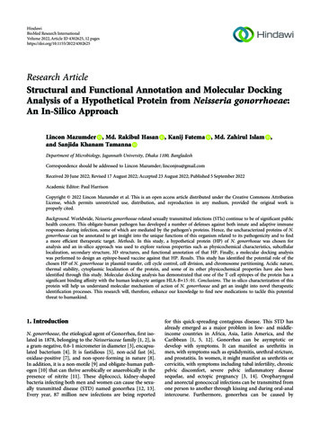
BioMed Research International9(a)(b)Figure 8: Docking analysis revealed by AutodockVina. (a) Three-dimensional structure of the predicted epitope, “AIYHLNVRY” and (b)visualization of binding interactions and residues after the docking of “AIYHLNVRY” with HLA-B 15 : 01.initial structural model was assessed for 3D structure errors.After energy minimization, ERRAT determined that themodel was of good quality with an overall quality factor of95.556 (Figure 6(b)), whereas it was 78.453% prior to energyminimization. After energy minimization, The Verify3Dshowed that (Figure 6(c)) 96.30 percent of the residues haveaveraged 3D-1D score 0.2, indicating that the model’senvironmental profile is good. A comparison of all the quality factors of the predicted structure before and after energyminimization is summarized in Table 6.3.6. Functional Annotation. Using the NCBI’s conserveddomain search tool, two functional domains of the HP wereidentified. The domain detected in the HP belongs to theMobA/MobL protein family (accession No. pfam03389).This family includes the MobA protein from the E. coli plasmid RSF1010 and the MobL protein from the Thiobacillusferrooxidans plasmid PTF1. These are mobilization proteins,which are required for particular plasmid transfer. Smc orchromosomal segregation ATPase is another superfamilythat involves cell cycle control, cell division, and chromosome partitioning. Plasmid transfer, cell division, cell cycleregulation, and chromosomal partitioning are essentialaspects of genetic engineering and the biotechnologicalapproach. Cell cycle regulation is critical for cell survivaland proliferation. Lack of cell cycle maintenance can resultin harmful mutations, leading to cell death and cancer[62]. This result was also cross-checked using INTERPRO,MOTIF, and Pfam. All produced similar findings, with positions ranging from 23 to 211 amino acid residues and an e-value of 3.5e-29.3.7. Active Site Detection. Several studies have documentedthat the discovery and identification of active sites on proteins are becoming highly significant. The position of theactive site on a protein is pivotal for a variety of purposes,including structural identification, functional site comparison, molecular docking, and de novo drug creation [26].Since the computed atlas of surface topography of proteins(CASTp) just employs the Cα atoms to represent the protein structure, it is quick and appropriate for usage withmodels and unreliable structures. The geometric potentialis a concept to quantitatively describe the shape of theprotein structure, which can be affected by the overallform of the structure and individual residue’s surroundings. About 85% of known binding sites may be reliablypredicted by CASTp with above 50% residue coverageand 80% specificity, and it often uses the geometric potential for this purpose [63]
Structural and Functional Annotation and Molecular Docking . lated in 1878, belonging to the Neisseriaceae family [1, 2], is a gram-negative, 0.6-1 micrometer in diameter [3], encapsu- . mycin and ceftriaxone as a dual therapy for the time being against this disease [12]. All these things have now made