Transcription
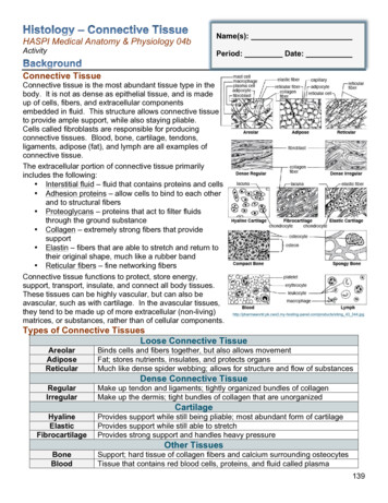
HASPI Medical Anatomy & Physiology 04bActivityName(s):Period: Date:Connective TissueConnective tissue is the most abundant tissue type in thebody. It is not as dense as epithelial tissue, and is madeup of cells, fibers, and extracellular componentsembedded in fluid. This structure allows connective tissueto provide ample support, while also staying pliable.Cells called fibroblasts are responsible for producingconnective tissues. Blood, bone, cartilage, tendons,ligaments, adipose (fat), and lymph are all examples ofconnective tissue.The extracellular portion of connective tissue primarilyincludes the following: Interstitial fluid – fluid that contains proteins and cells Adhesion proteins – allow cells to bind to each otherand to structural fibers Proteoglycans – proteins that act to filter fluidsthrough the ground substance Collagen – extremely strong fibers that providesupport Elastin – fibers that are able to stretch and return totheir original shape, much like a rubber band Reticular fibers – fine networking fibersConnective tissue functions to protect, store energy,support, transport, insulate, and connect all body tissues.These tissues can be highly vascular, but can also beavascular, such as with cartilage. In the avascular tissues,they tend to be made up of more extracellular (non-living)matrices, or substances, rather than of cellular anel.com/products/stimg 43 344.jpgTypes of Connective TissuesLoose Connective TissueAreolarAdiposeReticularBinds cells and fibers together, but also allows movementFat; stores nutrients, insulates, and protects organsMuch like dense spider webbing; allows for structure and flow of substancesRegularIrregularMake up tendon and ligaments; tightly organized bundles of collagenMake up the dermis; tight bundles of collagen that are unorganizedDense Connective s support while still being pliable; most abundant form of cartilageProvides support while still able to stretchProvides strong support and handles heavy pressureOther TissuesBoneBloodSupport; hard tissue of collagen fibers and calcium surrounding osteocytesTissue that contains red blood cells, proteins, and fluid called plasma139
Connective Tissue Disorders & DiseaseThere are several diseases and disorders that specificallytarget connective tissues. Collagen and elastin are the mostcommon components that are affected. These tissues maysimply become injured through trauma, such as a sprain,or they may be caused by genetic or environmental factors.Often it is the body’s own immune system that causes aninflammation in these tissues.Symptoms tend to be disease-specific, but the mostcommon symptoms associated with connective tissuehttp://www.jfponline.org/images/5910/5910JFP Article2-fig2.jpgdisorders include fatigue, fever, muscle pain, stiffness,weakness, and joint pain. A few examples of connective tissue disorders include:DisorderWhat is ndromeGenetic disease that causes thedeterioration of collagenFragile and stretchy skin, fatty lumps,extremely flexible joints1 in 5,000Genetic disease that causes abnormalfibrillin productionTall, slender, loose joints,disproportioned skeleton, scoliosis,dislocation of lens (eyes),cardiovascular disease, ectasia, lesselastic alveoliShort, bones easily fracture, bluesclera, deafness, scoliosis, kyphosis,bowed bonesSwollen joints, joint pain, morningstiffness, firm bumps under skin,fatigue, weight loss, feverSwelling, itching, tenderness of theskin, calcinosis, heartburn, Raynaud’sphenomenon, increased bloodpressure, renal crisis, bowel diseaseVaried; fever, fatigue, muscle ache,alopecia, arthritis, ulcers,photosensitivity, pleuritis, pericarditis,Raynaud’s sSclerodermaSystemic lupuserythematosusAKA brittle bone disease; Insufficientcollagen production, causing aninability to build bone structureAutoimmune disease that causes aninflammation of cartilage and jointsAutoimmune disease causing a buildup of scar tissue in the skin, bloodvessels, and organsInflammation of connective tissues1 in 5,0001 in 20,0009.8 in 1,0001 in 9061 in 1,000Shiel, W.C. and Stoppler, M.C. 2012. Connective Tissue Disease. www.medicinenet.com.Connective tissue charts (12)Computer/internet OR connective tissue slides and a microscopePart A. Becoming Familiar with Connective TissuesFigure 1In Part A of this lab you will have the opportunity to familiarize yourselfwith the different types of connective tissues. Posters with the 10main types of tissues have been placed throughout the room. Visiteach poster and record the description, function, and location in thefollowing chart. Draw and label an example in the right columnfor each picture. An example drawing can be seen in Figure 1 tothe s/thumb/7/75/Transverse Section Of Bone.png/250px-Transverse Section Of Bone.png140
a. Connective Tissue Proper: loose connective tissue, areolarDescription (write or draw)Draw an example. Use colored pencils and label ifnecessary.FunctionLocationb. Connective Tissue Proper: loose connective tissue, adiposeDescription (write or draw)Draw an example. Use colored pencils and label ifnecessary.FunctionLocationc. Connective Tissue Proper: loose connective tissue, reticularDescription (write or draw)Draw an example. Use colored pencils and label ifnecessary.FunctionLocation141
d. Connective Tissue Proper: dense connective tissue, dense regularDescription (write or draw)Draw an example. Use colored pencils and label ifnecessary.FunctionLocatione. Connective Tissue Proper: dense connective tissue, dense irregularDescription (write or draw)Draw an example. Use colored pencils and label ifnecessary.FunctionLocationf. Cartilage: hyalineDescription (write or draw)FunctionLocation142Draw an example. Use colored pencils and label ifnecessary.
g. Cartilage: elasticDescription (write or draw)Draw an example. Use colored pencils and label ifnecessary.FunctionLocationh. Cartilage: fibrocartilageDescription (write or draw)Draw an example. Use colored pencils and label ifnecessary.FunctionLocationi. Others: compact bone (osseous tissue)Description (write or draw)Draw an example. Use colored pencils and label ifnecessary.FunctionLocation143
j. Others: bloodDescription (write or draw)Draw an example. Use colored pencils and label ifnecessary.FunctionLocationk. Others: spongy boneDescription (write or draw)Draw an example. Use colored pencils and label ifnecessary.FunctionLocationl. Others: lymphDescription (write or draw)FunctionLocation144Draw an example. Use colored pencils and label ifnecessary.
Part B. Identify the Connective TissueIn Part B of this activity, use what you have just learned to identify the following connective tissues.Write your answers on the line in each box.A.B. C.D.E.G.H. I.F.J. K.L.M.O.N.A. ki/images/39 Fibrocartilage Connective Tissue.jpgB. http://lifesci.rutgers.edu/ babiarz/Histo/Blood/Smear1.jpgC. es/t19.jpgD. http://www.jeremyswan.com/anatomy/203/lab 03 tissues/Areolar-ct.jpgE. ards/1151100/png/elastic cartilage connective tissue 31328663923618.pngF. /24Bone1 100X rev.jpgG. alinecart.jpgH. -tissue-h-e-stain-lm-x100 i-G-64-6473-HCQH100Z.jpgI. http://dtc.pima.edu/blc/160Mars/160Mimages/Dense connectivex.jpgJ. tiveTissueSkin.jpgK. ix/slide007atyp1.jpgL. ki/images/32 Reticular Connective Tissue.jpgM. SYSTEM.jpgN. ilage%20400X.JPGO. http://medcell.med.yale.edu/histology/bone lab/images/trabecular bone.jpg145
Part C. Practice, Practice, PracticeYour instructor will either have slides available to view with the microscope OR you can use acomputer and the following website to choose slides to /virtual nrml/nrml lst.htmFor each type of connective tissue, observe the slide and identify the connective tissue. REMEMBER there are multiple tissue types on many of the slides. Start by searching through the slide for images similar to those in your drawings from Part A. You may need to move the slide around to find a good example! You may need to look up/research the organ function if it is unfamiliar.a. Connective tissue proper: loose connective tissue, areolarSlide Choices: areolar tissue slide, under epithelium, mucous membranes,surrounding capillariesOrganDraw an example. Use colored pencils.Organ FunctionTissue Functionb. Connective tissue proper: loose connective tissue, adiposeSlide Choices: Fat or adipose slide, under skin, around kidneys, around eye, breast,abdomenOrganOrgan FunctionTissue Function146Draw an example. Use colored pencils.
c. Connective tissue proper: loose connective tissue, reticularSlide Choices: Reticular tissue slide, lymph nodes, bone marrow, spleenOrganDraw an example. Use colored pencils.Organ FunctionTissue Functiond. Connective tissue proper: dense connective tissue, dense regularSlide Choices: Dense regular tissue slide, tendons, ligaments, aponeurosesOrganDraw an example. Use colored pencils.Organ FunctionTissue Functione. Connective tissue proper: dense connective tissue, dense irregularSlide Choices: Dense irregular tissue slide, dermis, submucosa, joint capsulesOrganDraw an example. Use colored pencils.Organ FunctionTissue Function147
f. Cartilage: hyalineSlide Choices: Hyaline cartilage, embryo skeleton, end of long bones, costal cartilage,nose, trachea, larynxOrganDraw an example. Use colored pencils.Organ FunctionTissue Functiong. Cartilage: elasticSlide Choices: Elastic cartilage, ear, epiglottisOrganDraw an example. Use colored pencils.Organ FunctionTissue Functionh. Cartilage: fibrocartilageSlide Choices: Fibrocartilage, intervertebral discs, knee joint, pubic symphysisOrganOrgan FunctionTissue Function148Draw an example. Use colored pencils.
i. Others: compact bone (osseous tissue)Slide Choices: BoneOrganDraw an example. Use colored pencils.Organ FunctionTissue Functionj. Others: bloodSlide Choices: Blood, red blood cell smearOrganDraw an example. Use colored pencils.Organ FunctionTissue Functionk. Others: spongy boneSlide Choices: Spongy boneOrganDraw an example. Use colored pencils.Organ FunctionTissue Function149
l. Others: lymphSlide Choices: Blood, bone marrow, lymphOrganDraw an example. Use colored pencils.Organ FunctionTissue FunctionAnalysis Questions - on a separate sheet of paper complete the following1. Create a concept map titled “Connective Tissues” with a short description OR a drawnexample including ALL of the following: loose connective tissue, dense connective tissue,areolar tissue, adipose tissue, reticular tissue, regular dense tissue, irregular dense tissue,cartilage, hyaline cartilage, elastic cartilage, fibrocartilage, other tissues, compact bone,spongy bone, blood, and lymph.2. What is the difference between loose and dense connective tissue?3. What is the difference between areolar, adipose, and reticular tissue?4. What is the difference between regular and irregular dense tissue?5. What is the difference between hyaline, elastic, and fibrocartilage?6. What is the difference between compact and spongy bone?7. CONCLUSION: In 1-2 paragraphs summarize the procedure and results of this lab.Review Questions - on a separate sheet of paper complete the following1. What three parts primarily make up connective tissue?2. What are fibroblasts?3. What parts of the body are classified as connective tissue?4. What is interstitial fluid?5. What are adhesion proteins?6. What are proteoglycans?7. What is the difference between collagen, elastin, and reticular fibers?8. What is the function of connective tissues?9. What is the difference between vascular and avascular tissue?10. Create a chart with a description, function, and location of all of the loose and denseconnective tissue types.11. What components of connective tissue are most commonly affected by disease and/ordisorder?12. Out of the disorders listed in the chart in the background section, which connective tissuedisorder is the most prevalent?13. Choose one of those disorders and provide a description and the symptoms.150
Part A. Becoming Familiar with Connective Tissues In Part A of this lab you will have the opportunity to familiarize yourself with the different types of connective tissues. Posters with the 10 main types of tissues have been placed throughout the room. Visit each poster and record the description, function, and location in the following chart.