Transcription
2010CVM 6100VeterinaryGross AnatomyGeneral Anatomy&Carnivore AnatomyLecture NotesbyThomas F. Fletcher, DVM, PhDandChristina E. Clarkson, DVM, PhD1
CONTENTSConnective Tissue chanics and Locomotion.12Serous Membranes and Cavities.15Formation of Serous Cavities.17Nervous System.19Autonomic Nervous System.23Abdominal Viscera.27Pelvis, Perineum and Micturition.32Female Genitalia.35Male Genitalia.37Head Features (Lectures 1 and 2).40Cranial Nerves.44
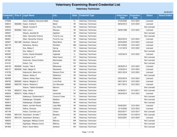
Connective Tissue StructuresHistologic types of connective tissue (c.t.):1] Loose areolar c.t. — low fiber density, contains spaces that can be filled with fat or fluid (edema)[found: throughout body, under skin as superficial fascia and in many places as deep fascia]2] Dense irregularly arranged c.t. — high density of collagen fibers, oriented in variable directions[found: dermis; deep fascia in some locations; periosteum; fibrous joint capsule]3] Dense regularly arranged c.t. — high density of parallel fibers, forming sheets, bands, or cords[found: aponeuroses; ligaments; tendons]Connective tissue structures identifiable in gross anatomy:Dermis [G. skin] — the physically tough/strong component of skin (deep to epidermis)Tendon — attaches muscle to bone (called aponeurosis when sheet-like)Ligament — attaches bone to bone (usually thickenings of fibrous joint capsules)[Note: visceral ligaments located in body cavities are entirely different structures]Fascia [L. band] — collagenous fibrous tissue that hold the body togethersuperficial fascia subcutaneous tissue between skin & muscles/bone (body wall)- regionally variable in amount (site for subcutaneous injection)- contains: cutaneous muscle, mammary tissue, fat (also edema fluid)[e.g., cutaneous trunci m.; superficial muscles of facial expression]deep fascia packing/binding tissue surrounding muscles, bones, & organs- compartmentalize skeletal muscles & gives rise to aponeuroses- forms several named structures, viz., named regional fascia, e.g., thoraco-lumbar fascia, fascia lata, etc.(fascia is named where it is thick & distinct (i.e., dense c.t. vs. looseareolar c.t.) retinaculum [L. rope or cable] fascia that binds passing tendons to thesurface of the carpus or tarsus (also, transverse humeral retinaculum) raphe [G. seam] fascia that joins right and left counterparts of a particular muscle at the midline (e.g., ventral abdomen linea alba) epimysium [G. on muscle] fascia covering the surface of a muscle,depending on the muscle, it may be thin (transparent) or dense (opaque& white); also,perimysium c.t. around muscle fascicles; andendomysium c.t. within muscle fascicles)Transverse section through askeletal muscle:1 epimysium;2 perimysium;3 endomysium
Axial section through metacarpus and digit:1 interosseus m.; 2 digital extensor tendon;3 metacarpal bone; 4 dorsal sesamoid bone;5 proximal phalanx; 6 proximal sesamoidbone; 7 metacarpal pad; 8 digital flexor tendons; 9 digital annular ligaments; 10 digitalpad; 11 unguis (nail)Transverse section through antebrachium (horse):1 superficial fascia; 2 cephalic vein; 3 radius (bone); 4 & 5 deep fascia (compartmentalizing muscles); Med. medial; Cr. cranial
Osteology.The dog has 321 bones.Bone FunctionsSupportbody shape & weightBone Classification SchemesLeversto perform workDevelopment:Endochondral bones — develop from cartilage precursorsProtection[most bones]of vulnerable organsIntramembranous bones — directly from mesenchyme (fascia)[bones of calvaria & face]Ca & PO4- Location:reservoir for ionsAxial skeleton — head, vertebral column ( including tail),ribs & sternumRed MarrowAppendicular skeleton — bones of limbs, includingsource of blood cellsscapula & os coxae(hip bone)Heterotopic bones — os penis [ carnivore; rodent ]os cardis [ cattle ]Bone CompositionShape:Long bones — length greater than diameterShort bones — approximately equivalent dimensionsFlat bones — e.g., scapula, os coxae, many bones of skullIrregular bones — short & multiple processes (vertebrae)Sesamoid bones — small “seed-like” within tendons,e.g., patella (knee cap)Collagen fibersby weight: 1/3 of boneby volume: 1/2 of boneHydroxyapatite crystals(Ca)10(PO4)6(OH)295% solid (vs. water)65% mineral; 35% organicStructure of a Long BoneRegionsof aLong Bonearticular foramenmetaphysisepiphysisphysis(epiphysealplate)5
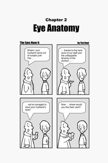
Mechanical ConsiderationsStrength amount of strain a bone canwithstand without breaking.Bone is best at withstanding compression,especially against the “grain”External ForceInternal Force (Stress)(compressing long axes of osteones)Tensile strength 1/2 of compression;comparable to tendons & ligamentsShear strength 1/4 of compression;most fracture are the result of shear forcesInternal Distortion (Strain)General Principle:Hollow-Shaft ConstructionForceBones are designed to provide adequatestrength with minimal material (minimal massor weight).ForceSuch an economy of bone mass/weightoffers evolutionary advantages; viz., fasterreaction capability; reduced metabolic requirements.compressivestraintensilestrainFlat Scapula Reinforced(transverse section)tensionSTRAIN 0compressionScapula strengthened with only a spineSesamoid bone — Patelladistancequadricepsfemorismuscle1] Eliminates tendon shear3] Increases TorqueFemurpatella2] Redirectslines of forceTorque F x dpatellaligamentTibia6
Arthrology(Joint Articulation Union of two or more bones)Classification:Fibrous joints — immobile joints, united by fibrous tissue, may ossify with age.Three types are recognized:1] Suture [L. seam] undulating seams between bones of the skull2] Gomphosis tooth in an alveolus, united by periodontal ligament3] Syndesmosis bones joined by ligaments, e.g., [radius & ulna] and [tibia & fibula]Cartilaginous joints — immobile joints, united by cartilage, ossify with age.Two types are recognized:1] Symphysis [G. grow together] fibrocartilage union,e.g., pelvic symphysis; mandibular symphysis; (also, intervertebral disk)2] Synchondrosis hyaline cartilage union, e.g., physisSynovial joints — mobile joints, fibrous tissue enclosing a synovial cavityClassified on the basis of.Number of bones:Simple joint formed by two bones, e.g., shoulder jointCompound joint formed by more than two bones, e.g.,elbow joint, carpal jointShape:Hinge (ginglymus) joint movement in one planeBall & socket (spheroid) joint capable of circumductionPlane joint gliding action, e.g., vertebral articular processesalso, Ellipsoid, Saddle, Condylar, TrochoidSynovial Joint Structure:[synovia G. with egg (white)]Joint features. articular (hyaline) cartilage covers the opposing surfaces of the bones synovial membrane lines a synovial cavity that separates the bones— the membrane secretes synovial fluid into the cavity fibrous (collagenous tissue) layer located external to synovial membrane— mechanically joins the bones, blends with periosteum— selectively thickened to form ligamentsNOTE: Joint Capsule fibrous layer and synovial membrane together.Additional features found in some synovial joints. meniscus fibrocartilage in the synovial cavity, interposed between the bones(one meniscus in temporomandibular joint; two semilunar menisci in stifle) internal ligaments that appear to be within the joint cavity (such ligaments areactually surrounded by synovial membrane and thus they are outside thesynovial cavity itself) fat pads between the fibrous & synovial layers produce synovial folds that mayprotrude into the joint cavity
MyologyThere are three categories of muscle tissue:1] smooth muscle not striated; associated with viscera (gut, vessels, glands, etc.)2] cardiac muscle striated; musculature of the heart3] skeletal muscle striated; generally attached to bone; usually under voluntary controlSkeletal MuscleSkeletal (striated) muscle is composed of elongate, multinucleated cells (muscle fibers).Different types of muscle fibers are found among the various skeletal muscles of the body, e.g.,— slow contracting, fatigue resistant, aerobic metabolism (Type I)— fast contracting, fatigue resistance, aerobic metabolism (Type 2A)— fast contracting, fatigue susceptible, anaerobic metabolism (Type 2B)Note: Skeletal muscle will not contract in the absence of a functional nerve supply (denervationatropy occurs). One neuron innervates a variable number of muscle fibers. The neuron plusthe muscle fibers it innervates constitute a motor unit. To produce a stronger contraction,the nervous system activates more motor units.Muscle-related connective tissue:Muscle fibers are within a connective tissue framework that is continuous with tendons. As aresult, passive muscles are able to serve as ties that reinforce joints & oppose forces on bones.Muscle associated fascia:1. epimysium loose or dense connective tissue surrounding an entire muscle2. perimysium loose connective tissue defining muscle fascicles3. endomysium small amounts of loose c.t. surrounding individual muscle fibersTendon protection:A. bursa synovial pocket inserted between a tendon and a bony prominenceB. tendon synovial sheath lubrication where tendons are bound, e.g., by retinaculum10
Muscle names:Muscle names may be latinized (flexor digitorum profundus) or anglicized (deep digitalflexor). Muscle are named (originally in the human) for their shape (deltoideus) or location (brachialis) or attachments (sternohyoideus) or structure (biceps) or function (supinator) or combinations ofthese (pronator quadratus; superficial digital flexor; serratus ventralis; flexor carpi radialis; etc.)Muscle roles within a given movement (classification of involved muscles):— agonist prime mover or principal muscle(s) executing the particular joint movement— antagonist muscle(s) that oppose the action of the agonist on the joint(s)— synergist muscle(s) that assist the agonist; e.g., fixators stabilize distant joints.Muscle architecture:Multiple muscles and multiple parts or heads (head a separate belly and origin) exist todistribute (as opposed to concentrate) stresses on bones and to provide movement diversity.Fascicle & fiber arrangement:Parallel arrangement, e.g., strap or spindle arrangement, fibers/fascicles arranged parallel tothe tendon of insertion. This results in a greater range of shortening and thus yields greater movement velocity (distance per time).Pennate arrangement fibers/fascicles arranged at an angle to the direction in which thetendon moves. This results in a greater area of muscle fibers along axes of contraction and producesmore strength (at the expense of a reduced range of contraction).Note: The amount of force that a muscle can generate is proportional to the area of musclefibers, i.e., number of contractile protein molecules, multiplied by the cosine of the muscle-tendon angle.Three types of pennate arrangement are:— unipennate, e.g., ulnar & radial heads of the deep digital flexor muscle;— bipennate, e.g., infraspinatus muscle;— multipennate, e.g., humeral head of the deep digital flexor muscle.Strap Muscle(Parallel Fibers)Pennate Muscle(Unipennate)BipennateMultipennate11
Muscle & Body BiomechanicsMuscle DynamicsMUSCLE FIBER ARRANGEMENTEFFECT ON STRENGTHThe amount of force that a muscle can generate is proportional to the cross-sectionalarea of muscle fibers (a.k.a. muscle cells) attaching to its tendon, i.e., the number ofcontractile proteins (actin and myosin) pulling on the tendon and contributing tomuscle force. pennation design increases the number of muscle fibers (cross sectional area) attachedto the tendon since force is a function of cross sectional area - a pennated muscle can generate moreforce than a comparable muscle with parallel fibers.EFFECT ON SHORTENINGIn this example, again consider two muscles - one with parallel fibers the other pennateAssume each muscle fiber will contract to 50% of its resting lengthTherefore: with parallel-arranged muscle fibers the entire muscle can contract by 50% with the pennate arrangement each individual muscle fiber is pulling at an angle,resulting in reduced overall shortening of the entire muscle belly.12
Animals are subject to the same physical laws as inanimate objects.An understanding of movement (e.g. motion of the joints and body) comes through anunderstanding of the forces acting upon the joint or the body that result in that movement.Forces can be simplified into linear and rotational componentsDEFINITIONS:LINEAR FORCE:Force can be broken down into various vectors. Vertical vectors (e.g. the downward forces due to body weight andthe upward forces of the supporting surface) Horizontal vectors (e.g. forces exerted to propel forward andbackward forces to brake forward motion)With adequate force and friction (traction) the body can propel itselfforward. (practical application dictates a need for good traction to allowthis forward motion – whoa to those leading a horse on ice)Horse’s weight vs.ground pushing upHorsepushing offvs. groundresistance(friction)Resultantvector (actionvs. reaction)ROTATIONAL FORCE (TORQUE)Rotational force force (F) x distance from fulcrum (d)Limb rotation muscle force (F) x distance from joint (d)Torque input (muscle generated) torque output (limb movement)Muscles generate forces which when applied to the skeleton will generaterotation about a joint.13
MUSCLE ATTACHMENT EFFECTSThe location of the muscle attachment (e.g. distance from joint) influences theresultant movement of that jointMECHANICAL ADVANTAGE VERSUS VELOCITY ADVANTAGEMuscles that attach further from the joint have a mechanical advantage over musclesattached closer to the jointIn the diagram below if muscles #1 and #2 were of equal strength (i.e., can generate thesame force) then muscle #2 could produce a greater rotational force because its attachmentis at a greater distance from the joint (rotational force muscle force X distance fromjoint).Conversely muscles that attach close to thepoint of rotation are able to produce fastermovement of the lever arm than muscle thatattach farther from the fulcrum.In the diagram to the right if muscle #1 andmuscle #2 both contract 10% during anidentical time period - muscle #1’s contractionwould result in a larger movement of the leverarm during that same frame of time thanmuscle #2. In other words, muscle #1 willresult in a more rapid rotation - it has a velocityadvantage.Muscles attaching close to the joint with their velocity advantage are termed “highgear” muscles and those with a more distal attachment resulting in a mechanicaladvantage are termed “low gear” muscles.It may be helpful to consider a similar gear analogy as in a car or bike. At low gears the output force isrelatively large – allowing the vehicle to climb up a steep hill. High gears on the other hand generates alot of speed – as would be advantageous in passing a vehicle.Note: the relationship between force and speed is inverse – one will increase at the sametime the other decreases14
JOINT POSITIONING EFFECTSTHE BODY’S LEVER SYSTEMUnique skeletal features result from functional adaptations over time.In the figure below - the upper diagram is an example of an animal that uses it’s frontlimbs for digging; the muscles attached to the point of the elbow (olecranon) arepositioned further from the elbow joint (fulcrum of movement) thereby generating largeforces for digging.The lower diagram with muscles attaching closer to the elbow joint is an runneradaptation that can result in a rapid rotation with muscle contraction (velocity advantage.For the same force andvelocity input (leftarrows), note therelative magnitude ofbolded arrows to theright of the diagrams –a large downwardforce (F) is generatedin upper diagram andrapid rotation (V;velocity) of movementis produced in thelower diagram.14A13
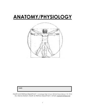
Serous Membranes & CavitiesBody CavitiesThe major cavities of the body are withinthe trunk. They contain visceral organs andserous membrane cavities:Thoracic cavity — is lined byendothoracic fascia.Abdominal & pelvic cavities — arelined by transversalis aphragmSerous Membrane Cavities— are lined by serous membrane— are normally empty (except for microscopic cells and a film of fluid)— function to preclude adhesions among organs, thereby allowing organs to movefreely relative to one another.A serous membrane consists of a single layer of flattened mesothelial cells applied to thesurface of a thin layer of collagenous tissue that attaches to underlying endothoracic/transversalis fascia.The mesothelium of the serous membrane forms the lining of a closed serous membrane cavity.Serous membrane lining the wall of a serous cavity is designated parietal while that coveringviscera is called visceral. Connecting serous membrane runs between parietal and visceral components.The serous membranes are:Peritoneum — the peritoneal cavity is found within the abdominal & pelvic body cavities.Connecting peritoneum forms:— mesentery— ligament.Pleura — two pleural cavities (separated by mediastinum) are found within the thoracic cavity.Parietal pleura is further subdivided into:— costal pleura— diaphragmatic pleura— mediastinal pleura & — pleural cupula.Connecting pleura forms the pulmonary ligament.Visceral pleura is also called pulmonary pleura.Pericardium — the pericardial cavity is found within the mediastinum of the thoracic cavity.Visceral pericardium is also called epicardium.Vaginal tunics — the cavity of the vaginal process begins at the vaginal ring and extends into thescrotum around the spermatic cord & testis.Connecting vaginal tunic forms: — mesorchium— mesoductus deferens.15
Peritoneal toneumgutperitonealcavityparietalperitoneumPleural (two) & Pericardial Cavitiescostalpleuravisceral cavitypleuralcavityLUNGparietalpericardiumvisceral pericardiumHEARTpericardial cavityligamentthoracicwall16
Formation of Body (Serous) CavitiesSerous cavities are cavities lined by serous membrane (mesothelium). In the adult, serouscavities are: the pericardial cavity, two pleural cavities, and the peritoneal cavity (including vaginalcavity extensions of the peritoneal cavity).Acquiring a three-dimensional understanding of howserous cavities are formed is a challenging exercise. Serouscavity formation may be summarized as follows: all of the serous cavities develop from a commonembryonic coelom and thus the cavities are continuous untilpartitions develop to separate them; the individual serous cavities are formed by inwardgrowth of tissue folds from the body wall (partitions) and byoutgrowth of coelomic cavity into the body wall (excavation).embryoCoelom elomembryoniccoelompericardiumhindgutDuring gastrulation, the space between ectoderm andcoelomendoderm (and between trophoblast and hypoblast) is filled byinflow of primary mesenchyme that becomes mesoderm.Cavitation occurs in lateral mesoderm, establishing theyolksaccoelom bounded by somatopleure and splanchnopleure.As head and tail processes develop and lateral bodyCoelomfolds merge medially (except at the umbilicus), embryonic and(Longitudinal View)extra-embryonic compartments of the coelomcan be differentiated. The former becomes theMesoderm somiteneural tubeserous cavities, the latter is filled by allantoicnotochord intermediatefetal membrane.foregut lateralFormation of the head process bringsLateralthe heart and pericardial coelom within theBodyembryo, positioned ventral to the foregut.FoldsRight and left sides of the embryonic coelomembryonicare separated by gut and by dorsal and ventralcoelommesenteries, the latter fails to develop at themesenterylevel of the midgut.Thus, the embryonic coelom featuresan anterior-ventral pericardial compartment, asomatopleureextra-embryoniccaudal peritoneal compartment, and bilateralcoelomsplanchnopleureyolkpleural compartments (channels) connectingsacthe pericardial and peritoneal compartments. Mesoderm lining the coelom forms mesothelium.Separation of Peritoneal and Pleural Cavities:In the adult, peritoneal and pleural cavities are separated by the diaphragm. The diaphragm isformed by a septum transversum, paired pleuroperitoneal folds, and somatic mesoderm. Diaphragmatic musculature is derived from somites in the cervical region (C5, 6, 7), where the diaphragm isinitially formed.17
Details of diaphragm formation include:Diaphragm Formation(Caudal View)— the septum transversum originates as mesoderm in front of theheart; as the heart shifts ventral to the foregut, the septum becomes incorporated into the ventral body wall and ventral mesentery caudal to the heart; itgrows dorsally and forms a transverse partition ventral to the level of the gutdegeneratingmesonephrospleuroperitonealfold— dorsal to the gut, bilateral pleuroperitoneal folds grow mediallyand meet at the dorsal mesentery— subsequent growth of the pleural cavity into somatic mesoderm(mesenchyme) will result in body wall mesoderm forming the marginalregions of the diaphragm (diaphragm musculature).septumtransversumSeparation of Pericardial and Pleural Cavities:In the adult, pericardial and pleural cavities are separatedby fibrous pericardium.Originally in the embryo, the pericardial coelomic cavitycommunicated with two dorsally positioned pleural cavities(canals). Subsequently, the cavities become partitioned by pairedpleuropericardial folds and then somatic mesoderm. Details ofthe separation include:crus agus— bilateral pleuropericardial folds (membranes), which accompanycommon cardinal veins as they join the heart, converge medially to unite withthe mediastinum (ventral mesentery) and partition the ventral pericardialcavity from the dorsal pleural canals;— subsequent ventrolateral growth of the pleural cavities into thebody wall incorporates somatic mesoderm (mesenchyme) into the futurefibrous pericardium.NOTE: Mediastinum is formed initially by dorsal and ventralmesenteries of the leGrowth of Pleural Cavities:Initially the pleural cavities are small canals into which the lung buds project. As the lungsgrow, the pleural cavities enlarge and appear to carve into the body wall (into somatic mesoderm/mesenchyme). As a result, somatic mesoderm forms partitions (fibrous pericardium and diaphragm)that wall off the pleural cavities.dorsal aortaesophaguslung budneural oldpleuralcoelomheartlungpleural cavitybody wallpericardialcoelompreicardial sacEarlyfibrouspericardiumPleural Cavity FormationLate18
Nervous SystemNeuron functional cell of Nervous System:— receives excitation (at a synapse or at a receptor);— conducts excitation (along an axon);— transmits excitation (via release of chemical at a synapse).Most neurons are multipolar — cell body is located where input excitation occursSensory neurons are unipolar — cell body is located along the axon (in a spinal ganglion)Multipolar NeuronDendritesSynapseCell bodyInputAXONconducts excitationtransmits excitation(via secretion)becomes excitedUnipolar NeuronSynapseAXON conducts excitationReceptorbecomes excitedCell body(in spinal ganglion)transmits excitationDefinitions:Nerve bundle of axons ensheathed by supporting cells and enveloped by connective tissueRoot nerve that is adjacent to the CNS and enveloped by meningesGanglion localized site where a nerve is enlarged due to a collection of cell bodies:Spinal ganglia — contain unipolar cell bodies (located on dorsal roots of spinal nn.)Autonomic ganglia — contain multipolar cell bodies that innervate viscera.Nervous System Divisions:Central (CNS):brain and spinal cordPeripheral (PNS): 12 pairs of cranial nerves (attached to brain);36 pairs of spinal nerves in the dog & cat (attached tospinal cord). [8 cervical; 13 thoracic; 7 lumbar; 3 sacral; & 5 caudal]Spinal Nerve:The spinal cord and spinal roots are located within the vertebralcanal of the vertebral column. Dorsal and ventral spinal roots unite toform a spinal nerve (bilaterally).Adjacent vertebrae combine to form an intervertebral foramen(dorsal to an intervertebral disc). The spinal nerve is found within theintervertebral foramen, from which it exits the vertebral canal.The spinal nerve is enveloped by connective tissue (epineurium,perineurium, & endoneurium). In contrast, the spinal cord and the dorsal and ventralspinal roots are surrounded by cerebrospinal fluid enclosed within meninges.Vertebra oramenprimarybranches of aspinal nervespinalnerve19
Spinal Nerve: typical pattern— short ( 1 cm); located at an intervertebral foramen— connected to the spinal cord by two roots (each comprised of rootlets):dorsal root — composed of afferent (sensory) axons; the site of a spinal ganglionventral root — composed of efferent axons that innervate muscle & gland— divides into four primary branches:meningeal branch — small; sensory to meningesramus communicans — connects to sympathetic trunk & innervates visceraventral branch — largest branch; hypaxial mm. & lateral and ventral cutaneous nn.dorsal branch — medial & lateral& dorsal cutaneous nn.y branches.; epaxial mm. pmeninges &subarachnoid spacecovering spinal corddorsal root spinal nerve(in meninges)dorsal branch:medial branchlateral branchventral branchspinalganglionsympathetic trunkramus communicansventral root(in meninges)meningrealmeningeal branchFour Primary Branches of a Spinal NerveFiber types: types of nerve fibers (axons) found in a spinal nerve and its branches Afferent (sensory) — axons associated with receptors and unipolar cell bodies in spinal gangliaGeneral Somatic Afferent (GSA): receptors in skin & muscles, tendons, jointsGeneral Visceral Afferent GVA): receptors in viscera Efferent (motor) — axons that innervate muscle & gland;cell bodies are located in the spinal cord (or in some cases autonomic ganglia)Somatic Efferent (SE): innervates skeletal muscleVisceral Efferent (VE): innervates cardiac m., smooth m., & glandNote: In older literature, SE & VE fiber types are designated GSE and GVE, preceded by the adjective General.Autonomic Nervous System (ANS) VE (sometimes the ANS includes GVA as well as VE fibers)The VE pathway, from the CNS to a visceral organ, is unique in that it is composed of twomultipolar neurons (the other efferent pathway and the two afferent pathways have just a single neuron).The first neuron in the pathway has its cell body in the CNS (brain or spinal cord). The cellbody of the second neuron is located within an autonomic ganglion in the peripheral nervoussystem. The autonomic nervous system operates largely at a subconscious level [auto-nomic self-rule].20
Cutaneous es of dorsal and ventral cutaneous nn.series of dorsal, lateral and ventral cutaneous nn.series of dorsal and lateral cutaneous nn.individually named cutaneous branches of regional nerves thatoriginate from nerve plexuses (brachial or lumbosacral) to the limbs.Face — named cutaneous branches of cranial nerves.C-1 is notcutaneousC-2 supplies dorsal headDorsal Cutaneous nn. of neck(from medial br. of dorsal br.)Dorsal cutaneous nn. of trunk(from lateral br. of dorsal br.)Great Auricular n.(from C-2 ventral branch)cutaneousnn. fromdorsal &ventralcaudalplexusesVentral Cutaneous nn. of neck(from ventral branch)Ventral cutaneous nn.(from ventral branches)Superficial radial n.(named cutaneous n.from named regional n.)Lateral cutaneous nn.(from ventral branches)first (intercostalbrachial) &last (lateral cutaneous femoral)are largestnamedcutaneousnerve:LateralCutaneousSural n.Brachial and Lumbosacral nerve plexuses:Individual muscles are composed of multiplemyotomes that overlap in forming the muscle.In the case of trunk muscles, which are generally broad, multiple dorsal or ventral branches of spinalnerves can be seen to serially innervate each individualmuscle. The innervations overlap within the musclebecause of myotome overlap.In the case of limb muscles, each muscle isinnervated by the branch of a single regional nerve.Because of multiple myotomes per muscle, the regionalnerves must contain axons from ventral branches ofmultiple spinal nerves. The exchange of axons amongventral branches as they form regional nerves producesa nerve plexus for each limb.Trunk mm.L1 L2L3Limb mm.L4L4 L5L5L6L6L7L7L4L5L6L7VentralbranchesPlexusRegional N.Overlappingmyotomes inseriallyinnervavatedmuscleSciatic N.L6,L7,S1Femoral N.L4,L5,L6From a Gross Anatomy perspective .MYOTOME muculature derived from one somite and thus innervated by a single spinal nerve per body half.DERMATOME skin derived from one somite and thus innervated by a single spinal nerve per body hal
1 2010 CVM 6100 Veterinary Gross Anatomy General Anatomy & Carnivore Anatomy Lecture Notes by Thomas