Transcription
CLINICAL STUDYTransradial Versus Transfemoral ArterialAccess in Liver Cancer Embolization:Randomized Trial to Assess PatientSatisfactionRicardo Yamada, MD, Stephen Bracewell, MD, Beatriz Bassaco, BS,Juan Camacho, MD, M. Bret Anderson, MD, Andrew Conrad, MD,Corie Lynn, BS, Paul Burns, BS, Heather Collins, PhD, andMarcelo Guimaraes, MD, FSIRABSTRACTPurpose: To determine whether transradial access (TRA) or transfemoral access (TFA) provides better patient satisfaction during intraarterial therapy (IAT) for liver cancer.Materials and Methods: This randomized, prospective, intra- and interpatient controlled trial compared TRA vs TFA accesses inpatients with primary or metastatic liver cancer undergoing IAT. After having one of each type of access (1 TRA and 1 TFA), all patientsselected their preferred access regardless of whether a third intervention was indicated. The primary endpoint was patient accesspreference; secondary endpoints were access-related complications, procedure time, contrast agent volume, and radiation doses to thepatient and operator. Patients were evaluated on postprocedure days 1 and 30.Results: Fifty-five patients with liver cancer (31 hepatocellular carcinoma, 24 metastatic disease) were enrolled, and 124 IAT procedures were performed. A total of 36 patients underwent at least 1 intervention each with TRA and TFA. Of those, 29 patients (81%)preferred TRA and 7 (19%) preferred TFA (ratio, 4:1; P .001). Median radiation exposure to the operator was significantly lower forTRA (5.5 mrem) vs TFA (13 mrem; P ¼ .01). Incidences of complications, procedure time, contrast agent volume, and radiationexposure to patients were similar between groups.Conclusions: TRA was the preferred access for the majority of patients and was associated with less radiation exposure to the operator.No differences were detected in incidence of adverse events, procedure time, contrast agent volume, or patient radiation exposure.ABBREVIATIONSHCC ¼ hepatocellular carcinoma, IAT ¼ intra-arterial therapy, TFA ¼ transfemoral access, TRA ¼ transradial accessTransradial access (TRA) has been used in cardiologyinterventions for the past three decades (1). Recent studies incardiology (2–6) present strong evidence favoring TRAversus transfemoral access (TFA), such as lower morbidityand mortality rates, shorter hospital admission, and asuperior cost/benefit profile. Patient preference and qualityof life were addressed in a randomized trial (7) favoringTRA over TFA during cardiac catheterization (87% ofpatients preferred TRA).Shiozawa et al (8) were the first to retrospectivelycompare TRA with TFA in hepatic intra-arterial therapy(IAT), and demonstrated comparable efficacy (98.3%From the Division of Vascular & Interventional Radiology, Department ofRadiology (R.Y., S.B., B.B., J.C., M.B.A., A.C., C.L., H.C., M.G.), Medical University of South Carolina, 25 Courtenay Dr., MSC 226, Charleston, SC 29425;and Department of Radiation Safety (P.B.), Medical University of South Carolina, Charleston, South Carolina. Received May 30, 2017; final revisionreceived August 28, 2017; accepted August 30, 2017. Address correspondence to M.G.; E-mail: guimarae@musc.eduM.G. received patent rights from Cook (Bloomington, Indiana) and is a paidconsultant for Boston Scientific (Marlborough, Massachusetts), Medtronic(Dublin, Ireland), Terumo (Tokyo, Japan), Guerbet (Villepinte, France), GeneralElectric (Chicago, Illinois), and Baylis Medical (Montreal, Quebec, Canada).None of the other authors have identified a conflict of interest.From the SIR 2017 Annual Meeting.Published by Elsevier, Inc., on behalf of SIR.J Vasc Interv Radiol 2018; 24
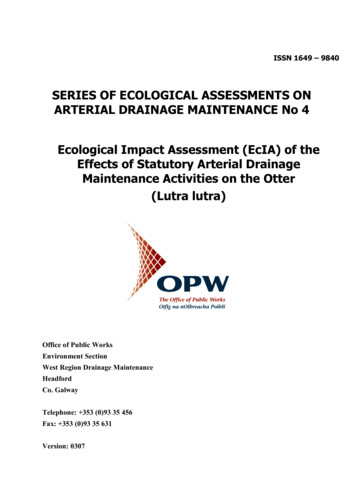
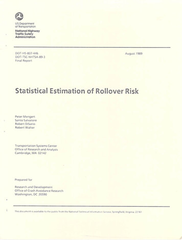
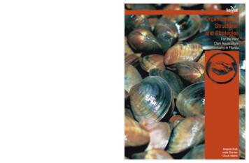
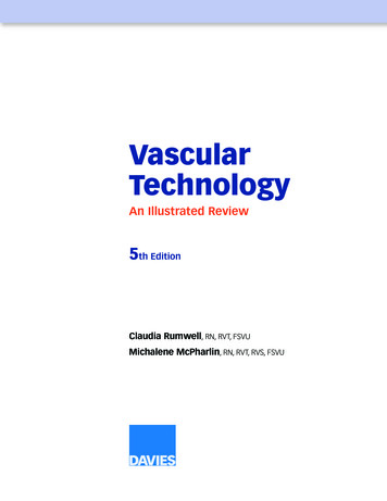
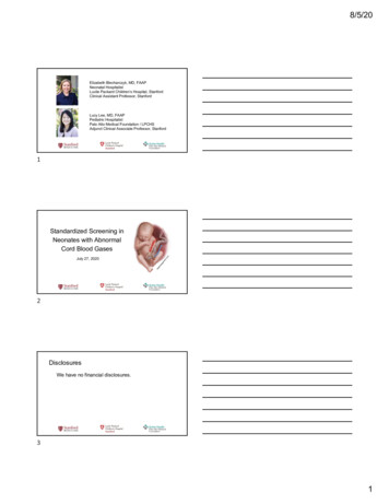
Volume 29 Number 1 January 201839technical success with TRA). Although patient preferenceregarding access site was not addressed in a randomized,prospective fashion, preference for TRA in patients who hadboth accesses was reported, and there was a lower incidence ofcomplications with TRA (4.5%) than with TFA (12.7%) (8).Recently, a large prospective cohort of more than 1,500 casesof noncoronary interventions via TRA confirmed the feasibility and safety of this approach, with a 98.2% technicalsuccess rate and an overall complication rate of less than 3%(9). Among the cases analyzed, 485 were transarterial chemoembolization procedures, confirming increasing use of thisapproach to perform IAT for liver malignancies.The purpose of the present study was to define patientpreference regarding arterial access for hepatic IAT andother potential benefits of TRA over TFA, includingcomplication rate, procedure time, contrast media volume,and radiation exposures to the patient and operator, in arandomized controlled trial.MATERIALS AND METHODSInclusion and Exclusion Criteria andPreoperative AssessmentThis study was a randomized prospective trial with intraand interpatient controls approved by the institutionalreview board (protocol ID code NCT 03163186). Inclusioncriteria were age 18 years; performance status 0/1; radialartery with anteroposterior diameter 2 mm; type A, B, orC waveforms on Barbeau test; and diagnosis of primary ormetastatic liver neoplasm amenable to transarterial blandembolization or chemoembolization. Patients were diagnosed with hepatocellular carcinoma (HCC) or metastaticneuroendocrine cancer. Patients were enrolled with theexpectation of undergoing at least two of the three plannedIAT procedures to obtain local tumor control. The Barbeautest was performed by using a pulse oximeter on the leftsecond digit (10). Anteroposterior, inner wall–to–inner walldiameter of the radial artery was measured within 2 cmproximally from the styloid process under ultrasound (US)visualization.Exclusion criteria were type D waveform on Barbeau test,radial artery anteroposterior diameter 2 mm, history ofstroke, presence of a heavily calcified aortic arch, andrequirement for additional procedures during hospitalization.A total of 55 patients with primary or metastatic hepatictumors were enrolled. A total of 124 procedures were performed, and 36 patients underwent at least two procedures,one with TRA and one with TFA. Nineteen patients hadonly one procedure and were therefore not included in theaccess preference analysis (Fig 1). Demographiccharacteristics of the 36 patients who had at least twoprocedures are presented in Table 1.Figure 1. Flowchart illustrating patient enrollment and allocation during the study.Table 1. Demographic DataCharacteristicValueSexMale28 (77.8)Female8 (22.2)RaceWhite26 (72.2)Black10 (27.8)TumorHCCNeuroendocrine29 (80.5)7 (19.5)Age (y)Mean62.81SD6.81Note–Values in parentheses are percentages.HCC ¼ hepatocellular carcinoma; SD ¼ standard deviation.to undergo the initial procedure via TRA or TFA. Thesecond procedure was performed via the alternate accessby default, ie, if the patient had a TRA first, it wasmandatory that the second procedure be performed viaTFA, or vice-versa. Randomization was constrained toensure equal numbers of patients in each arm and that eachphysician treated the same number of patients in each arm.There were two primary operators. The study design wascounterbalanced so that 50% of patients underwent TFA firstand 50% underwent TRA first.Hepatic Arterial Embolization TechniquesRandomizationThe randomization process is demonstrated in Figure 2.Patients were randomly assigned by the study coordinatorFemoral access. —Percutaneous access to the right common femoral artery was obtained under US guidance with amicropuncture kit (Cook, Bloomington, Indiana). A 5-F
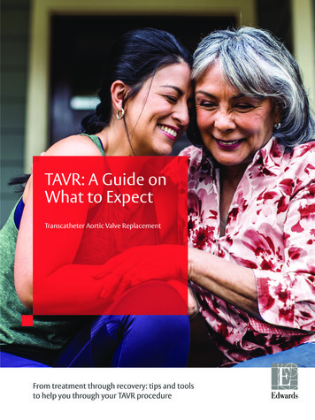
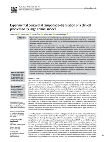
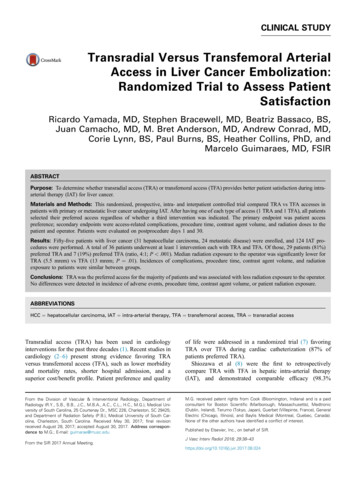
40 Transradial vs Transfemoral Access in Liver Cancer EmbolizationYamada et al JVIRFigure 2. Randomization process.sheath (Terumo, Somerset, New Jersey) was used to secure theaccess. Over a 0.035-inch polytetrafluoroethylene-coated Jwire (Boston Scientific, Marlborough, Massachusetts), theceliac trunk was catheterized with a 5-F Mikaelson Catheter(Boston Scientific) and superselective catheterization wasperformed with a 2.4- or 2.8-F Progreat microcatheter(Terumo). A femoral arteriogram was obtained beforearterial closure with AngioSeal (Terumo). Radiationprotection was provided with a lead shield skirt underneaththe angiographic table between the radiation source andoperator. No vasodilator or anticoagulant agents were usedvia TFA.Radial access. —The patient’s left arm was abducted to75 –90 . The left radial artery was accessed with single-wallpuncture under US guidance with the use of a RadiofocusTransradial kit (Terumo). The kit includes a 21-gauge shortneedle, a 0.021-inch nitinol wire, and a 5-F hydrophilicsheath. When access had been secured, 2,000–3,000 IU ofheparin was administered systemically through a peripheralintravenous access and 200 μg of nitroglycerin was instilledthrough the sheath. An additional 1,000 IU of heparin wasadministered every 30 minutes. A forearm angiogram wasobtained to delineate the arterial anatomy. Under directfluoroscopic visualization, a 0.035-inch angled J-tipGlidewire (1.5-mm radius) and a 5-F, 110-cm Jackycatheter (Terumo) were advanced coaxially into thedescending aorta, and selective catheterization of the celiactrunk was performed. For superselective catheterization ofthe hepatic artery, a 2.4 or 2.8-F Progreat microcatheter(130 or 150 cm in length; Terumo) was used. Afterembolization, the catheter was removed over the wire andan additional 200 μg of nitroglycerin was instilled intraarterially through the sheath, and a completion forearmangiogram was again obtained. Patent hemostasis techniquewas applied by using an external compression pneumaticdevice (TR Band; Terumo), and the reversed Barbeau testwas performed to rule out excessive radial artery occlusivecompression (confirmed by the presence of oximetry pulsewaveform during compression of the ulnar artery). The TRBand insufflation volume and time were communicated tonursing staff in the recovery area. Band deflation started1 hour after initial placement, and one fourth of the totalvolume was removed every 15 minutes. According to themanufacturer’s instructions, the band was removed after 2hours. A lead shield mounted on wheels, measuring 7 feetin height and 4 feet wide, was positioned on the left sideFigure 3. Postcatheterization questionnaire administered to allpatients 1 and 30 days after each transarterial (chemo-)embolization procedure.of the angiographic table, immediately caudally from thepatient’s left arm.EmbolizationSuperselective treatment was performed whenever possible.If that was not achievable, segmental or lobar treatment waspursued. For transarterial bland embolization, a combinationof 300–500-μm polyvinyl alcohol particles (Cook), 5 mL ofLipiodol (Guerbet, Villepinte, France), 10 mL of salinesolution, and 10 mL of contrast medium (Omnipaque 350; GEHealthcare, Milwaukee, Wisconsin) was administered. Forconventional transarterial chemoembolization, 20 mg of thechemotherapy agent mitomycin was added to the aforementioned solution. Patients were admitted for overnight observation. The following morning, all patients were assessed forcomplications, and blood samples were collected.All patients were evaluated in the clinic 30 days after theprocedure, including physical examination, US of the radialartery, collection of blood sample, and application of apostprocedural questionnaire.EndpointsPrimary endpoint. —The primary endpoint was patientpreference regarding arterial access. This was addressed bya postprocedural questionnaire (Fig 3) that was administeredto patients on day 1 after intervention and at the 30-day
Volume 29 Number 1 January 2018clinic visit for every procedure performed. Questionnaireswere analyzed by an independent observer, and primaryoperators had no access to them.41Table 2. Comparison between TRA and TFADetailTRATFAP Value297 .00116 (71)8 (53).1100NAMedian57.4953.9SD49.5620.4Patient preferenceSecondary endpoints. —Adverse events were definedaccording to Common Terminology Criteria for AdverseEvents, version 4.0. Blood loss was assessed objectively bycomplete blood count before intervention and on day 1 afterthe procedure. Access site hematoma/bruising was determined by physical examination. At each 30-day follow-upvisit, radial artery patency was evaluated by physical andUS examinations.Radiation dose analysis was performed by intrapatientcomparison. Given differences in fluoroscopy equipment(ie, flat-panel vs image intensifiers), only 52 of 124 procedures were included in the analysis: 26 with TRA and 26with TFA. The analysis was also counterbalanced so that50% of patients had TRA first and 50% had TFA first.Radiation dose to the patient was measured by air kerma andobtained from the fluoroscopy unit. Radiation dose to theoperator was obtained from a Landauer (Glenwood, Illinois)dosimeter badge placed on the operator’s waist (outside thelead apron). Procedure time was defined as the duration ofmoderate sedation.Statistical AnalysisThe primary endpoint analysis was based on a one-sample,two-sided exact binomial test. The null hypothesis was that50% of patients would prefer TRA to TFA. The alternativehypothesis was that 75% of patients would prefer TRA toTFA. The expectation was that no more than 10 patientswould report no preference or undergo only one of the twoprocedures. As a result, the goal was to enroll at least 50patients, providing 90% power. The two-sided a-value wasprespecified at 0.025. A binomial test was used to assesscomplication rates for the femoral versus the radialapproach, and a two-sided exact P value was reported. A2 2 repeated-measures analysis of variance ([procedure:femoral vs radial] [time: before vs after the procedure])was used to evaluate potential changes in hemoglobinlevels. Post-hoc comparisons were conducted with the Sidakprocedure to account for multiple comparisons. Repeatedmeasures t tests were used to assess differences betweenradial and femoral approaches with respect to procedureduration. Two-tailed P values were reported. The aforementioned analyses were conducted on the first two procedures. A c2 test (Fisher exact test) was used to evaluatedifferences in preference for TRA or TFA as a function ofthe attending physician, and a two-sided exact P value wasreported. Statistical significance was considered at thea ¼ .05 threshold for all analyses.RESULTSEfficacy and Survey ResultsThere was equal distribution in access, with 18 patients eachundergoing TRA and TFA as their first access. AfterBruisingConversionProcedure time (min).70Radiation dose to patient(air kerma; tion dose to lues in parentheses are percentages.NA ¼ not applicable; SD ¼ standard deviation; TFA ¼ transfemoral access; TRA ¼ transradial access.undergoing treatment via both accesses, 29 of 36 patients(81%) preferred TRA and only 7 (19%) preferred TFA(P .001). There was no difference in patient preference as afunction of primary operator, with 87.5% and 72.2%preferring the radial approach for operators 1 and 2,respectively (P ¼ .41). There was no difference in the volumeof contrast medium between TRA and TFA, with respectivemeans of 114.9 mL 49.56 (standard deviation) and 119.9mL 34.23 (P ¼ .66). There was no difference in procedureduration between TRA and TFA, with respective means of57.5 minutes 49.56 and 54.0 minutes 20.40 (P ¼ .70).There was no access conversion in either group. Medianradiation exposure (as air kerma) to the patient was alsosimilar between accesses: 1,200.5 mGy (range, 107–3,479mGy) for TFA and 811 mGy (range, 80–5,458 mGy) forTRA (P ¼ .229). Median radiation exposure to the operatorwas lower for TRA (5.5 mrem; range, 1–43 mrem) than forTFA (13 mrem; range, 1–121 mrem; P ¼ .01; Table 2).SafetyThere were 25 acute minor complications documentedamong 18 patients in 124 interventions. Most patients(n ¼ 12; 66.7%) experienced complications after only oneprocedure, with five patients (27.8%) experiencing complications on two occasions and only one patient (5.6%)experiencing complications on three occasions. Acute minorcomplications were limited to access-site bruising/pain onpostprocedure day 1 in 17 of 71 procedures with TRA(22%) and 8 of 53 procedures with TFA (15%), whichshowed no significant difference (P ¼ .11). Two patientsexperienced the late minor complication of partial radialartery thrombosis (less than one third of radial arterydiameter) identified on US at 30-day follow-up; these wereclinically silent. Resolution was noted on the day of asubsequent procedure (4–6 wk later) or on later follow-up
42 Transradial vs Transfemoral Access in Liver Cancer Embolizationconsultation (12 wk) as part of standard care. No majoradverse events occurred with either access type.Although there was a decrease in mean hemoglobin levelafter procedures performed via both accesses (TRA, from12.42 g/dL 1.81 to 11.84 g/dL 2.11; 95% confidenceinterval, 0.0312–0.858; TFA, from 12.5 g/dL 1.79 to 11.87g/dL 1.67; 95% confidence interval, 0.382–0.879;P .001), this did not have any clinical significance. Overall,there was no difference in hemoglobin level between thefemoral versus the radial approach (P ¼ .78). There was noclinically evident stroke or hand ischemia at 30 days.DISCUSSIONThe present randomized and prospective trial with intra- andinterpatient control compared TRA versus TFA for IAT forliver malignancies and demonstrated that the majority ofpatients preferred the radial approach (4:1 ratio). In addition,there was no increased incidence of complications with TRA,and procedure durations were similar for both methods.Radiation exposure to the patient was similar for bothaccesses, but the operator exposure was lower with TRA.According to most patients, TRA was the preferred optionbecause of less pain and discomfort. Also, limited bedrestriction made a major impact on the postproceduralexperience. Patients could ambulate earlier, allowing themto go to the restroom instead of using a urinal. Furthermore,in case of abdominal pain and nausea/vomiting, patientscould position themselves according to their needs and notbe worried about leg straightening. This result is consistentwith the cardiology literature, which has shown improvedquality of life after TRA compared with TFA for coronaryinterventions, with 87% of patients favoring TRA (7). Theprevious study (7) had no intrapatient comparison becausepatients had one of the two accesses, whereas the presentstudy was a randomized trial specifically designed toaddress patient experience regarding access site. For visceralinterventions, the authors of another study (11) had a similarobservation in their patient population, although not as theirprimary endpoint.There were no major complications in either group in thepresent study, including stroke or hand ischemia, two of themost worrisome complications of TRA. Similarly, large caseseries of visceral interventions performed via TRA (9,11)showed low incidences of major complications: 0.13% inthe study of Posham et al (9) and 0.17% in the study ofBishay et al (11). In the cardiology literature, a prospectiverandomized trial (12) demonstrated no difference in theincidences of silent cerebral infarcts with TRA versus TFA(11.7% vs 17.5%). Ratib et al (13) followed more than 5,000patients for 5 years and reported a minimal rate of strokeafter TRA of 0.11%–0.20%. Left radial access to avoidcrossing most of the supra-aortic vessels, J-shaped soft-tipguide wire and catheter advancement under direct fluoroscopic visualization (thereby avoiding inadvertent cannulation of the left vertebral artery or ascending aortic arch), andpatient selection can minimize the risk of stroke. ThoroughYamada et al JVIRpreprocedural screening with the Barbeau test and USmeasurement of the radial artery is mandatory to avoidischemic complications of the hand.Minor complications in the present study were limited tosuperficial bruising (22%) and partial thrombosis of theradial artery (4.2%). The incidence of superficial bruisingwas higher than previously described by Bishay et al (11),who reported an incidence of 2.3% during hepatic embolization via TRA. This could be associated with subjectiveclinical assessment. Nonetheless, none of the complicationsin the present study were clinically relevant, and no radialartery occlusion was encountered. Administration of systemic heparin and nitroglycerin through the sheath immediately after its placement and immediately before sheathremoval, as well as achieving patent hemostasis whileapplying the pneumatic compression device, are key factorsin the prevention of radial artery occlusion (14).A previous meta-analysis of 24 randomized trials of patients undergoing coronary catheterization (15) demonstratedincreased fluoroscopy time and radiation exposure to patientswith TRA (fluoroscopy time, 38 min 12 for TRA and 35min 13 for TFA). The weighted mean difference of kerma–area product for coronary interventions was 0.55 Gy cm2favoring TFA (95% confidence interval, 0.08–1.02 Gy cm2;P ¼ .02). In the present study, despite the longer distancebetween the radial artery and celiac trunk and the need tomanipulate the wire and catheter into the descending aorta,the procedure durations were similar for both approaches.This was also observed in a recent cardiology prospectivetrial (16) in which fluoroscopy time was not statisticallydifferent with TRA versus TFA (12.2 min vs 9.8 min; P ¼.507). Operator experience and device choice can influencethe procedure duration and the use of an angled hydrophilic Jtip wire (1.5 mm radius), and the angle of the 5-F diagnosticcatheter tip allowed easy access to the descending aorta.Radiation exposure as measured by air kerma to the patient did not differ between accesses, similar to what wasobserved in the previously mentioned cardiology study (16).Another study of coronary interventions (17) in whichpatients were randomized to TRA or TFA showed no differences in radiation exposure to patients in centers with highprocedure volumes (air kerma, 652 mGy for TRA vs 621mGy for TFA; P ¼ .403), suggesting that, similar to fluoroscopy time, radiation exposure can be influenced byoperator experience. Finally, in the present study, radiationexposure to the operator was lower with TRA as a result ofthe positioning of the radiation shield and the longer distancebetween the operator and radiation source. We found nopublished studies in the literature that assessed this outcome.TRA demonstrated distinct advantages over TFA, whichhas made TRA the default access for most visceralinterventions at the authors’ institution. TRA not only provides a safe alternative access, but it also offers a newpatient-centric model aligned with the best practices conceptin the ever-changing health care landscape. Recent European Society of Cardiology guidelines (18) recommendedthat TRA become the preferred approach in coronary
Volume 29 Number 1 January 2018interventions. Studies have demonstrated decreased costsassociated with TRA versus TFA, primarily because there isno need for arterial closure devices and there is a decreasedincidence of readmission for bleeding complications(4,7,19). The use of TRA has facilitated shorter hospitalstays, partly as a result of the “radial lounge” concept (20).In this model, postprocedural monitoring after TRA generally requires a decreased nursing ratio, and patients may situp in a chair and ambulate immediately. Moreover, it hasbeen postulated that TRA may cause less hematoma inpatients with coagulopathies, a common underlying condition in HCC (21).The present study is limited by several factors. Therewere limitations inherent to subjectivity in the patient survey, as patients could have had inherent bias in favor ofTRA over TFA before enrollment. Also, patient preferenceis a subjective assessment, involving justifications such asless discomfort and limited bed restriction. Time to ambulate, which would be an objective assessment, was notanalyzed in this study. TRA in female patients can betechnically more difficult as a result of anatomic andphysiologic factors. As 78% of patients in the present studywere male, it is possible that TRA in this patient populationwas more feasible; however, sex was not an exclusion criterion, and patients were enrolled according to hospital/referral demand.Radiation dose reduction to the operator with TRA was beachieved with only the specific patient positioning andshielding used in this study. Different techniques such as leftarm adduction may not show similar benefit. As mentionedbefore, the sample size was powered to identify patientpreference only and not the other secondary endpoints,including complications. Therefore, interpretation of theresults regarding the secondary endpoints is limited, and alarger sample size would be required for accurate assessment.Finally, transarterial (chemo-)embolization procedurescan vary significantly, given differences in tumor burden,number of selective catheterizations, and time to deliverembolic material. Despite the randomization processincluding intra- and interpatient comparison, those variablesmight have not been completely counterbalanced, leading tobiased results.In conclusion, for the majority of patients with primary ormetastatic liver tumors undergoing transarterial (chemo-)embolization, TRA was the access of choice. TRA was notassociated with an increased incidence of adverse events orincreased procedure time. In addition, radiation exposure tothe operator was lower with TRA.REFERENCES1. Campeau L. Percutaneous radial artery approach for coronary angiography. Cathet Cardiovasc Diagn 1989; 16:3–7.43 P, et al; MATRIX Investigators. Radial2. Valgimigli M, Gagnor A, Calabroversus femoral access in patients with acute coronary syndromes undergoing invasive management: a randomised multicentre trial. Lancet2015; 385:2465–2476.3. Romagnoli E, Biondi-Zoccai G, Sciahbasi A, et al. Radial versus femoralrandomized investigation in ST-segment elevation acute coronary syndrome: the RIFLE-STEACS (Radial Versus Femoral Randomized Investigation in ST-Elevation Acute Coronary Syndrome) study. J Am Coll Cardiol2012; 60:2481–2489.4. Mitchell MD, Hong JA, Lee BY, et al. Systematic review and cost–benefitanalysis of radial artery access for coronary angiography and intervention.Circ Cardiovasc Qual Outcomes 2012; 5:454.5. Mehta SR, Jolly SS, Cairns J, et al. Effects of radial versus femoral arteryaccess in patients with acute coronary syndromes with or without STsegment elevation. J Am Coll Cardiol 2012; 60:2490–2499.6. Jolly SS, Yusuf S, Cairns J, et al; RIVAL trial group. Radial versus femoralaccess for coronary angiography and intervention in patients with acutecoronary syndromes (RIVAL): a randomised, parallel group, multicentretrial. Lancet 2011; 377:1409–1420.7. Cooper CJ, El-Shiekh RA, Cohen DJ, et al. Effect of transradial access onquality of life and cost of cardiac catheterization: A randomized comparison. Am Heart J 1999; 138:430–436.8. Shiozawa S, Tsuchiya A, Endo S, et al. Transradial approach for transcatheter arterial chemoembolization in patients with hepatocellular carcinoma: comparison with conventional transfemoral approach. J ClinGastroenterol 2003; 37:412–417.9. Posham R, Biederman DM, Patel RS, et al. Transradial approach fornoncoronary interventions: a single-center review of safety and feasibilityin the first 1,500 cases. J Vasc Interv Radiol 2016; 27:159–166. re MM. Evaluation of10. Barbeau GR, Arsenault F, Dugas L, Simard S, Lariviethe ulnopalmar arterial arches with pulse oximetry and plethysmography:comparison with the Allen’s test in 1010 patients. Am Heart J 2004; 147:489–493.11. Bishay VL, Biederman DM, Ward TJ, et al. Transradial approach forhepatic radioembolization: initial results and technique. AJR Am JRoentgenol 2016; 207:1112–1121. D, et al. Silent cerebral infarcts after cardiac12. Hamon M, Lipiecki J, Carriecatheterization: a randomized comparison of radial and femoral approaches. Am Heart J 2012; 164:449–454.13. Ratib K, Mamas MA, Routledge HC, et al. Influence of access site choiceon incidence of neurologic complications after percutaneous coronaryintervention. Am Heart J 2013; 165:317–324.14. Pancholy S, Coppola J, Patel T, Roke-Thomas M. Prevention of radialartery occlusion-patent hemostasis evaluation trial (PROPHET study): arandomized comparison of traditional versus patency documentedhemostasis after transradial catheterization. Cathet Cardiovasc Interv2008; 72:335–340.15. Plourde G, Pancholy SB, Nolan J, et al. Radiation exposure in relation tothe arterial access site used for diagnostic coronary angiography andpercutaneous coronary intervention: a systematic review and metaanalysis. Lancet 2015; 386:2192–2203.16. Becher T, Behnes M, Ünsal M, et al. Radiation exposure and contrastagent use related to radial versus femoral arterial access during percutaneous coronary intervention (PCI)—results of the FERARI study.Cardiovasc Revasc Med 2016; 17:505–509.17. Jolly SS, Cairns J, Niemela K, et al; RIVAL Investigators. Effect of radialversus femoral access on radiation dose and the importance of proceduralvolume: a substudy of the multicenter randomized RIVAL trial. JACCCardiovasc Interv 2013; 6:258–266.18. Montalescot G, Crea F. The year in cardiology 2015: acute coronarysyndromes. Eur Heart J 2016; 37:221–228.19. Le Corvoisier P, Gellen B, Lesault PF, et al. Ambulatory transradialpercutaneous coronary intervention: a safe, effective, and cost-savingstrategy. Cathet Cardiovasc Interv 2013; 81:123.20. Brewster S, Khimdas K, Cleary N, et al. Impact of a dedicated “radiallounge” for percutaneous coronary procedures on same-day dischargerates and bed utilization. Am Heart J 2013; 165:299–302.21. Fischman AM, Swinburne NC, Patel RS. A technical guide describing theuse of transradial access technique for endovascular interventions. TechVasc Interv Radiol 2015; 18:58–65.
review board (protocol ID code NCT 03163186). Inclusion criteria were age 18 years; performance status 0/1; radial artery with anteroposterior diameter 2 mm; type A, B, or C waveforms on Barbeau test; and diagnosis of primary or metastatic liver neoplasm amenable to transarterial bland embolization or chemoembolization. Patients were diag-