Transcription
Turk J Surg 2018 34 (3): 205-211Original ArticleDOI: 10.5152/turkjsurg.2018.4181Experimental pericardial tamponade–translation of a clinicalproblem to its large animal modelGábor Bari2, Szilárd Szűcs1ABSTRACT, Dániel Érces1, Mihály Boros1, Gabriella Varga1Objectives: Pericardial tamponade is a life-threatening medical emergency, when the hemodynamic consequencesof low cardiac output severely disturb the perfusion of the peripheral tissues. Our aim was to design a reliable largeanimal model to reproduce the clinical scenario with the relevant pathophysiological consequences of pericardialtamponade -induced cardiogenic shock.Material and Methods: Anesthetized Vietnamese mini pigs were used (n 12). Following laparotomy, a cannulawas fixed into the pericardium through the diaphragm without thoracotomy. A sham-operated group (n 6) served as control, while in the second group (n 6) pericardial tamponade was induced by intra-pericardial injectionof heparinized own blood. Throughout the 60-min pericardial tamponade and the 180-min reperfusion, macrohemodynamics, renal circulation and the mesenteric macro- and micro-circulatory parameters were monitored.Myeloperoxidase activity was measured to detect neutrophil leukocyte accumulation and in vivo histology wasperformed by confocal laser scanning endomicroscopy to observe the structural changes of the intestinal mucosa.Results: PT increased the central venous pressure, heart rate, and decreased mean arterial pressure. The mesentericartery flow (from 355.5 112.4 vs 182.0 59.1 mL/min) and renal arterial flow (from 159.63 50.7 vs 35.902 27.9mL//min) and the micro-circulation of the ileum was reduced. The myeloperoxidase activity was elevated (from3.66 1.6 to 7.01 1.44 mU/mg protein) and manifest injury of the ileal mucosa was present.ORCID IDs of the authors:G.B. 0000-0003-4167-5736;S.S. 0000-0001-8478-6386;D.E. 0000-0002-4283-2441;M.B. 0000-0003-1410-1999;G.V. 0000-0003-1888-8629.Cite this paper as:Bari G, Szűcs S, Érces D, BorosM, Varga G. Experimentalpericardial tamponade–translation of a clinicalproblem to its large animalmodel. Turk J Surg 2018 34(3): 205-211.1Institute of Surgical Research,Szeged University, Szeged, Hungary2Department of Cardiac Surgery,Szeged University, Szeged, HungaryThis study was presented at the17th Congress of the EuropeanShock Society, 13-15/09/2017,Paris, France.Corresponding AuthorGabriella ved: 06.04.2018Accepted:18.06.2018 Copyright 2018by Turkish Surgical AssociationAvailable online atwww.turkjsurg.comConclusion: This experimental model suitably mimics the hemodynamics and the pathology of clinical pericardialtamponade situations, and on this basis, it provides an opportunity to study the adverse macro- and micro-circulatory effects and biochemical consequences of human cardiogenic shock.Keywords: Animal model, cardiogenic shock, mesenteric ischemia, mini pig, pericardial tamponadeINTRODUCTIONThis article will explore the line between patients and experimental subjects, in an attempt to provide aproper view on the role of classical surgical research in contemporary cardiac surgery. According to theproverbial quote of Francis D. Moore, a “surgical investigator must be a bridge tender, channeling knowledge from biological science to the patient’s bedside and back again” (1). This traditional approach emphasizes the great potential of biomedical sciences to provide help for unsolved problems in surgery. Anequally important aspect of this type of research is that it develops new data, or more comprehensibleknowledge on a specific area of clinical practice. In this sense, the science of surgery, like any other areaof biomedical sciences, has four main methodological steps: observation of a clinical problem, formulation of a hypothesis, and performance of test procedures to reach a conclusion, supporting or refutingthe initial hypothesis. In this analytical circle, models are indispensable tools to check the validity ofhypotheses and to generate clinically useful data. In pragmatic terms differences are always presentbetween the clinical reality and the models and therefore these dissimilarities should be reduced bychoosing the experimental setup as accurate as possible. Understandably, the demand is huge for developing high fidelity models to decrease existing limitations and to obtain better applicable results forhuman medical science and healthcare (2).A specific problem addressed by our study is cardiogenic shock due to cardiac tamponade. Pericardial tamponade is one of the most dreadful complication of cardiac surgery and invasive cardiology.Tamponade can develop after myocardial infarction, it can be the corollary of intra- or post-operativecomplications, or the consequence of non-cardiac surgical procedures (NCSP) as well (3). During tamponade, fluid will accumulate within the pericardial space, which can be transudate or pus, howeverthe most common cause is bleeding (4). The increasing pressure in the closed pericardial sac blocks thevenous inflow and decreases stroke volume. The subsequently reduced cardiac output (CO) results intissue hypoxia and multi-organ damage. The long-term systemic consequences of pericardial tamponade are pronounced and serious even after the primary cause has been diagnosed and the basic problem was solved, i.e., the pericardial pressure was decreased via pericardiocentesis, re-operation or otherintervention (5). The main reason for late complications is ischemia-reperfusion injury of distant organs,including the lungs, kidneys, and splanchnic tissues. This circulatory condition results in an antigen-205
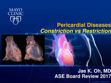
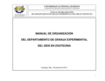
Bari et al.Experimental model of pericardial tamponadeFigure 1. The insertion of the pericardial cannula via thetransphrenic route (1 diaphragm, 2 pericardium, 3 liver,arrow: the cannula fixed into the pericardium)independent inflammatory reaction with cytokine storm andincreased endothelial permeability, which finally leads to anoverwhelmingly severe systemic immune response (6).According to previous experiences, cardiogenic shock can bemodelled if an animal is anaesthetized, mechanically ventilated, and the pericardial sac is accessible through thoracotomy(7). The disadvantage of the model is thoracotomy itself withsignificantly impaired lung function, extended wound surfacewith a high risk of bleeding, lung injury, and tissue damage.Based on the above, our aim was to develop an experimentalprocedure where thoracotomy can be avoided and thus to design a new model of iatrogenic cardiogenic shock, where thesituation is realistic, similar to the clinical appearance.MATERIAL AND METHODSAnimalsThe experiments were performed in accordance with NationalInstitutes of Health guidelines on the handling and care ofexperimental animals and European Union Directive 2010/63for the protection of animals used for scientific purposes. Thestudy was approved by the Animal Welfare Committee of theUniversity of Szeged (approval number: V/148/2013).206Surgical interventionsAnesthesia was induced by a mixture of ketamine (20 mg kg 1im.) and xylazine (2 mg kg 1 im.) and maintained by a continuous infusion of propofol (6 mg kg 1h 1 iv.). The animals wereplaced on heating pad in a supine position; the body temperature was kept between 36-37 C. After endotracheal intubation, mechanical ventilation was started with a tidal volumeof 10 mlkg 1. The left jugular vein was cannulated for fluid anddrug administration and the left femoral artery for the measurement of mean arterial pressure (MAP), heart rate (HR) andcardiac output (CO) by transpulmonary thermodilution (PICCOCatheters; PULSION Medical Systems, Feldkirchen, Germany).MAP, HR, CO and SMA flow (SMAF) data were recorded, pres-sure signals (BPR-02 transducer; Experimetria Ltd, Budapest,Hungary) and superior mesenteric artery (SMA), renal artery(RA) flow signals (T206 Animal Research Flowmeter; TransonicSystems Inc., Ithaca, NY, USA) were measured continuouslyby surgically placing a urinary catheter in the bladder via thefemoral incision. and registered with a computerized data-acquisition system (SPEL Advanced HAEMOSYS 1.17; Experimetria Ltd, Budapest, Hungary). Ringer’s lactate was given at therate of 10 mlkg 1h 1. After a median laparotomy, the SMA wasdissected free and a flow probe was placed around the SMA(Transonic Systems Inc., Ithaca, NY, USA). The RA was dissectedfree and a flow probe was placed around it (Transonic SystemsInc., Ithaca, NY, USA). The wound of the abdominal wall wastemporarily closed thereafter with clips. In addition, intra-vitalexamination of the intestinal microcirculation was carried outby an orthogonal polarization spectral imaging system (OPS;Cytoscan A/R, Cytometrics, Philadelphia, PA, USA), and theextent of damage of the gastric mucosa was evaluated by invivo histology (Five1, Optiscan Pty. Ltd., Melbourne, Victoria,Australia). The diaphragm was accessed through a medianlaparotomy and a 3-cm incision was made at the sternal part,avoiding the muscular region of the diaphragm. The pericardium was opened and a cannula was fixed into the pericardial cavity with a pledgeted purse string suture (Figure 1). Thetamponade was induced for 60 min by intra-pericardial administration of heparinized own blood (100 50 mL), and theMAP was kept between 40–45 mmHg. During the observationperiod and between microcirculatory imaging the abdominalwall was closed by surgical clips.Experimental ProtocolThe animals were randomly allocated into two experimentalgroups. Group 1 (n 6) served as sham-operated control, withthe same surgical interventions, time-frame, and sampling asin group 2 (n 6) but without the induction of a tamponade. Inboth groups, the diaphragm was accessed through a medianmini-laparotomy. A 3-cm incision was performed at the sternalpart, avoiding the muscular region of the diaphragm. The pericardium was opened and a cannula was fixed into the pericardial cavity with a pledgeted purse string suture. In Group2, after the end of 60-min tamponade the blood was releasedfrom the pericardial sac and the animals were monitored for180 min. Blood gas and hemodynamic parameters were measured in every 30 min. In vivo histological examination on theileal mucosa and determination of neutrophil granulocyteswas performed at baseline, 30 min after the relief of tamponade (90 min) and at the end of the experiments (240 min). Myeloperoxidase enzyme activity was measured at the 5; 30; 60;90 and 240 min of observation period.Hemodynamic MeasurementsCentral venous pressure (CVP), mesenteric and renal arteryblood flow (AMSF, ARF) signals were monitored continuouslyand registered with a computerized data acquisition system(SPELL Hemosys; Experimetria, Budapest, Hungary). The MAP,CO, and HR were measured with the PICCO Plus monitoringsystem (PULSION Medical Systems; Munich, Germany).Myeloperoxidase enzyme activityBlood samples (0.5 mL) were taken from the left jugular veininto precooled, heparizined (100 U mL 1) polypropylene tubes,centrifuged at 1.200 g at 4 C for 15 minutes, and stored at
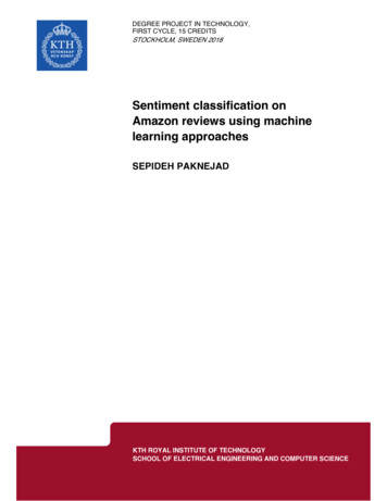
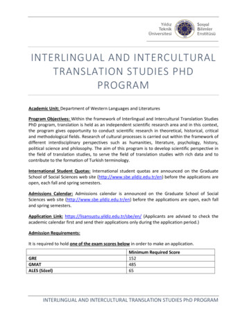
Turk J Surg 2018 34 (3): 205-211120100Mean arterial pressure(mmHg)Cardiac output(I (min de0.5a8090120 150Time (min)ControlTamponade20180210Tamponade02403060b90 120 150Time (min)18021024016010Heart rate(1 min-1)Central venous de0306090120150180210240Figure 2. a-d. Changes in cardiac index (a), mean arterial pressure (b), heart rate (c), and central venous pressure (d) in control(white circle–solid line) and tamponade groups (black diamond–solid line). The plots demonstrate the median and the 25th(lower whisker) and 75th (upper whisker) percentiles*p 0.05 for groups vs baseline values, xp 0.05 for cardiac tamponade vs control group values 70 C until assay. Plasma MPO enzyme activity, a marker ofpolymorphonuclear leukocyte activation, was determined bythe modified method of Kaszaki et al. (7).Microcirculatory measurementAn intra-vital orthogonal polarization spectral imaging technique (Cytoscan A/R; Cytometrics, Philadelphia, PA) was usedfor non-invasive visualization of the mucosal microcirculationof ileum. This technique utilizes reflected polarized light atthe wavelength of the isosbestic point of oxy- and deoxyhemoglobin (548 nm). As polarization is preserved in reflection,only photons scattered from a depth of 200 to 300 μm contribute to image formation. A 10 objective was placed ontothe mucosal surface of the small intestine, and microscopicimages were recorded by a S-VHS video recorder 1 (PanasonicAG-TL 700; Matsushita Electric Ind, Osaka, Japan). Quantitativeassessment of the microcirculatory parameters was accomplished off-line by frame-to-frame analysis of the videotapedimages. Red blood cell velocity (RBCV, μm/s) changes in thecapillary were determined in three separate fields by means ofa computer-assisted image analysis system (IVM Pictron, Budapest, Hungary). All microcirculatory evaluations were performed by the same investigators (GB and GV).In vivo detection of mucosal damageThe extent of damage of the ileal mucosa was evaluated bymeans of fluorescence confocal laser scanning endomicroscopy (CLSEM) developed for in vivo histology. The analysis wasperformed twice, separately by two investigators (GV and GB).The mucosal surface of the ileum was surgically exposed andlaid flat for examination. The ilaeal mucosal structure was recorded after the topical application of the fluorescent dye acriflavin (Sigma-Aldrich Inc, St. Louis, MO, USA). The surplus dyewas washed off the mucosal surface of the ileum with saline 2min before imaging. The objective of the device was placedonto the mucosal surface of the ileum and confocal imaging207
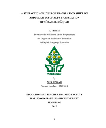
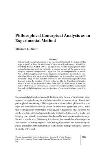
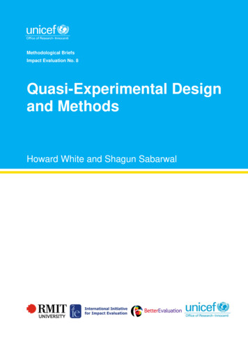
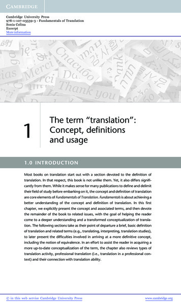
Bari et al.Experimental model of pericardial tamponade200500Renal artery flow(mL min-1)Superior mesenteric artery flow(mL e300aControlTamponadeTamponade90 120 150 180 210 240Time (min)600b306090 120 150Time (min)180 210240Figure 3. a-b. Changes in mesenteric artery (a) and renal artery flow (b) in control (white circle–solid line) and tamponade(black diamond–solid line) groups. The plots demonstrate the median and the 25th (lower whisker) and 75th (upper whisker)percentiles.*p 0.05 for groups vs baseline values, xp 0.05 for cardiac tamponade vs control group valuesControlTamponade600Red blood cell velocity(um s-1)Myeloperoxidase enzyme activity (mU(mg de020830Tamponade6090Time (min)024090Time (min)240Figure 4. Changes in myeloperoxidase enzyme activity incontrol (white box) and tamponade (gray striped box to theright side) groups. The plots demonstrate the median andthe 25th (lower whisker) and 75th (upper whisker) percentiles*p 0.05 for groups vs baseline values, xp 0.05 for cardiactamponade vs control group valuesFigure 5. Changes in red blood cell velocity in control (whitebox) and tamponade (gray striped box to the right side)groups. The plots demonstrate the median and the 25th(lower whisker) and 75th (upper whisker) percentiles.*p 0.05 for groups vs baseline values, xp 0.05 for cardiactamponade vs control group valueswas performed 5 min after dye administration (1 scan/image,1024x512 pixels and 475x475 μm per image). The changesin the mucosal architecture were examined by using a semiquantitative scoring system as described previously (8). (I. oedema (0 no oedema, 1 moderate epithelial swelling, 2 severe oedema); and II. epithelial cell outlines (0 normal, clearly,well-defined outlines, 1 blurred outlines, 2 lack of normalcellular contours).Friedman repeated measures analysis of variance on ranks wasapplied within groups. Time-dependent differences from thebaseline for each group were assessed by Dunn’s method. Thedifferences between groups were analyzed with Mann-Whitneyprobe. In the figures, median values and 75th and 25th percentiles are given; p 0.05 were considered significant.Statistical AnalysisData analysis was performed with a statistical software package(SigmaStat for Windows, Jandel Scientific, Erkrath, Germany).Changes in Hemodynamic ParametersThe average duration of the surgical preparation phase was70 20 min. The MAP remained at 40–45 mmHg as previouslyRESULTS
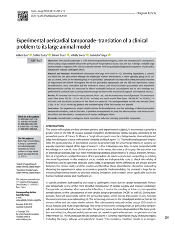
Turk J Surg 2018 34 (3): 205-211abcdefFigure 6. a-f. In vivo histology (CLSEM technique) showing the changes of the epithelial surface of the terminal ileum after acriflavinstaining. Mucosal surface of control group (a, c, e); and structure of mucosal surface in tamponade group (b, d, f) are shown209
Bari et al.Experimental model of pericardial tamponadeplanned throughout the 60 min of pericardial tamponade,accompanied by a concomitantly decreased CO. The CO increased to baseline levels after the reversal of tamponade (Figure 2a) but the MAP remained significantly depressed (Figure2b). The elevation of CO was accompanied by a compensatoryelevation of HR (Figure 2c). The decreased venous return wasevidenced by a significant elevation of CVP throughout thetamponade phase.Significant deterioration of artery mesenteric flow was observed during tamponade, but no significant differences werefound in the post–tamponade phase as compared to the baseline values and the control group (Figure 3a). The renal arteryflow was significantly and permanently reduced during tamponade and the post-tamponade period as well (Figure 3b).Leukocyte accumulationThe plasma MPO activity increased during the tamponade period and its peak was reached 30 minutes after tamponade. Atthe end of the observation period the MPO activity decreasedtowards the control level (Figure 4).Changes in microcirculationThe RBCV did not change in the control group, while in thetamponade group, a significant decrease from the baselineand control group was detected but at the end of the observation period (Figure 5).In vivo HistologyThe mucosal morphology was examined by intra-vital CLSEMtechnique in real time. The epithelial morphology of the smallintestinal mucosa presented normal morphological patternsin sham-operated animals and in the control samples of thetamponade group while 30 min after tamponade inductionlongitudinal fissures appeared and partial epithelium defectswere detected (Figure 6a-e). The lack of epithelium was extended by the end of observation (Figure 6f).DISCUSSIONAnimal models have important, sometimes, decisive roles intoday’s medical research. Large animal models have many advantages for studying human pathophysiology and by theirsize they can provide additional benefits for the developmentof new surgical techniques and therapies. Experimental pericardial tamponade models using thoracotomy can be suitableto examine the complication of open cardiac surgical procedures or to monitor the hemodynamic changes (7, 9). Our purpose was to develop a suitable model based on experimentalsurgical standards for the investigation of cardiogenic shock,which evolves after NCSP where the duration, extent, characteristics, and outcome variables of impaired systemic circulation can be determined exactly. According to our resultsthe presented experimental model can be used to study theacute pathophysiological consequences of low cardiac outputstates. Moreover, it may well reflect the signs of cardiogenicshock during NCSP. In addition, this pericardial tamponadesetup can be useful for the practical training of surgical residents for the real-life demonstration and management of pericardiocentesis or emergency thoracotomy.210Increased pressure in the pericardium effects the low-pressureareas, like the superior and inferior caval veins and the rightatrium. During the heart cycle, tension changes dynamicallyinside the atria and ventricles, thus the pericardial pressureblocks the right atrial inflow and consecutively the right ventricular diastolic filling. The high venous pressure leads to thedistension of the jugular veins (Kussmaul-sign) the cardiacsounds are “muffled.” In addition, the peripherial pulse cannot be palpated while cardiac auscultation detects heartbeats,this is referred as pulsus paradoxus. The ECG signs reveal thelower amplitude of R waves due to the increased resistanceof intra-pericardial liquids (5, 10). The development of cardiogenic shock activates numerous compensation mechanismsto maintain the perfusion pressure to the vital organs. Besidesthe enhanced contractility, the heart rate is increased to maintain CO. Additionally, the body redistributes the circulatingvolume by systemic and local vasoactive responses, including the remarkable vasoconstriction of the muscle-, skin-, andmesenteric vessels (5, 10-12).The decreasing diuresis could be the first sign of accumulating pericardial fluid and thriving pericardial tamponade oncardiac surgical ICU. In our experiments, the renal blood flowremained significantly depressed despite the resolving CO.The deteriorating renal function is a challenging problem inclinical practice, thus our porcine model could be an important tool for studies aiming to study the improvement of renalfunction.CONCLUSIONOur model using anesthetized mini pigs reflects the local andsystemic hemodynamic and inflammatory changes of the clinical picture correctly. In the absence of thoracotomy, the surgical procedure is relatively easy and fast while at the same timethe experimental setup allows for extensive hemodynamicmonitoring, tissue biopsies, and biochemical measurements.We tried to make the model as realistic as possible, thus theanimal’s own blood is used to fill the pericardium. Leakage ofblood was not detected and therefore the amount of intrapericardial fluid could be standardized. The MAP level can beprecisely controlled and the pressure response can be exactlymodified throughout the experiments. Another advantage isthat there is no explicit need for mechanical ventilation, andthe lack of thoracotomy allows for longer duration or evenchronic experiments with prolonged data collection. Besides,the transphrenic thoracal approach is definitely less stressful(refinement), and considering that the reduced invasivenessenables a reduction in animal numbers, this new model facilitates the adherence to the 3R principles as well.Ethics Committee Approval: Ethics committee approval was received for this study from the Ethics Committee of Szeged University(V/148/2013).Informed Consent: N/A.Peer-review: Externally peer-reviewed.Author Contributions: Concept - G.B., D.É., M.B., G.V.; Design - B.G.,S.S., É.D., G.V.; Supervision - M.B. Resource - M.B., G.V.; Materials - B.G.,S.S., É.D., G.V.; Data Collection and/or Processing - B.G., S.S., É.D., G.V.;Analysis and/or Interpretation B.G., S.S., É.D., G.V.; Literature Search- B.G., S.S., É.D., G.V.; Writing Manuscript - G.B., G,V.; Critical Reviews- D.É., M.B.
Turk J Surg 2018 34 (3): 205-211Acknowledgments The authors are grateful to Ágnes Fekete, CsillaMester, Nikolett Beretka, Éva Nagyiván, Károly Tóth, and Péter Sárkányfor their skillful assistance.Conflict of Interest: The authors have no conflicts of interest to declare.Financial Disclosure: This study was funded by Hungarian NationalResearch, Development and Innovation Office NKFIH-K120232 andNKFIH-K116861 and GINOP-2.3.2-15-2016-00015 grants.References1.2.3.4.5.Moore FD. The university in American surgery. Surgery 1958; 44: 1-10.Osuchowski MF, Remick DG, Lederer JA, Lang CH, Aasen AO,Aibiki M, et al. Abandon the Mouse Research Ship? Not Just Yet!.Shock 2014; 41: 463-475. [CrossRef]Orbach A, Schliamser JE, Flugelman MY, Zafrir B. Contemporaryevaluation of the causes of cardiac tamponade: Acute and longterm outcomes. Cardiol J 2016; 23: 57-63. [CrossRef]Bodson L, Bouferrache K, Vieillard-Baron A. Cardiac tamponade:Curr Opin Crit Care 2011; 17: 416-424. [CrossRef]Carmona P, Mateo E, Casanovas I, Peña JJ, Llagunes J, Aguar F, etal. Management of Cardiac Tamponade After Cardiac Surgery. JCardiothorac Vasc Anesth 2012; 26: 302-311. [CrossRef]6.Rivera-Nieves J, Gorfu G, Ley K. Leukocyte adhesion moleculesin animal models of inflammatory bowel disease. Inflamm BowelDis 2008; 14: 1715-1735. [CrossRef]7. Kaszaki J, Nagy S, Tárnoky K, Laczi F, Vecsernyés M, Boros M.Humoral changes in shoc induced by cardiac tamponade. CircShock 1989; 29: 143-153.8. Kovács T, Varga G, Erces D, Tőkés T, Tiszlavicz L, Ghyczy M, etal. Dietary Phosphatidylcholine Supplementation AttenuatesInflammatory Mucosal Damage in a Rat Model of ExperimentalColitis. Shock 2012; 38: 177-185. [CrossRef]9. Érces D, Nógrády M, Nagy E, Varga G, Vass A, Süveges G, et al.Complement C5A Antagonist Treatment Improves the Acute Circulatory and Inflammatory Consequences of Experimental Cardiac Tamponade. Crit Care Med 2013; 41: e344-e351. [CrossRef]10. Spodick DH. Acute cardiac tamponade. N Engl J Med 2003; 349:684-690. [CrossRef]11. Ashikhmina EA, Schaff HV, Sinak LJ, Li Z, Dearani JA, Suri RM, etal. Pericardial Effusion After Cardiac Surgery: Risk Factors, Patient Profiles, and Contemporary Management. Ann Thorac Surg2010; 89: 112-118. [CrossRef]12. Kuvin JT, Harati NA, Pandian NG, Bojar RM, Khabbaz KR. Postoperative cardiac tamponade in the modern surgical era. Ann ThoracSurg 2002; 74: 1148-1153. [CrossRef]211
performed by confocal laser scanning endomicroscopy to observe the structural changes of the intestinal mucosa. Results: PT increased the central venous pressure, heart rate, and decreased mean arterial pressure. The mesenteric artery flow (from 355.5 112.4 vs 182.0 59.1 mL/min) and renal arterial flow (from 159.63 50.7 vs 35.902 27.9