Transcription
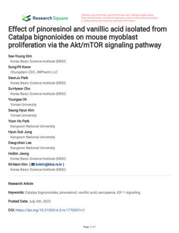
Effect of pinoresinol and vanillic acid isolated fromCatalpa bignonioides on mouse myoblastproliferation via the Akt/mTOR signaling pathwaySeo-Young KimKorea Basic Science Institute (KBSI)Sung-Pil KwonChungdam CDC JNPharm LLCSeonJu ParkKorea Basic Science Institute (KBSI)Su-Hyeon ChoKorea Basic Science Institute (KBSI)Youngse OhYonsei UniversitySeung Hyun KimYonsei UniversityYoon Ho ParkKangwon National UniversityHyun Suk JungKangwon National UniversityDeug-chan LeeKangwon National UniversityHoibin JeongKorea Basic Science Institute (KBSI)Kil-Nam Kim ( knkim@kbsi.re.kr )Korea Basic Science Institute (KBSI)Research ArticleKeywords: Catalpa bignonioides, pinoresinol, vanillic acid, sarcopenia, IGF-1 signaling.Posted Date: July 6th, 2022DOI: https://doi.org/10.21203/rs.3.rs-1770207/v1Page 1/17
License: This work is licensed under a Creative Commons Attribution 4.0 International License.Read Full LicensePage 2/17
AbstractSarcopenia is a disease in which skeletal muscle decreases with age. Stimulating the proliferation anddifferentiation of muscle cells may help prevent sarcopenia. To discover effective natural substancesenabling to treat muscle loss without side effects, we evaluated muscle growth with several compoundsextracted from Catalpa bignonioides Walt. Among these compounds, pinoresinol and vanillic acidincreased C2C12, a mouse myoblast cell line, proliferation the most without cytotoxicity. Thesesubstances activated the Akt/mammalian target of rapamycin (mTOR) pathway, which positivelyregulates the proliferation of muscle cells. In addition, they strongly bound to insulin-like growth factor 1receptor (IGF-1R), which is an upstream of the Akt/mTOR pathway, indicating that both pinoresinol andvanillic acid stimulate myoblast proliferation through direct interaction with IGF-1R. These results suggestthat pinoresinol and vanillic acid may improve the proliferation of skeletal muscle via IGF-1R/Akt/mTORsignaling and thus alleviate diseases such as sarcopenia.1. IntroductionSarcopenia refers to the gradual loss of skeletal muscle mass and strength associated with age.Sarcopenia is inevitable in most people and is considered a major cause of disability and fragility in olderadults, reducing their quality of life. It begins at the age of 40 years, with a decrease in muscle mass of upto 8% decennially; this rate may double by the age of 70 years [1]. Loss of skeletal muscle mass andfunction is caused by impaired myogenesis [2]. Skeletal muscle formation is achieved throughproliferation and differentiation of muscle fibers, and the insulin-like growth factor 1 (IGF1)/Akt/mammalian target of rapamycin (mTOR) signaling pathway, which stimulates them, is consideredto be a master regulator of skeletal myogenesis [3, 4]. Although IGF-1 upregulation inhibited sarcopenia inanimal studies, the administration of IGF-1 itself had a minor effect in older adults [5], encouragingfurther studies on other compounds that can achieve the desired effect. Therefore, compounds thatstimulate skeletal myogenesis and IGF-1 can help overcome age-related skeletal muscle loss.Steroidal androgens of the protein anabolic steroid class are drugs typically used to stimulate muscleenhancement. The biological efficacy of these drugs is demonstrated by muscle mass increase, growthspurts in premature children, and bone loss attenuation in older adults [6, 7], These compounds areprescribed for various therapeutic purposes. However, long-term or excessive usage may cause sideeffects, such as skin diseases [7] or reproductive and endocrine functional deterioration [8, 9]. Aiming totreat sarcopenia via metabolic changes, Belli et al. reported that trimetazidine, a metabolic modulatordrug, may induce myoblast differentiation in a cell line and increase muscle strength in mice [10].Although trimetazidine is used in the treatment of angina, its long-term use has been associated withgastrointestinal disturbances, vomiting, and nausea [11]. Therefore, there is a need for a compound thatprevents skeletal muscle mass loss without increasing side effect risks.Catalpa bignonioides Walt. (Bignoniaceae) is a bean tree native to southeastern America. It has beenused in traditional medicinal practices for respiratory diseases, scrofulous ulcers, and helminthicPage 3/17
infections, among others. A previous study on the bioactivity of C. bignonioides extracts [12] has shownthat its flowers, leaves, and capsule valves have antioxidant activity [13]. Other studies revealed thatcatalpic acid, a conjugated triene fatty acid abundant in C. bignonioides, may improve insulinhomeostasis by decreasing fat accumulation in the adipose tissue of mice [14]. Recently, we identifiedthe constituents of the methanol extract of C. bignonioides fruits through phytochemical analysis,revealing that some compounds had properties stimulating α-glucosidase inhibition and insulin secretion[15]. Although studies evaluating the biological activities of compounds isolated from C. bignonioides areon-going, the impact of C. bignonioid-derived substances on muscle diseases, such as sarcopenia,remains unclear. Herein, we aimed to identify components from the fruits of C. bignonioides extracts thatpromote muscle proliferation and differentiation via Akt/mTOR signaling.2. Materials And Methods2.1. MaterialsDulbecco modified eagle medium (DMEM) was purchased from Welgene (Gyeongsangbuk-do, Korea).Fetal bovine serum (FBS) was purchased from Omega Scientific, Inc. (Tarzana, CA, USA). Penicillin andstreptomycin were purchased from Invitrogen (Carlsbad, CA). Horse serum (HS), m bromide (MTT), dimethyl sulfoxide (DMSO), and radioimmunoprecipitationassay buffer were purchased from Sigma-Aldrich (St. Louis, MO, USA). NuPAGE 4–12% Bis-Tris gel waspurchased from Life Technologies (Carlsbad, CA, USA). Ployvinylidine fluoride membranes werepurchased from Bio-Rad Laboratories (Hercules, CA). Rabbit anti-mouse phosphoSmad2(Ser465/467)/3(Ser423/425) (Cat#8828), rabbit anti-mouse Smad4 (Cat#46535), rabbit antimouse phospho-Akt (Ser473, Cat#4060), rabbit anti-mouse phospho-mTOR (Ser2448, Cat#5536), rabbitanti-mouse phospho-ribosomal protein S6 kinase (p70S6K) (Thr421/Ser424, Cat#9204), rabbit antimouse phospho-eukaryotic initiation factor 4E-binding protein 1 (4E-BP1) (Thr37/46, Cat#2855), goatanti-rabbit IgG (Cat#7074), and goat anti-mouse IgG (Cat#7076) were purchased from Cell SignalingTechnology (Danvers, MA, USA). Mouse anti-mouse myoblast determination protein 1 (MyoD) (Cat#sc377460), mouse anti-mouse myogenin (Cat#sc-12732), and mouse anti-mouse β-actin (Cat#sc-47778)were purchased from Santa Cruz Biotechnology (Dallars, TX, USA).2.2. Plant materialC. bignonioides fruits were collected from the Arboretum of Seoul National University in Suwon, Korea in2021 and authenticated by Dr. Rack-Seon Seong, a director of the Center of Natural Resources Research,Jeonnam Bioindustry Foundation. A voucher specimen (CB202106) was deposited at the Korea BasicScience Institute (Chuncheon, Korea).2.3. Extraction and isolationThe dried fruits of C. bignonioides (1.3 kg) were extracted with MeOH (5 L 3 times) under sonication at30 for 4 h to yield an extract (91.0 g), which was then dissolved in H2O and successively partitionedPage 4/17
using CHCl3 and EtOAc to obtain CHCl3 (CB1, 16.0 g), EtOAc (CB2, 2.5 g), and H2O (CB3, 71.0 g) extractsafter removing the solvents in vacuo.The CHCl3 fraction was subjected to silica gel CC and eluted with a gradient of hexane : acetone (40:1 2.5:1, v/v) and CHCl3 : MeOH (20:1 2.5:1, v/v) to yield nine sub-fractions, CB1A (3.0 g), CB1B (2.4 g),CB1C (1.0 g), CB1D (1.5 g), CB1E (1.0 g), CB1F (1.2 g), CB1G (0.8 g), CB1H (1.0 g), and CB1I (0.5 g). TheCB1F fraction was applied to a YMC RP-18 column, which was eluted with MeOH : H2O (1.3:1, v/v),yielding four smaller fractions, CB1F1 (58.2 mg), CB1F2 (41.5 mg), CB1F3 (18.4 mg), and CB1F4 (16.5mg). The CB1F1 fraction was subjected to HPLC using a J’sphere ODS H-80 250 mm 20 mm column,eluted with 28% MeCN in H2O at a flow rate of 3 mL/min to yield 1 (6.8 mg) and 2 (7.1 mg). The CB1F2fraction was subjected to the same HPLC conditions, except that the elution solvent was 40% MeCN inH2O, to afford 3 (8.1 mg).The H2O fraction (CB3, 71.0 g) was chromatographed on a Diaion HP-20 column and eluted with H2Ocontaining increasing concentrations of MeOH (25, 50, and 100%) to obtain three subfractions, CB3A(10.0 g), CB3B (13.0 g), and CB3C (6.0 g). The CB3B fraction was subjected to silica gel CC and elutedwith a gradient of CHCl3 : MeOH (10:1 2.5:1, v/v) to yield three sub-fractions, CB3B1 (2.0 g), CB3B2(2.7 g), and CB3B3 (2.0 g). The CB3B1 fraction was applied to a silica gel column and eluted with CHCl3 :MeOH : H2O (5:1:0.1, v/v), CB3B11 (31.0 mg), CB3B12 (96.0 mg), CB3B13 (82.0 mg), CB3B14 (150.4 mg),CB3B15 (66.7 mg), and CB3B16 (213.8 mg). The CB3B14 fraction was subjected to HPLC purificationunder 40% MeCN to yield 13 (26.2 mg) and 14 (6.8 mg). The CB3B16 fraction was subjected to the sameHPLC conditions, except that elution with 23% MeCN in H2O afforded 15 (140.0 mg). The CB3C fractionwas subjected to silica gel CC and eluted with a gradient of CHCl3 : MeOH (10:1 2.5:1, v/v) to yieldthree sub-fractions, CB3C1 (0.4 g), CB3C2 (1.5 g), and CB3C3 (1.0 g). The CB3C1 fraction was applied toa YMC RP-18 column and eluted with MeOH : H2O (1:1, v/v), yielding three smaller fractions, CB3C11 (0.2g), CB3C12 (55.5 mg), and CB3C13 (14.0 mg). The CB3C11 fraction was subjected to HPLC using aJ’sphere ODS H-80 250 mm 20 mm column, eluted with MeCN : H2O (18:82), and a flow rate of 3mL/min to yield 4 (55.9 mg), 5 (7.3 mg), and 6 (14.3 mg). The CB3C2 fraction was applied to a YMC RP18 column, which, when eluted with MeOH : H2O (1:1, v/v), yielded three smaller fractions, CB3C21 (0.2 g),CB3C22 (0.6 g), and CB3C23 (0.2 g). The CB3C21 fraction was subjected to HPLC using a J’sphere ODSH-80 250 mm 20 mm column, eluted with MeCN : H2O (30:70), and a flow rate of 3 mL/min to yield 7(35.5 mg), whereas the CB3C23 fraction gave 8 (30.1 mg), 9 (18.5 mg), and 10 (6.3 mg). The CB3C3fraction was applied to a YMC RP-18 column, which when eluted with MeOH : H2O (1.4:1, v/v), yieldedfour smaller fractions, CB3C31 (42.8 mg), CB3C32 (0.1 g), CB3C33 (30.8 mg), and CB3C34 (18.6 g). TheCB3C32 fraction was subjected to HPLC using a J’sphere ODS H-80 250 mm 20 mm column, elutedwith MeCN : H2O (25:75), at a flow rate of 3 mL/min to yield 11 (11.3 mg). The CB3C34 fraction wassubjected to the same HPLC conditions, except that the eluding solvent was MeCN : H2O (23:77), toafford 12 (7.1 mg).2.4. Cell culture and differentiationPage 5/17
C2C12 cells (mouse myoblast cell line) were maintained in DMEM supplemented with 10% FBS, penicillin(100 U/mL), and streptomycin (100 µg/mL). For differentiation, when the cell confluence reachedapproximately 80%, the medium was replaced with DMEM containing 2% HS. DMEM containing 2% FBSwas replaced every other day, and differentiation proceeded for 6 days. All cell cultures were maintainedat 37 in a 5% CO2 incubator.2.5. Cytotoxicity assayThe cytotoxicity of compounds extracted from C. bignonioides was assessed using a colorimetric assay.In total, 1 105 cells/mL were seeded in 96-well plates and incubated with the test compounds for 24 h.Thereafter, 100 µg/mL MTT was added to each well. After 2.5 h incubation at 37 , the supernatantswere aspirated, and cells were treated with DMSO to dissolve the formazan crystals. The absorbance ofthe colored solution was determined at 540 nm using a SpectraMax M2/M2e spectrophotometer(Molecular Devices, San Jose, CA, USA).2.6. Cell proliferation activityTo measure skeletal muscle cell proliferation activity, C2C12 cells were seeded at a concentration of 5 104 cells/mL in a 96-well plate, and the medium was replaced with DMEM containing 2% HS two dayslater to induce differentiation. The compounds were added whenever the medium (DMEM with 2% HS)was changed every other day. Six days after differentiation induction, cell proliferation was assessedusing a 5-bromo-2’-deoxyuridine (BrdU) assay kit (Millipore, Billerica, MA, USA).2.7. Western blotCell lysates were prepared using radioimmunoprecipitation assay buffer. Quantified protein lysates wereloaded onto NuPAGE 4–12% Bis-Tris gels, which were then blotted onto a polyvinylidene fluoridemembrane. Primary antibodies, including rabbit anti-mouse phospho-Smad2/3, rabbit anti-mouseSmad4, rabbit anti-mouse phospho-Akt, rabbit anti-mouse phospho-mTOR, rabbit anti-mouse phosphop70S6K, rabbit anti-mouse phospho-4E-BP1, mouse anti-mouse MyoD, mouse anti-mouse myogenin, andmouse anti-mouse β-actin antibodies were diluted at 1:1000 and incubated overnight at 4 . Secondaryantibodies, including goat anti-rabbit IgG and goat anti-mouse IgG, were diluted at 1:3000 and incubatedfor 1.5 h at 25 . Signals were developed using the SuperSignal West Femto Trial Kit (Thermo FisherScientific; Waltham, MA, USA), and images were acquired using Fusion FX (Vilber Lourmat Ste, Collegien,France) or VISQUE InVivo Smart-LF (Vieworks. Co, Ltd., Anyang-si, Korea).2.8. In silico molecular docking simulationTo verify the potential active chemicals that could act as IGF-1 receptor (IGF-1R) agonists, a moleculardocking study was performed. First, the crystal structure of IGF-1R (PDB ID: 1IGR) was obtained from theProtein Data Bank (PDB, http://www.pdb.org; accessed on January 1, 2022). A docking simulation wasperformed using AUTODOCK VINA to investigate whether the potential active chemicals bind to IGF-1R[16] and LIGPLOT to analyze IGF-1R and chemical interactions [17]. The 2-dimensional interaction mapshows hydrogen bonds in green and labeled non-ligand residues involved in hydrophobic contact in red.Page 6/17
2.9. Statistical analysisVariables were compared using two-tailed one-way ANOVA with Tukey’s post-hoc test using Prismsoftware (Version 4.00; GraphPad Inc.; La Jolla, CA, USA). Findings were considered statisticallysignificant at p-values of 0.05.3. Results3.1. Pinoresinol and vanillic acid facilitated C2C12 cellproliferationTo examine the effect of 15 constituents from C. bignonioides fruit extracts on skeletal muscle growth,we measured cell proliferation activity in cell line C2C12 (Fig. 1) [15]. Prior to examining the effect on cellproliferation, the viability of C2C12 myoblasts was evaluated using MTT colorimetric assay. Weconfirmed that none of the compounds, except compound 9 (6-O-trans-feruloyl catalpol), showed toxicityto C2C12 cells at a concentration of 50 µM (Fig. 2A).Next, the proliferation effect of skeletal muscle cells was measured during the differentiation period byBrdU cell proliferation assay, except for compound 9, which was excluded because of its toxicity.Compared to the control conditions, most compounds triggered approximately 1.2-fold increase in cellproliferation (Fig. 2B). Among them, compound 3 (pinoresinol) and compound 5 (vanillic acid) showed a1.8-fold increase.To determine effects at low concentrations, cell proliferation was measured in a dose-dependent mannerusing pinoresinol and vanillic acid, which had the greatest effect among the compounds. Pinoresinol andvanillic acid showed cell proliferation activity during the differentiation phase even at concentrations of6.25 µM and 12.5 µM, respectively (Fig. 3).3.2. Pinoresinol and vanillic acid stimulated Akt/mTORsignaling pathway in C2C12 cellsMyogenesis, differentiation, and maturation of skeletal muscle cells are regulated by signaling pathwaysactivated by the transforming growth factor beta (TGF-β) superfamily [18]. To determine whether cellproliferation activity of pinoresinol and vanillic acid was mediated by TGF-β signaling, we performedwestern blotting for Smad proteins in C2C12 differentiated cells treated with either compound. We foundthat p-Smad2 and p-Smad3 were not significantly downregulated by pinoresinol or vanillic acids (Fig. 4).Furthermore, the expression level of Smad4 was mildly decreased by these compounds.IGF-1 signaling is a positive regulator of muscle cell proliferation and differentiation [19]. To examine theactivation of IGF-1 signaling by pinoresinol and vanillic acid, we analyzed the phosphorylation levels ofAkt, mTOR, and p70S6K using western blotting. We found that p-Akt, p-mTOR, and p-p70S6K levels wereincreased by these compounds (Fig. 4). In addition, these compounds decreased the phosphorylation ofPage 7/17
4E-BP1, indicating suppression of its growth inhibitory function. Furthermore, essential regulators thatinduce muscle differentiation, such as MyoD and myogenin, were significantly increased in C2C12myoblasts treated with pinoresinol or vanillic acid. These results suggest that pinoresinol and vanillicacid stimulate myogenic differentiation by regulating Akt/mTOR signaling in mouse muscle cells.3.3. Pinoresinol and vanillic acid were docked into IGF-1receptor through in silico analysisWe showed that the levels of protein expression involved in IGF-1 signaling were highly upregulated inpinoresinol- or vanillic-acid-treated C2C12 cells. To determine the potential of these two activecompounds to bind to the IGF-1R, we simulated the docking of pinoresinol, vanillic acid, and IGF-1R.Docking of the ligand-protein complexes was successful, as both compounds stably posed to the activesites of IGF-1R (Fig. 5A to 5C). In addition, dihydrotestosterone (DHT), used as a positive control [20], wasshown in the docking simulation to stably pose the active site of IGF-1R with a similar low binding energyvalue as pinoresinol or vanillic acid (Fig. 5D and Table 1).Table 1Results of docking simulations of two activechemicals (pinoresinol and vanillic acid) with IGF-1R(PDB ID: 1IGR). DHT is dihydrotestosterone used aspositive control.ReceptorLigand(Binding site)Binding energy(kcal/mol)IGF-1RPinoresinol-6.7(1IGR-active site)Vanillic acid-5.2DHT-7.04. DiscussionIn this study, we demonstrated the effects on myoblast proliferation of two compounds extracted from C.bignonioides: pinoresinol and vanillic acid. Pinoresinol is a biologically active ligand mainly found inmedicinal plants, such as Styrax sp. and Forsythia suspense, and in olive oil [21, 22]. Pinoresinolpossesses antioxidant, anti-inflammatory, and antifungal activities, and has been used in traditionalmedicine for a long time [21–23]. A recent study demonstrated that the effect of defatted sesame seedsin alleviating hypoglycemia is mediated by the inhibitory function of α-glucosidase activity of pinoresinol[24]. In a study on anticancer activity, pinoresinol was found to induce apoptosis and to suppressmigration in human liver cancer cells [25]. Vanillic acid, an oxidative form of vanillin and phenoliccompound, has also been used in folk medicine. Vanillic acid is abundant in the root of Angelica sinensis,also known as female ginseng, and exhibits antioxidant, anticancer, and cardioprotective activities [26–28]. However, no studies have examined the effect of vanillic acid or pinoresinol on muscle cells orPage 8/17
muscle-related diseases. This study is first to show that pinoresinol and vanillic acid extracted from anedible plant promote proliferation in myoblasts.Skeletal muscles play pivotal roles in physical activity and energy metabolism. Growth and maintenanceof skeletal muscle are essential for the management of sarcopenia, an age-related disease in whichskeletal muscle decreases. The growth and differentiation of skeletal muscle are regulated by negativeand positive regulators, specifically, TGF-β and IGF-1/Akt/mTOR pathways, respectively. TGF-β familymembers, including myostatin, are known to inhibit myogenic differentiation in cultured primarymyoblasts or myoblast cell lines, such as C2C12 [29–31]. C2C12 myoblasts lacking the TGB-β1 signaldue to mutation of TβR II, a component of the TGF-β receptor, cannot form myotubes [32]. IGF-1 binds toIGF-1R, phosphorylates insulin receptor substrate-1 (IRS-1), an adaptor protein inside the cell, andsequentially phosphorylates phosphoinositide 3-kinase (PI3K) and Akt. Meanwhile, mTOR, which is adownstream target of Akt, phosphorylates p70S6K to promote protein synthesis and 4E-BP1 to inducetranslation initiation [33]. The Akt/mTOR/p706K pathway mediates downstream signaling of IGF-1 topromote protein synthesis and body growth and promotes both cell proliferation and differentiation incultured myoblasts [34]. IGF-1 also induces cell differentiation by inducing the expression of myogenicregulatory factors (MEFs) such as MyoD and myogenin during myogenic differentiation [35]. In the L6E9cell line (rat-derived myoblast) in the process of differentiation, when IGF-1 was overexpressed, myogeninlevels increased, and myotubes became enlarged [36]. We demonstrated that natural substances withmuscle proliferation activity, pinoresinol and vanillic acid, increased the expression of MyoD andmyogenin, and phosphorylation of downstream targets of IGF-1, including Akt, mTOR, and p70S6K.Furthermore, through in silico docking analysis, we showed the potential of these substances to bind tothe active site of IGF-1R.Although TGB-β is known to negatively regulate myoblast differentiation by suppressing the expressionof two MRFs (MyoD and myogenin) through Smad3 [18, 37–39], our study confirmed that the myogeniceffects of pinoresinol and vanillic acid were unlikely achieved through the TGB-β/Smad pathway.5. ConclusionsIn summary, this study demonstrated that pinoresinol and vanillic acid isolated from C. bignonioidesstimulated the Akt/mTOR pathway to promote proliferation and differentiation of myoblasts, suggestingthat they bind to IGF-1R, upstream of Akt/mTOR. These findings may provide fundamental data forpinoresinol and vanillic acid as novel therapeutic agents to treat muscle-related diseases such assarcopenia by inhibiting muscle loss.DeclarationsFundingPage 9/17
This research was financially supported by the Ministry of Small and Medium-sized Enterprises (SMEs)and Startups (MSS), Korea, under the “Regional Specialized Industry Development Plus Program (R&D ,S3092691)” supervised by the Korea Institute for Advancement of Technology (KIAT).Data availabilityData is available upon request.Authors' contributionsSeo-Young Kim: Conceptualization, Data curation, Formal analysis. Sung-Pil Kwon: Conceptualization,Funding acquisition, Investigation. SeonJu Park: Formal analysis. Su-Hyeon Cho: Formal analysis,Resources. Youngse Oh: Formal analysis, Resources. Seung Hyun Kim: Formal analysis. Yoon Ho Park:Formal analysis, Visualization. Hyun Suk Jung: Formal analysis, Visualization. Deug-chan Lee: Fundingacquisition. Hoibin Jeong: Data curation, Investigation, Project administration, Validation, Writing original draft. Kil-Nam Kim: Conceptualization, Data curation, Funding acquisition, Project administration,Supervision.Ethics approvalNot applicable.Consent to participateNot applicable.Consent for publicationNot applicable.Conflicts of InterestThe authors declare no conflict of interest.References1. Newman AB et al (2003) Strength and muscle quality in a well-functioning cohort of older adults: theHealth, Aging and Body Composition Study. J Am Geriatr Soc 51(3):323–3302. Kwak JY, Kwon KS (2019) Pharmacological Interventions for Treatment of Sarcopenia: CurrentStatus of Drug Development for Sarcopenia. Ann Geriatr Med Res 23(3):98–1043. Laplante M, Sabatini DM (2012) mTOR signaling in growth control and disease. Cell 149(2):274–2934. Ge Y, Chen J (2012) Mammalian target of rapamycin (mTOR) signaling network in skeletalmyogenesis. J Biol Chem 287(52):43928–43935Page 10/17
5. Sakuma K, Yamaguchi A (2012) Sarcopenia and age-related endocrine function. Int J Endocrinol2012:1273626. Grunfeld C et al (2006) Oxandrolone in the treatment of HIV-associated weight loss in men: arandomized, double-blind, placebo-controlled study. J Acquir Immune Defic Syndr 41(3):304–3147. Melnik B, Jansen T, Grabbe S (2007) Abuse of anabolic-androgenic steroids and bodybuilding acne:an underestimated health problem. J Dtsch Dermatol Ges 5(2):110–1178. Matsumoto AM (1990) Effects of chronic testosterone administration in normal men: safety andefficacy of high dosage testosterone and parallel dose-dependent suppression of luteinizinghormone, follicle-stimulating hormone, and sperm production. J Clin Endocrinol Metab 70(1):282–2879. Meriggiola MC et al (2002) Higher testosterone dose impairs sperm suppression induced by acombined androgen-progestin regimen. J Androl 23(5):684–69010. Belli R et al (2019) Metabolic Reprogramming Promotes Myogenesis During Aging Front Physiol10:89711. Chrusciel P, Rysz J, Banach M (2014) Defining the role of trimetazidine in the treatment ofcardiovascular disorders: some insights on its role in heart failure and peripheral artery disease.Drugs 74(9):971–98012. Munoz-Mingarro D et al (2003) Biological activity of extracts from Catalpa bignonioides Walt.(Bignoniaceae). J Ethnopharmacol 87(2–3):163–16713. Dvorska M et al (2007) Antioxidant activity of Catalpa bignonioides. Fitoterapia 78(6):437–43914. Hontecillas R et al (2008) Catalpic acid decreases abdominal fat deposition, improves glucosehomeostasis and upregulates PPAR alpha expression in adipose tissue. Clin Nutr 27(5):764–77215. Oh Y et al (2021) The Chemical Constituents from Fruits of Catalpa bignonioides Walt. and Theiralpha-Glucosidase Inhibitory Activity and Insulin Secretion Effect.Molecules, 26(2)16. Trott O, Olson AJ (2010) AutoDock Vina: improving the speed and accuracy of docking with a newscoring function, efficient optimization, and multithreading. J Comput Chem 31(2):455–46117. Wallace AC, Laskowski RA, Thornton JM (1995) LIGPLOT: a program to generate schematicdiagrams of protein-ligand interactions. Protein Eng 8(2):127–13418. Liu D, Black BL, Derynck R (2001) TGF-beta inhibits muscle differentiation through functionalrepression of myogenic transcription factors by Smad3. Genes Dev 15(22):2950–296619. Ahmad SS et al (2020) Implications of Insulin-Like Growth Factor-1 in Skeletal Muscle and VariousDiseases.Cells, 9(8)20. Kim SY et al (2021) The Effects of Marine Algal Polyphenols, Phlorotannins, on Skeletal MuscleGrowth in C2C12 Muscle Cells via Smad and IGF-1 Signaling Pathways.Mar Drugs, 19(5)21. Jung HW et al (2010) Pinoresinol from the fruits of Forsythia koreana inhibits inflammatoryresponses in LPS-activated microglia. Neurosci Lett 480(3):215–220Page 11/17
22. Owen RW et al (2000) The antioxidant/anticancer potential of phenolic compounds isolated fromolive oil. Eur J Cancer 36(10):1235–124723. Hwang B et al (2010) Antifungal effect of ( )-pinoresinol isolated from Sambucus williamsii.Molecules 15(5):3507–351624. Wikul A et al (2012) ( )-Pinoresinol is a putative hypoglycemic agent in defatted sesame (Sesamumindicum) seeds though inhibiting alpha-glucosidase. Bioorg Med Chem Lett 22(16):5215–521725. Lopez-Biedma A et al (2016) Phytoestrogen ( )-pinoresinol exerts antitumor activity in breast cancercells with different oestrogen receptor statuses. BMC Complement Altern Med 16:35026. Yao X et al (2020) Vanillic Acid Alleviates Acute Myocardial Hypoxia/Reoxygenation Injury byInhibiting Oxidative Stress. Oxid Med Cell Longev, 2020: p. 834803527. Jung Y et al (2017) Vanillic acid attenuates testosterone-induced benign prostatic hyperplasia in ratsand inhibits proliferation of prostatic epithelial cells. Oncotarget 8(50):87194–8720828. Prince PS, Dhanasekar K, Rajakumar S (2011) Preventive effects of vanillic acid on lipids, bax, bcl-2and myocardial infarct size on isoproterenol-induced myocardial infarcted rats: a biochemical and invitro study. Cardiovasc Toxicol 11(1):58–6629. McPherron AC, Lawler AM, Lee SJ (1997) Regulation of skeletal muscle mass in mice by a new TGFbeta superfamily member. Nature 387(6628):83–9030. Massague J et al (1986) Type beta transforming growth factor is an inhibitor of myogenicdifferentiation. Proc Natl Acad Sci U S A 83(21):8206–821031. Yablonka-Reuveni Z, Rivera AJ (1997) Proliferative Dynamics and the Role of FGF2 DuringMyogenesis of Rat Satellite Cells on Isolated Fibers. Basic Appl Myol 7(3–amp4):189–20232. Filvaroff EH, Ebner R, Derynck R (1994) Inhibition of myogenic differentiation in myoblastsexpressing a truncated type II TGF-beta receptor. Development 120(5):1085–109533. Gruner S et al (2016) The Structures of eIF4E-eIF4G Complexes Reveal an Extended Interface toRegulate Translation Initiation. Mol Cell 64(3):467–47934. Engert JC, Berglund EB, Rosenthal N (1996) Proliferation precedes differentiation in IGF-I-stimulatedmyogenesis. J Cell Biol 135(2):431–44035. Yoshida T, Delafontaine P (2020) Mechanisms of IGF-1-Mediated Regulation of Skeletal MuscleHypertrophy and Atrophy.Cells, 9(9)36. Musaro A, Rosenthal N (1999) Maturation of the myogenic program is induced by postmitoticexpression of insulin-like growth factor I. Mol Cell Biol 19(4):3115–312437. Brennan TJ et al (1991) Transforming growth factor beta represses the actions of myogenin througha mechanism independent of DNA binding. Proc Natl Acad Sci U S A 88(9):3822–382638. Goulding M, Lumsden A, Paquette AJ (1994) Regulation of Pax-3 expression in the dermomyotomeand its role in muscle development. Development 120(4):957–97139. Liu D, Kang JS, Derynck R (2004) TGF-beta-activated Smad3 represses MEF2-dependenttranscription in myogenic differentiation. EMBO J 23(7):1557–1566Page 12/17
FiguresFigure 1Chemical structures of compounds 1-15. 1, isolariciresinol; 2, ( )-lariciresinol; 3, pinoresinol; 4, 4hydroxybenzoic acid; 5, vanillic acid; 6, trans-p-coumaric acid; 7, catalposide; 8, specioside; 9, 6-O-transferuloyl catalpol; 10, minecoside; 11, ide; 12, zoyl-β-D-glucopyranosyl-(1 2)-β-D-glucopyranosyl-(1 2)]-βD-glucopyranoside; 13, des-p-hydroxybenzoy
that its owers, leaves, and capsule valves have antioxidant activity [13]. Other studies revealed that catalpic acid, a conjugated triene fatty acid abundant in C. bignonioides, may improve insulin homeostasis by decreasing fat accumulation in the adipose tissue of mice [14]. Recently, we identied