Transcription
Herrero-Sánchez et al. Journal of Hematology & Oncology (2016) 9:113DOI 10.1186/s13045-016-0343-5RESEARCHOpen AccessTargeting of PI3K/AKT/mTOR pathway toinhibit T cell activation and prevent graftversus-host disease developmentMª Carmen Herrero-Sánchez1,2,3, Concepción Rodríguez-Serrano1,2,3, Julia Almeida2,3,4, Laura San Segundo2,3,Susana Inogés5, Ángel Santos-Briz2,6, Jesús García-Briñón2,7, Luis Antonio Corchete1,2,3, Jesús F. San Miguel8,Consuelo del Cañizo1,2,3 and Belén Blanco1,2,3*AbstractBackground: Graft-versus-host disease (GvHD) remains the major obstacle to successful allogeneic hematopoieticstem cell transplantation, despite of the immunosuppressive regimens administered to control T cell alloreactivity.PI3K/AKT/mTOR pathway is crucial in T cell activation and function and, therefore, represents an attractivetherapeutic target to prevent GvHD development. Recently, numerous PI3K inhibitors have been developedfor cancer therapy. However, few studies have explored their immunosuppressive effect.Methods: The effects of a selective PI3K inhibitor (BKM120) and a dual PI3K/mTOR inhibitor (BEZ235) on human T cellproliferation, expression of activation-related molecules, and phosphorylation of PI3K/AKT/mTOR pathway proteinswere analyzed. Besides, the ability of BEZ235 to prevent GvHD development in mice was evaluated.Results: Simultaneous inhibition of PI3K and mTOR was efficient at lower concentrations than PI3K specific targeting.Importantly, BEZ235 prevented naïve T cell activation and induced tolerance of alloreactive T cells, while maintainingan adequate response against cytomegalovirus, more efficiently than BKM120. Finally, BEZ235 treatment significantlyimproved the survival and decreased the GvHD development in mice.Conclusions: These results support the use of PI3K inhibitors to control T cell responses and show the potential utilityof the dual PI3K/mTOR inhibitor BEZ235 in GvHD prophylaxis.Keywords: Hematopoietic stem cell transplantation, Graft-versus-host disease, T cell, PI3K/AKT/mTOR pathway,PI3K inhibitorBackgroundAllogeneic hematopoietic stem cell transplantation(allo-HSCT) remains the only curative option formany hematologic malignancies. Unfortunately, a seriouscomplication is frequently developed after allo-HSCT:graft-versus-host disease (GvHD). GvHD occurs whendonor T lymphocytes recognize as foreign and destroypatient’s healthy tissues. Despite the immunosuppressiveregimens administered, GvHD remains the major cause of* Correspondence: belen blancod@yahoo.es1Servicio de Hematología, Hospital Universitario de Salamanca, Paseo de SanVicente 58-182, 37007 Salamanca, Spain2Instituto de Investigación Biomédica de Salamanca (IBSAL), Paseo de SanVicente 58-182, 37007 Salamanca, SpainFull list of author information is available at the end of the articlemorbidity and mortality after allo-HSCT. Thus, newtherapeutic strategies are needed.One of the key signaling pathways involved in T cellactivation and function is phosphatidylinositol 3-kinase/AKT/mammalian target of rapamycin (PI3K/AKT/mTOR) [1]. This pathway controls numerous cellularprocesses, including proliferation, survival, migration,and metabolism [2]. In particular, PI3K activation in Tcells promotes survival [3] and cell cycle progression [4],modulates differentiation [5, 6], and controls the acquisition of effector and memory phenotypes [7]. Thus, inhibitors of PI3K/AKT/mTOR pathway can interfere withT cell activation and function.The use of PI3K/AKT/mTOR inhibitors has beenscantily explored in the allo-HSCT context. Only the 2016 The Author(s). Open Access This article is distributed under the terms of the Creative Commons Attribution 4.0International License (http://creativecommons.org/licenses/by/4.0/), which permits unrestricted use, distribution, andreproduction in any medium, provided you give appropriate credit to the original author(s) and the source, provide a link tothe Creative Commons license, and indicate if changes were made. The Creative Commons Public Domain Dedication o/1.0/) applies to the data made available in this article, unless otherwise stated.
Herrero-Sánchez et al. Journal of Hematology & Oncology (2016) 9:113Page 2 of 14utility of the mTORC1 inhibitor rapamycin (Sirolimus)has been extensively studied, providing promising results[8]. In addition, it has been suggested that the beneficialeffects observed in patients with chronic GvHD treatedwith tyrosine kinase inhibitors could be due, in part, totheir ability to inhibit PI3K signaling in T cells [9]. However, few studies have evaluated the immunosuppressiveeffect of PI3K inhibitors on T lymphocytes [10–12] andtheir ability to prevent GvHD development [13, 14].Herein, we have analyzed the effects of two novel antitumor drugs, the pan-class I PI3K inhibitor BKM120 andthe dual PI3K/mTOR inhibitor BEZ235, on T cell activation and evaluated the utility of BEZ235 in a murinemodel of GvHD.stimulated or not with plate-bound anti-CD3 (5 μg/ml)and soluble anti-CD28 (2.5 μg/ml) monoclonal antibodies (mAbs) (BD Biosciences, San Jose, CA, USA)and treated with different doses of BKM120 orBEZ235 (0–10 μM).MethodsDrugsBEZ235 was kindly provided by Novartis Pharma (Basel,Switzerland). BKM120 was purchased from SelleckChemicals (Houston, TX, USA). For in vitro studies,BKM120 and BEZ235 were reconstituted in DMSO at10 mM and stored frozen at 20 C until use. For invivo assays, BEZ235 solution was prepared freshbefore administration. In brief, BEZ235 was dissolved inone volume of N-methyl-2-pyrrolidone (Sigma-Aldrich,St. Louis, MO) and then nine volumes of polyethyleneglycol 300 (Sigma-Aldrich) were added. The applicationvolume was 10 ml/kg body weight.Western blot analysisUnstimulated or stimulated T cell-enriched PBMCs weretreated with different concentrations of BKM120 orBEZ235. After 48 h, cells were lysed and cell extractswere electrophoresed, transferred onto PVDF membrane(Millipore, Bedford, MA, USA), and immunoblottedwith antibodies against caspase 3, phosphorylated AKT(p-AKT) (T308 and S473), total AKT, phosphorylated4E-BP1 (p-4E-BP1) (T37/46), total 4E-BP1, phosphorylated RPS6 (p-RPS6) (S235/236), total RPS6, phosphorylated p38 MAPK (p-p38) (T180/Y182), total p38 MAPK,phosphorylated ERK1/2 (p-ERK1/2) (T202/Y204) (allfrom Cell Signaling Thechnology , Leiden, Netherlands),or total ERK2 (Santa Cruz Biotechnology, Heidelberg,Germany); antibodies to GAPDH (Cell Signaling Thechnology ) and calnexin (Enzo Life Science, PlymouthMeeting, PA, USA) were used as loading controls. Antirabbit or anti-mouse antibodies conjugated to horseradish peroxidase (GE Healthcare, Buckinghamshire, UK)were used as secondary antibodies. Proteins were visualized with an ECL detection system (GE Healthcare).Cell isolation and culturePeripheral blood mononuclear cells (PBMCs) were isolated from buffy coats of volunteer healthy donors bydensity gradient centrifugation using Ficoll–Paque solution (GE Healthcare Bio-Sciences, Uppsala, Sweden). Buffycoats were provided by the Centro de Hemoterapia yHemodonación de Castilla y León, after written informedconsent obtention. The research was approved by theClinical Research Ethics Committee (CEIC) of “Area deSalud de Salamanca” (2012/11/132).For Western blot analysis, PBMCs were allowed to adhere to tissue culture flasks (Corning, NY, USA) O/N at37 C and thereafter, non-adherent cells (T cell-enrichedPBMCs) were collected. For cell cycle, apoptosis, andcytokine secretion assays, T cells were isolated fromPBMCs by immunomagnetic selection, using the Pan TCell Isolation Kit (Miltenyi Biotec, Bergisch Gladbach,Germany). The purity of isolated populations was routinely 95 %.PBMCs or isolated T cells were cultured in well plates(Greiner Bio-One, Frickenhausen, Germany) at a densityof 1 106 cells/ml in RPMI 1640 medium supplementedwith 2 mM L-glutamine, 100 U/ml penicillin, 100 μg/mlstreptomycin (all from Gibco-Invitrogen, Paisely, UK),and 10 % human AB serum (Sigma-Aldrich). Cells wereProliferation assaysPBMCs were stained with PKH-67 green fluorescentdye (Sigma-Aldrich) following manufacturer’s instructions. Thereafter, PKH-stained cells were seeded inthe absence or in the presence of anti-CD3/anti-CD28mAbs as described above and treated with differentconcentrations of the drugs. After 5 days, cells werestained with 7-amino-actinomycin D (7AAD) andanti-CD3-APC and acquired in a FACSCalibur flowcytometer (all from BD Biosciences). Percentage ofproliferating T cells (CD3 PKHlow) was calculated usingthe Infinicyt software (Cytognos, Salamanca, Spain).Cell cycle analysisUnstimulated or anti-CD3/anti-CD28 stimulated isolatedT cells were cultured in the presence of different concentrations of BKM120 or BEZ235. After 4 days, cells werestained with propidium iodide, using the CycleTEST PLUS DNA Reagent Kit (BD Biosciences) and acquired ona FACSCalibur flow cytometer. The distribution of cellsalong the cell cycle phases was analyzed using ModFit LT Macintosh program (Verity Software House, Topsham,ME, USA).
Herrero-Sánchez et al. Journal of Hematology & Oncology (2016) 9:113Apoptosis assessmentUnstimulated or anti-CD3/anti-CD28 stimulated isolatedT cells were cultured in the presence of different concentrations of the compounds. After 2 days, cells were stainedwith Annexin V-PE (BD Pharmingen , San Diego, USA).Samples were acquired on a FACSCalibur flow cytometerand analyzed using the Infinicyt software.Cytokine assaysIsolated T cells were stimulated with anti-CD3/antiCD28 mAbs in the presence of several concentrations ofBKM120 or BEZ235. After 48 h, concentration of different cytokines in culture supernatants was analyzed on aFACSCalibur flow cytometer using the Human Th1/Th2Cytokine Cytometric Bead Array (CBA) kit and BD CBAsoftware (all from BD Biosciences).Immunophenotypic analysisUnstimulated or anti-CD3/anti-CD28 stimulated PBMCswere treated with different concentrations of BKM120or BEZ235. After 48 h, cells were stained with thefollowing combination of mAbs: .5/anti-CD25-PE-Cy7/anti-Granzyme B-Alexa Fluor 647/anti-CD4-APC-AlexaFluor 750/anti-CD27-Brillant Violet 421/anti-CD3-BrillantViolet 510. For intracellular staining of IFN-γ and granzyme B, brefeldin A (10 μg/ml) (Sigma-Aldrich) was addedfor the last 4 h prior to acquisition and the IntraStain Kit(Dako Cytomation, Denmark) was used. Acquisition wasperformed on a FACSCanto flow cytometer (BD) and analyzed using the Infinicyt software.Enzyme-linked immunospot (ELISPOT) assaysPBMCs from cytomegalovirus (CMV)-positive donors(responder cells) were stimulated with irradiated (25 Gy)PBMCs from a second donor (allogeneic cells) in a 2:1ratio. Different doses of BKM120 or BEZ235 were added.After 96 h, responder cells were collected and cultured,in the absence of drugs, in an IFN-γ ELISpot plate(Mabtech, Nacka Strand, Sweeden): (a) in the absence ofstimulation (control), (b) re-stimulated with allogeneiccells from the same donor of the primary culture, or (c)re-stimulated with CMV-pp65 recombinant protein(Miltenyi Biotec). After 36 h, ELISPOT was performedfollowing manufacturer’s instructions. Spots corresponding to IFN-γ secreting cells were quantified using anImmunospot ELISPOT reader (CTL, Aale, Germany).The percentage of IFN-γ secreting cells was determinedby subtracting, from the number of spots counted inallogeneic or CMV-pp65 re-stimulated wells, the background spots in the corresponding unstimulated (control)wells. These values were normalized with respect to thoseobtained from the samples pre-stimulated in the absencePage 3 of 14of drugs (0 μM) and re-stimulated with allogeneic cells orCMV-pp65, respectively, considered as 100 %.GvHD murine modelFemale recipient Balb/c (H2d) and male donor C57BL/6(H2b) mice (12 weeks old) were purchased from HarlanLaboratory (Carshalton, UK). Animal experimentswere approved by the ethical committee of SalamancaUniversity (N 201300004045).Balb/c mice received total body irradiation (8.5 Gy intwo fractions) from a Cs137 source and then an intravenous injection of 10 106 C57BL/6 bone marrow cellswithout (BM group) or with 5 106 splenocytes. Micereceiving splenocytes were either untreated (GvHDgroup) or treated orally with BEZ235 (GvHD BEZ235group) once a day, from day 1 to day 60 posttransplantation. Different concentrations of BEZ235(5–50 mg/kg/day) were tested and, finally, the bestresults were observed at a dose of 5 mg/kg/day.Four experiments were performed with at least twomice per group. A control mouse receiving irradiationwithout stem cell support (Total Body Irradiation, TBIgroup) was also included in each experiment.Balb/c mice were monitored daily for survival and forthe following clinical signs of GvHD: weight loss, posture (hunching), activity, fur texture and skin integrity.Each parameter received a score of 0 (normal), 1 (mildto moderate), or 2 (severe) and a clinical GvHD indexwas generated subsequently by summation of the fivecriteria scores (maximum index 10), as previously described [15]. All moribund mice were humanely killed.For histopathological analysis of GvHD target organs(large intestine, skin and liver), at least one animal of eachgroup was killed in the third week post-transplantationand once treatment was completed. Tissues were fixedin 10 % neutral-buffered formalin (Sigma-Aldrich),embedded in paraffin, and sectioned and stained withhematoxylin and eosin (both from Merck KGaA,Darmstadt, Germany). Slides were examined under aBX41 light microscope and images were capturedusing a DP50 digital camera and the software Cell A2.6 (all from Olympus Optical Co. Ltd., Tokyo, Japan).Details of the scoring system are summarized in Table 1.Statistical analysisMost statistical analyses were performed using IBMSPSS Statistics 20 (Chicago, IL, USA). Differences between the effects of different doses of a drug and between both drugs were analyzed by the Kruskal–Wallismultiple-comparison Z value test. Pairwise comparisonswere performed using the Mann–Whitney test withBonferroni correction. Survival curves were plottedusing Kaplan–Meier estimates and a log-rank test wasused to compare survival rates.
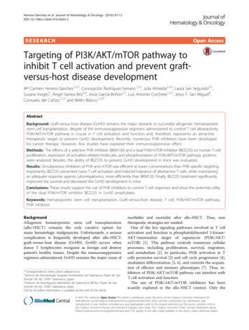
Herrero-Sánchez et al. Journal of Hematology & Oncology (2016) 9:113Page 4 of 14Table 1 Histologic criteria for GvHD scoreScoreSkinLiverLarge intestine0NormalNormalNormalFocal mild portal lymphoid infiltrateOccasional or rare necrotic cellsin glands or crypts0.51Basal vacuolar changeWidespread mild portal lymphoidinfiltrateIsolated apoptotic epithelial cells,without crypt loss2Dyskeratotic cells in the epidermis and/orfollicle, dermal lymphocytic infiltrateFocal bile duct invasion or cellularinjuryIndividual crypt loss. Regenerationchanges3Fusion of basilar vacuoles to form cleftsand microvesiclesMultiple foci of bile duct injury andregenerationContiguous area of multiplecrypt loss4Separation of the epidermis from the dermisWidespread injury and destructionof bile ductsExtensive crypt dropout withdenudation of epitheliumFor statistical analysis of weight variation and clinicalscores, curves of each mouse along time from transplantation were fitted by cubic splines using the program“Compare” in the SIMFIT statistical package (http://simfit.org.uk), applying unweighted least square fitting.Areas under the curve (AUCs) from the replicate curvesof mice in each group were compared by a one-wayANOVA followed by the Tukey’s post hoc test.Statistical significance in all tests was concluded forvalues of p 0.05.ResultsEffect of BKM120 and BEZ235 on PI3K/AKT/mTOR pathwayFirst, the effect of BKM120 and BEZ235 on the phosphorylation of PI3K/AKT/mTOR pathway proteins instimulated T cells was assessed (Fig. 1). BKM120effectively reduced AKT phosphorylation at T308 atall the tested doses, and so did BEZ235, althoughwith less efficacy. The treatment with both inhibitorsalso reduced AKT-S473 phosphorylation, observing acomplete abrogation at 1 μM for BEZ235 and 10 μMfor BKM120.The amount of phosphorylated 4E-BP1 and RPS6proteins was also diminished in the presence of bothdrugs. However, while this decrease was clear only atthe highest concentration of BKM120 (10 μM), thelowest dose of BEZ235 (0.5 μM) was sufficient tocompletely abolish the presence of the phosphorylatedforms of 4E-BP1 and RPS6. Of note, this completeabrogation was accompanied by the strong reductionin RPS6 and 4E-BP1 expression.As 4E-BP1 expression can be negatively regulated bythe mitogen-activated protein kinases (MAPKs) ERKand p38 [16], their phosphorylation was assessed. Bothdrugs reduced ERK1/2 phosphorylation on stimulatedT cells. However, BKM120 (10 μM) and, especially,BEZ235 (0.5–10 μM) increased p38 phosphorylation,correlating with the reduction in RPS6 and 4E-BP1 expression (Fig. 1).Effect of BKM120 and BEZ235 on T cell proliferation andapoptosis inductionThereafter, we assessed the effect of both inhibitors onstimulated T cell proliferation. The percentage of proliferating T cells significantly decreased at high concentrations of both drugs, although at low doses BEZ235 wasmuch more effective than BKM120 (Fig. 2a).Next, we investigated whether this reduction in proliferation was related to cell cycle arrest, to an increase inapoptosis or to both. The percentage of cells in synthesisand G2/mitosis phases significantly decreased amongstimulated T cells in the presence of both inhibitors, although, at low doses, BEZ235 was more efficient thanBKM120 (Fig. 2b).Regarding apoptosis, the addition of inhibitors did notsignificantly change the percentage of annexin V cells,neither among unstimulated nor among stimulated Tcells (Fig. 2c, d). However, the amount of cleaved caspase 3 in stimulated cells decreased in the presence ofBEZ235 and at high concentrations of BKM120 (Fig. 2e).Effect of BKM120 and BEZ235 on T cell cytokine secretionBoth inhibitors induced, in general, a dose-dependentdecrease in Th1/Th2 cytokine secretion. The effect ofBEZ235 was greater than that of BKM120 at low/intermediate doses (Fig. 3). As an exception, and despite aninitial dose-dependent decrease, IL-2 concentrationstarted a tendency to recover the levels of stimulated untreated cells at BEZ235 concentrations 1 μM.Effect of BKM120 and BEZ235 on stimulated T cellphenotypeAnother interesting point was to evaluate whether theinhibitors impaired the expression of T cell activationmarkers. For this purpose, cell surface expression of CD25and intracellular expression of IFN-γ and granzyme B wereanalyzed on CD4 and CD8 stimulated T cells.In both populations, increasing doses of the inhibitorsinduced a clear trend toward a decrease in the percentageof IFN-γ and granzyme B cells (Fig. 4a, b). However,
Herrero-Sánchez et al. Journal of Hematology & Oncology (2016) 9:113Stimulation:Inhibitors:Concentration (µM):-Page 5 of g. 1 Effect of BKM120 and BEZ235 on phosphorylation of PI3K/AKT/mTOR and MAPK pathway proteins. T cell-enriched PBMCs were stimulatedfor 48 h with anti-CD3 and anti-CD28 mAbs in the presence of different concentrations of BKM120 or BEZ235. Analysis of phosphorylation andexpression of different proteins belonging to PI3K/AKT/mTOR and MAPKs pathways was performed. Western blot representative of at least threeindependent experimentsonly the diminution of IFN-γ cells in BEZ235 (5–10 μM)treated samples, both among CD4 and CD8 populations, was significant. Similar results were observed regarding the percentage of CD25 cells among CD8 population, being significantly reduced only in thecase of BEZ235 10 μM. On the contrary, the percentage of CD25 cells among CD4 population remainedelevated (Fig. 4c), although median fluorescence
Herrero-Sánchez et al. Journal of Hematology & Oncology (2016) 9:113Page 6 of 14baBKM120BEZ235% proliferating T cells10060##20#20% S-G2/M T cells8040BKM120BEZ23525# #1510#000.10.2510.55010Inhibitor concentration [µM]c0.10.25d510Stimulated100% annexin V T cellsUntreatedBKM120BEZ2358010.5Inhibitor concentration [µM]Unstimulated100% annexin V T .515010Stimulation:-Inhibitors:-Concentration (µM):0.10.51510Inhibitor concentration [µM]Inhibitor concentration [µM]e##5anti-CD3/anti-CD28-BKM1200.5Cleaved caspase 31BEZ235100.511019KDa17KDaGAPDHFig. 2 Effect of BKM120 and BEZ235 on T cell proliferation and apoptosis induction. PBMCs (a) or isolated T cells (b) were stimulated and treatedwith different concentrations of BKM120 or BEZ235. a Percentage of T cells that had undergone one or more cell divisions (CD3 PKHlow) after5 days of culture. b Percentage of T cells in synthesis and G2/mitosis (S-G2/M) phases after 4 days of culture. #p 0.05 with respect to untreatedcontrol (0 μM). Percentage of annexin V cells among unstimulated (c) or stimulated (d) isolated T cells. Data pooled from six independentexperiments. Outliers are represented by circles (values 1.5 IQR) and stars (values 2 1.5 IQR). IQR, interquartile range. e Western blotanalysis of cleaved caspase 3 in T cell-enriched PBMCs. Results representative from three independent experimentsintensity (MFI) of CD25 was reduced with both inhibitors (Fig. 4d).In addition, T cells were classified into different maturation subsets based on the expression of CD27 andCD45RA [17]: naïve (CD45RA CD27 ), early effector (TEE;CD45RA CD27high), central memory (TCM; CD45RACD27 ), effector memory (TEM; CD45RA-CD27 ), and effector/TEMRA (effector/terminally differentiated effector
Herrero-Sánchez et al. Journal of Hematology & Oncology (2016) 9:113Page 7 of 14Fig. 3 Effect of BKM120 and BEZ235 on Th1/Th2 cytokine secretion. Concentration of IL-2, IFN-γ, TNF-α, IL-4, IL-10, and IL-6 in the culture supernatant of isolated T cells stimulated in the presence of different concentrations of BKM120 or BEZ235. Concentration values corresponding to unstimulated untreated samples are also shown. Data represent the mean SEM of at least three independent experiments. #p 0.05 with respectto stimulated untreated samples (0 μM)memory CD45RA cells; TE/T; CD45RA CD27 ) T cells.The effect of both inhibitors on the different T cell subsetswas analyzed.Stimulation in the absence of treatment gave rise,among both CD4 and CD8 T cells, to a population ofTEE cells, which was significantly reduced in the presence of BEZ235 (Fig. 5a). By contrast, stimulation induced a non-significant decrease in the percentage ofnaïve cells, which was reversed by the addition of the inhibitors (Fig. 5b).The percentage of the other subpopulations hardlychanged in the presence of the drugs, except the percentage of TCM cells among CD4 population, whichshowed a trend to decrease with stimulation, and to recover the value of unstimulated control when the drugswere added (Additional file 1: Figure S1).Regarding the percentage of CD25, IFN-γ and granzyme B-positive cells in the different CD4 and CD8 Tcell maturation subsets, the drugs exerted, in general, asimilar effect to that observed in CD4 and CD8 wholepopulations (Additional file 1: Figure S2 and S3). Asan exception, the percentage of granzyme B cellsamong TE/T cells remained high in the presence ofthe inhibitors, although the treatment induced a trendto reduce the intensity of expression of this molecule.Moreover, BEZ235 10 μM reduced it significantly(Additional file 1: Figure S4).Effect of BKM120 and BEZ235 on T cell tolerizationNext, we assessed whether the drugs were able to induceanergy on alloreactive T cells without hampering the immune response against pathogens. To address this question, PBMCs were stimulated with allogeneic PBMCs inthe presence of BKM120 or BEZ235, and, subsequently,with these allogeneic cells or with CMV-pp65 protein inthe absence of drugs.As shown in Fig. 6, BKM120 (10 μM) and BEZ235(1 μM) induced a non-significant decrease in IFN-γ response to re-stimulation with allogeneic cells, whilemaintaining a high percentage of IFN-γ secreting cells in
Herrero-Sánchez et al. Journal of Hematology & Oncology (2016) 9:113Page 8 of 14Fig. 4 Effect of BKM120 and BEZ235 on expression of T cell activation markers. Percentage of IFN-γ (a), granzyme B (b), and CD25 (c) cellsamong CD4 and CD8 T cells, unstimulated or stimulated in the presence of different concentrations of BKM120 and BEZ235. Data represent themean SD from five independent experiments. d MFI of CD25 expression was calculated from four independent experiments. #p 0.05 withrespect to stimulated untreated samples (0 μM)response to re-stimulation with CMV-pp65 protein.However, only BEZ235 10 μM induced a significantdecrease in IFN-γ secreting cells in response to allogeneic cells.Effect of BEZ235 in a murine model of GvHDBased on the results obtained in vitro, BEZ235 was selected to evaluate its potential utility in GvHD prophylaxis in a murine model.The administration of BEZ235 significantly increased survival (p 0.002) with respect to GvHD untreated mice (Fig. 7a). BEZ235 did not significantlyameliorate the weight loss suffered as a consequenceof transplantation (Fig. 7b) but reduced the severityof the other GvHD clinical signs evaluated (Fig. 7c).Histopathological analysis of GvHD target organs wasperformed at the third week post-transplantation andonce treatment was completed ( 60 days). Damagesin the skin, large intestine, and liver were observed inuntreated mice at the third week, and the only mousethat survived beyond day 60 also showed evidentGvHD signs in these organs. BEZ235 treatment modestly reduced tissue damage by week 3; however, onlymild portal lymphoid infiltrate was observed inBEZ235-treated mice that survived beyond day 60post-transplantation. The score of GvHD-associatedtissue damage in the different groups is summarizedin Table 2.
Herrero-Sánchez et al. Journal of Hematology & Oncology (2016) 9:113Page 9 of 14Fig. 5 Effect of BKM120 and BEZ235 on the percentage of T cell maturation subsets. Percentage of a early effector and b naïve cells among CD4 andCD8 cells unstimulated or stimulated in the presence of different concentrations of BKM120 or BEZ235. Mean SEM of five differentexperiments. #p 0.05 with respect to stimulated untreated samples (0 μM); n.s. non-significant differences with respect to stimulateduntreated samples (0 μM)DiscussionIn the last decade, numerous class I PI3K inhibitors withvarious profiles, such as pan-Class I PI3K, isoformspecific PI3K or dual PI3K/mTOR inhibitors, have beendeveloped for clinical applications, especially in the fieldof oncology. In the current study, we have evaluated theability of the pan-Class I PI3K inhibitor BKM120 andthe dual PI3K/mTOR inhibitor BEZ235 to block T cellactivation and shown for the first time the potential utility of BEZ235 in the context of allo-HSCT.Although largely demonstrated on tumor cells [18, 19],the antiproliferative effect of these drugs on T cells hadbeen scantily evaluated [11, 20]. We have confirmed theability of BKM120 and BEZ235 to inhibit activated T cellproliferation and, in the case of BEZ235, we have shownfor the first time the induction of cell cycle arrest in G0/G1 phase.Cell proliferation is strongly associated with PI3K/AKT/mTOR pathway, since components such as AKT, RPS6and 4E-BP1 drive the synthesis and activity of cell cyclerelated proteins [21–26]. In this regard, we have shownthat the degree of T cell proliferation correlated to phosphorylation levels of AKT, 4E-BP1 and RPS6, and thatsimultaneous inhibition of PI3K and mTOR was effectiveat lower concentrations than PI3K inhibition alone.On the other hand, the inhibitors reduced not onlyRPS6 and 4E-BP1 phosphorylation, but also theirexpression. It has been reported that the expression ofribosomal protein genes, such as RPS6, is cell cycledependent and, therefore, levels of RPS6 remain lowduring the G0 phase [27], as we observed in resting Tcells. Thus, the decrease of RPS6 expression could belinked to the antiproliferative effect of the inhibitors. Regarding 4E-BP1, it has been shown that its expression isdownregulated by the activity of MAPKs ERK and p38[16]. Interestingly, both drugs induced an increase inp38 phosphorylation, that could contribute to the 4EBP1 expression decrease. However, as we had previouslyshown for BKM120, neither of the drugs enhanced ERKphosphorylation on stimulated T cells [11].On the other hand, BKM120 and BEZ235 induced adose-dependent decrease in Th1/Th2 cytokine secretion,according to studies performed with PI3K [10, 12] andmTOR inhibitors [28, 29]. Once more, BEZ235 wasmore potent than BKM120 at intermediate doses. Thiscould be due to the direct effect of BEZ235 on mTORkinase, which regulates the activity of T-bet and GATA-3,key transcription factors in Th1/Th2 cytokine production[5, 6, 30]. Nevertheless, despite an IL-2 decrease at lowdoses of BEZ235, concentrations 1 μM led to a tendencyto recover IL-2 secretion. Probably, the potent inhibitionof PI3K/mTOR signaling by BEZ235 leads to upregulationof other pathways that drive IL-2 synthesis, such asCa2 /calcineurin/NFAT, NF-κB or RAS pathways. In
Herrero-Sánchez et al. Journal of Hematology & Oncology (2016) 9:113Re-stimulation:Allogeneic cellsCMV-pp65% INF- secreting 0n.s.n.s.#001100110Inhibitor concentration [ M]Fig. 6 Effect of BKM120 and BEZ235 on T cell tolerization. Percentageof IFN-γ secreting cells among lymphocytes pre-stimulated withallogeneic cells in the presence of different doses of BKM120 orBEZ235 and re-stimulated, in the absence of drugs, with the sameallogeneic cells or with CMV-pp65. Every value was normalized to thenumber of IFN-γ secreting cells that had been pre-stimulated in theabsence of drugs (0 μM) and subjected to the corresponding kind ofre-stimulation. Results are means SEM of three i
Samples were acquired on a FACSCalibur flow cytometer and analyzed using the Infinicyt software. Cytokine assays Isolated T cells were stimulated with anti-CD3/anti-CD28 mAbs in the presence of several concentrations of BKM120 or BEZ235. After 48 h, concentration of differ-ent cytokines in culture supernatants was analyzed on a