Transcription
CARDIOVASCULARULTRASOUNDLung ultrasound: a new tool for the cardiologistGarganiGargani Cardiovascular Ultrasound 2011, /9/1/6 (27 February 2011)
Gargani Cardiovascular Ultrasound 2011, /9/1/6REVIEWCARDIOVASCULARULTRASOUNDOpen AccessLung ultrasound: a new tool for the cardiologistLuna GarganiAbstractFor many years the lung has been considered off-limits for ultrasound. However, it has been recently shown thatlung ultrasound (LUS) may represent a useful tool for the evaluation of many pulmonary conditions incardiovascular disease. The main application of LUS for the cardiologist is the assessment of B-lines. B-lines arereverberation artifacts, originating from water-thickened pulmonary interlobular septa. Multiple B-lines are presentin pulmonary congestion, and may help in the detection, semiquantification and monitoring of extravascular lungwater, in the differential diagnosis of dyspnea, and in the prognostic stratification of chronic heart failure and acutecoronary syndromes.BackgroundSonographic prejudices: the history of lung ultrasoundFor many years ultrasound has not been employed forthe evaluation of the lung [1]. All diagnostic ultrasoundmethods are based on the principle that ultrasound isreflected by an interface between media with differentacoustic impedance. In normal conditions, with aeratedlungs, the ultrasound beam finds the lung air and noimage is visible, because no acoustic mismatch mayreflect the beam, which is rapidly dissipated by air [2].The only detectable structure is the pleura, visualized asa hyperechoic horizontal line, moving synchronouslywith respiration (see additional file 1). When the aircontent decreases - as in pulmonary edema, pulmonaryfibrosis, etc. - the acoustic mismatch needed to reflectthe ultrasound beam is created, and some imagesappear. In the presence of extravascular lung water(EVLW), the ultrasound beam finds subpleural interlobular septa thickened by edema. The reflection of thebeam creates some comet-tail reverberation artifacts,called B-lines or ultrasound lung comets. A B-line is adiscrete, laser-like, vertical, hyperechoic image, thatarises from the pleural line, extends to the bottom ofthe screen without fading, and moves synchronouslywith respiration. Multiple B-lines are the sonographicsign of lung interstitial syndrome, and their numberincreases along with decreasing air content (see additional file 2). When the air content is further decreased,such as in lung consolidations, the acoustic window onCorrespondence: gargani@ifc.cnr.itInstitute of Clinical Physiology, National Research Council of Pisa, Italythe lung becomes completely open, and the lung may bedirectly visualized as a solid parenchyma, as the liver orthe spleen (figure 1). Consolidations may be then measured and followed-up.There are some anectodical reports on B-lines since theeighties [3,4]. In 1994, Targetta firstly described the presence of B-lines in diseased lungs [5]. But it was DanielLichtenstein, a French intensivist, who established forthe first time the 2 main structural correlates of B-lines,comparing ultrasound findings with chest computedtomography (CT) [6]. CT data showed that B-lines werecorrelated to the thickening of subpleural interlobularsepta in pulmonary interstitial edema, and to the fibroticthickening in pulmonary fibrosis. The modern era oflung ultrasound (LUS) was born. It is true indeed, thatLUS had already been employed since many years forthe evaluation of pleural effusion (PE), but the acknowledgement of the information provided by artifacts represented a completely new approach. In 2004, Picano andJambrik, in our laboratory, brought LUS from the Intensive Care Unit to the Cardiology Department, describingthe correlation between EVLW assessed by chest X-ray,and the number of B-lines detected by LUS [7]. In thefollowing years, experimental [8,9], clinical [10-14], andmethodological [15] validation of B-lines have beenprovided.MethodologyLUS examination can be performed using any commercially available 2-D scanner, with any transducer (phasedarray, linear-array, convex, microconvex). There is noneed for a second harmonic or Doppler imaging mode. 2011 Gargani; licensee BioMed Central Ltd. This is an Open Access article distributed under the terms of the Creative CommonsAttribution License (http://creativecommons.org/licenses/by/2.0), which permits unrestricted use, distribution, and reproduction inany medium, provided the original work is properly cited.
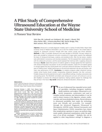
Gargani Cardiovascular Ultrasound 2011, /9/1/6Page 3 of 9“GREY”NormalMild/moderateinterstitial edemaSe ereSevereinterstitial edema/alveolar edemaConsolidationNo B-linesOne B-lineThree B-linesFive B-linesTwo B-linesAir100%0%Figure 1 Physical basis of lung ultrasound. The less air is in thelung, the easier is the detection of lung abnormalities larPara- AnterioraxillaryMidaxillaryleft sideright sideThe examination can be performed with any type of echographic platform, from fully equipped machines to pocketsize ones [15]. Patients can be in the near-supine, supineor sitting position, as clinically indicated [16]. All the chestcan be easily scanned by ultrasound, just laying the probealong the intercostal spaces. However, some specific methods have been proposed: ultrasound scanning of the anterior and lateral chest may be obtained on the right and lefthemithorax, from the second to the fourth (on the rightside to the fifth) intercostal spaces, and from the parasternal to the axillary line, as previously described [7,17]; (figure 2). Other approaches have been proposed, for instanceby Volpicelli et al. [10], with evaluation of 8 scanning sites,4 on the right and 4 on the left hemithorax. When assessing B-lines - the most informative LUS sign for the cardiologist - the sum of B-lines found on each scanning siteyields a score, denoting the extent of extravascular fluid inthe lung. In each scanning site, B-lines may be countedfrom zero to ten. Zero is defined as a complete absence ofB-lines in the investigated area; the full white screen in asingle scanning site is considered, when using a cardiacprobe, as corresponding to 10 B-lines (figure 3). Sometimes B-lines can be easily enumerated, especially if theyare a few; whereas, when they are more numerous, it isless easy to clearly enumerate them, since they tend to beconfluent. In this situation, in order to obtain a semiquantification of the sign, one can consider the percentage ofFull white screen 10 B-linesFigure 3 How to enumerate B-lines. Each hyperechogenic verticalstripe, spreading from the pleural line and extending to the edge ofthe screen, is a B-line. When using a cardiac probe, a whole whitescreen is considered as corresponding to a plateau value of 10B-lines.the scanning site occupied by B-lines (i.e. the percentageof white screen compared to black screen) and then divideit by ten (figure 3). For clinical purposes, B-lines maybe categorized from mild to severe degree, similar towhat is done for most echocardiographic parameters [16],(table 1). B-lines have a very satisfactory intraobserver andinterobserver variability, around 5% and 7%, respectively [7].Clinical applicationsHeart failureDiagnosisIn a 1994 review on the assessment of EVLW, Langestated that «The possibility to detect pulmonary edemabefore it becomes clinically apparent, is so inherentlyattractive that the effort to develop and validate suchtechnique still continues after many years of tireless andrelatively unrewarding attempts» [18]. Chest X-rayremains by far the best and most used screening test forthe detection of pulmonary edema, but it is often difficult to interpret and imprecise, and with high interobserver variability [19]. The absence of chest X-rayfindings does not exclude the presence of a high pulmonary capillary wedge pressure (PCWP) [20]. According to recent 2009 AHA/ACC guidelines, serial chest Xrays are not recommended in the assessment of pulmonary congestion in chronic heart failure (HF), sincethey are too insensitive to detect but the most extremeTable 1 Scoring of B-linesIIIIVVFigure 2 Methodology for lung ultrasound evaluation. Thoracicscanning areas for semiquantitative assessment of B-lines. (Modifiedfrom Jambrik et al, 2004 [7]).ScoreNumber of B-linesExtravascular lung water0 5No sign126 - 1516 - 30Mild degreeModerate degree3 30Severe degree(Modified from Picano et al, 2006 [16]).
Gargani Cardiovascular Ultrasound 2011, /9/1/6changes in the fluid status [21]. Direct measurement ofPCWP via catheterization is the gold standard to evaluate hemodynamic congestion, but its invasive naturelimits clinical utility. Thus, because the current technology for measuring pulmonary edema can be inaccurate(chest X-ray), cumbersome (nuclear medicine and radiology techniques), or invasive (indicator dilution), thereis great potential for a technology that could quantifypulmonary edema non-invasively in real time, with aradiation-free and portable method.B-lines have been proposed as a reliable ultrasoundtechnique for the assessment of pulmonary congestionin HF patients. The number of B-lines increases withworsening New York Heart Association (NYHA) functional class [14]. Sonographic B-lines are related toradiographic Kerley B-lines and lung water score onchest X-ray [7], to EVLW measured invasively by thethermodilution method [9], and to the severity of diastolic dysfunction, for any given level of systolic dysfunction [14]. B-lines are useful for the differential diagnosisof cardiogenic versus non-cardiogenic dyspnea. Lichtenstein et al., firstly described that B-lines could differentiate acute cardiogenic pulmonary edema fromexacerbation of chronic obstructive pulmonary disease(COPD), since B-lines were present in all patients withcardiogenic edema, whereas 24 of the 26 patients withexacerbation of COPD had no B-lines, with a sensitivityof 100% and a specificity of 92% [12]. These data werefurther confirmed by our group, as we found that Blines are reliable in predicting the cardiogenic origin ofdyspnea, with an accuracy comparable to natriureticpeptides [11]. B-lines could be a plausible alternative inacute settings where natriuretic peptide analysis is notavailable, or when there is no time enough to performit, as in patients with rapidly developing acute respiratory failure. Moreover, they could aid when natriureticpeptides levels are in the “grey zone”.B-lines are very dynamic, as shown by their rapidincrease after exercise, both in patients with and withoutleft ventricular dysfunction [22]. An “alveolar-capillarystress echo” is possible by evaluation of B-lines changesduring stress. They can be easily added to wall motionscore index and valvular heart disease assessment duringstress echocardiography, providing the additional information of the appearance of EVLW, not inferable by anyother echocardiographic parameter. The presence of Blines at peak stress can distinguish patients with stressinduced high left ventricular filling pressures but withoutfailure of the alveolar-capillary membrane (hemodynamiccongestion), from patients with stress-induced high leftventricular filling pressures and failure of the alveolarcapillary membrane, that leads to redistribution of fluidwithin the lungs (pulmonary congestion) (figure 4).Page 4 of 9Peak E/E’Peak B-linesnormalno monarycongestionFigure 4 Alveolar-capillary membrane stress echo. Theadditional value of B-lines evaluation during stressechocardiography.In less than ten years, the proposal to use B-lines toevaluate pulmonary congestion in HF patients, hasmoved from the research setting to the clinical arena,and it is now entering recommendation papers [23,24].Recently, it has been endorsed by a scientific statementby the Acute Heart Failure Committee of the Heart Failure Association of the European Society of Cardiologyas future direction for assessing and grading congestionin acute HF [24].In HF patients, LUS may also enable the detection of PE.Evaluation of PE is the more established application ofLUS [1]. The effusion should firstly be sought in dependent zones, i.e. lateral and posterior chest. In presence of aradiopacity on chest X-ray, LUS is able to better differentiate PE from atelectasis, consolidations, masses or an elevated hemidiaphragm, and can be repeated serially atbedside. LUS has a better sensitivity and reliability thanbedside chest X-ray for the diagnosis of PE [25]. Bedsidechest X-ray rarely detects small effusions and can alsomiss effusions of up to 500 mL [26]. LUS may detect theeffusion, evaluate its extension, and indicate the appropriate area for an eventual thoracentesis.TreatmentThe recognition, quantification and monitoring of pulmonary congestion is important for the clinician at allstages of care of the HF patient. Accurate assessmentof effectiveness of medical treatment is mandatory inthese patients [27]. Chest X-ray is the most usedscreening test for in-hospital follow-up of pulmonarycongestion, although showing the above mentionedlimitations. Another way to monitor congestion isthrough monitoring body weight. However, it has alimited reliability as a predictor of congestion status, asbody weight fluctuations may not always reflectchanges in intravascular volume, and weight gain mayreflect normal fluctuations in time, and weight loss
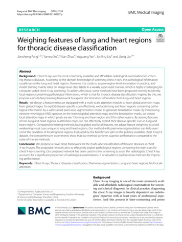
Gargani Cardiovascular Ultrasound 2011, /9/1/6due to loss of muscle/fat (cardiac cachexia) mayobscure increased fluid retention [28].B-lines have been proposed as a bedside, easy-to-use,alternative diagnostic tool for clinically monitoring pulmonary congestion in HF patients [13], as they clearafter adequate medical treatment. Since B-lines can bedissolved in a few minutes by an acute diuretic load,they may represent a useful bedside tool to monitor, ina real-time fashion, diuretic therapy response [29]. Thedynamic behaviour of B-lines is highlighted also by theirsignificant reduction after dialysis [30]. B-lines resolution occurs real-time as fluid is removed from the body[31], suggesting that this method could be easilyemployed in all situations where a dynamic evaluationof fluid changes is of importance.The simplicity and low-techology of this examinationmakes it appealing also for an out-hospital office monitoring of HF patients. Pharmacological therapy could betailored as soon as the patient, although asymptomatic,shows a significant increase in B-lines number. Thiscould, at least in theory, prevent some new hospitalizations for worsening dyspnea, since symptoms wouldappear with some days of delay [32]. The possibility toassess B-lines with light, portable, hand-held devices,could also allow the cardiologist to evaluate the degreeof decompensation at patients’ home [15].PrognosisPersistent hemodynamic congestion, that is not adequately recognised and treated before discharge, is associated with adverse clinical outcome in HF patients [27].On the other hand, post-discharge freedom of pulmonary congestion is associated with a better prognosis [33].It has been demonstrated that in patients admitted tothe hospital with dyspnea and/or chest pain, the presence of B-lines identifies a subgroup at higher risk ofexperiencing events: the higher the number of B-lines,the worse the outcome. The 16-month event-free survival showed a significantly better outcome for patientswithout B-lines, whereas a worse outcome was observedin patients with a severe degree of B-lines. In regard tofuture HF hospitalizations alone, and not as part of thecombined end-point, the rate of new hospitalization forprogression of HF was also higher in patients withsevere B-lines and lower in patients without B-lines [34].In patients with acute coronary syndromes, the numberof B-lines, associated to some very easy echocardiographicparameters of left and right cardiac function, provide aclear prognostic stratification in a composite end-pointincluding death, non-fatal myocardial infarction and newadmission for acute decompensated HF [35].From a practical point of view, B-lines assessment maybe useful at all stages of HF management: in outpatients,to monitor increasing EVLW as a sign of impendingdecompensation, that is more reliable than body weightPage 5 of 9changes; for the primary diagnosis of acute HF syndromes, in patients admitted with acute dyspnea to theEmergency Room, where even an old, low-technologyechographic device may allow B-lines detection; duringhospitalization for risk stratification and to titrate therapies, and for prognostic stratification at discharge.Acute Respiratory Distress SyndromeAcute respiratory distress syndrome (ARDS) is a common syndrome of diffuse lung injury with a high mortality rate [36]. Differential diagnosis between acutecardiogenic pulmonary edema and ARDS may often bedifficult. In ARDS, LUS may provide a very early detection of pulmonary edema [8]. LUS showed a sensitivityof 98% and a specificity of 88% in diagnosing the presence of the interstitial syndrome as seen at CT, performing better than both auscultation and chest X-ray[37]. Being a condition of pulmonary edema, althoughnon-cardiogenic, the sonographic pattern of multiple Blines is present in ARDS as well as in cardiogenic pulmonary edema. However, there are some clues that mayhelp to differentiate the two conditions, since they areoften found in ARDS, but are not present in cardiogenicpulmonary edema: alterations of the pleura, due to smallsubpleural consolidations; “spared areas”, defined asareas of normal sonographic lung appearance surrounded by areas of multiple B-lines; large consolidations of various size [38]. In patients with acutedyspnea, multiple B-lines associated to pleural alterations, represented by subpleural consolidations, arehighly suggestive of non-cardiogenic pulmonary edema(Figure 5).Although not frequently, the cardiologist may sometimes need to differentiate ARDS from cardiogenic pulmonary edema, especially in Intensive Care Unit, inAcute cardiogenicpulmonary edemaChronicheart failureALI/ARDSPulmonaryfibrosisClinical settingacutechronich iacutechronich iB-lines number / / / / B-linesdistributionmultiple, diffuse,bilateral(white lung)multiple, diffuse,bilateral, followingdecubitant regions(black and white lung)non-homogeneousdistribution, presenceof spared areasmore frequentlyposterior at lungbasisOther LUS signspleural effusionpleural effusionpleural effusion, pleuralalterations,parenchymalconsolidations ofvarious sizepleural thickeningEchocardiogramabnormalabnormallikely normallikely normalALI acute lung injury; ARDS acute respiratory distress syndrome; LUS lung ultrasound.Figure 5 How to distinguish different etiologies of interstitialsyndrome by lung ultrasound.
Gargani Cardiovascular Ultrasound 2011, /9/1/6Page 6 of 9patients after cardiothoracic surgery. Bedside chest Xrays are often very difficult to interpret, whereas LUS ismuch less affected from being performed at bedside.Moreover, LUS may be of great help in resource-limitedsettings, where an early diagnosis of ARDS can be lifesaving, as in high altitude pulmonary edema [39,40] andafter apnea diving [41].In ARDS, LUS is useful not only in the diagnosis, butalso in the follow-up: bedside LUS is able to adequatelyestimate lung recruitment induced by positive endexpiratory pressure (PEEP) [42], with a high significantcorrelation between CT and ultrasound lung reaerationscores [43].PneumothoraxPneumothorax (PTX) can occur after cardiothoracic surgery. Bedside chest X-ray may misdiagnose up to 30% ofcases [44]. Radiographically “occult” PTX may rapidlyprogress to tension PTX, if its diagnosis is missed ordelayed, especially in patients receiving mechanical ventilation [45]. Cardiologists may be able to diagnose PTXwhile performing an echocardiogram. Being a nondependent condition, in the supine patient PTX should besought at first at the least gravitationally dependentareas, progressing more lateral. Absence of lung slidingis a basic and initial step for the diagnosis [46]. Lungsliding is the dynamic horizontal movement of thepleural line, synchronized with respiration (see additional file 1). For objectifying and documenting normallung sliding, M-mode yields a simple pattern, the seashore sign (Figure 6, panel A). The presence of lung sliding allows PTX to be confidently discounted because thenegative predictive value is 100% [44]. The abolition oflung sliding can be also evaluated by M-mode, whichshows a characteristic pattern, the stratosphere signABCFigure 6 A. Normal lung pattern on M-mode: the seashore sign.The motionless superficial layers generate horizontal lines (thewaves). The deep artifacts follow the lung sliding, hence the sandypattern. B. Exclusively horizontal lines are displayed, indicatingcomplete absence of dynamics at the level of, and below, thepleural line, a pattern called the stratosphere sign. C. M-modeevaluation of the lung point: a sudden change from the seashore tothe stratosphere sign is clearly visible (arrow). (Modified fromLichtenstein et al, 2000 [48]).(Figure 6, panel B), opposed to the normal seashore sign.However, absent lung sliding does not always meanPTX. Many other situations yield abolished lung sliding,such as high-frequency ventilation, massive atelectasis,pleural adherences, severe fibrosis, etc. Another condition needed for a LUS diagnosis of PTX is the absenceof B-lines: the slightest B-line allows prompt ruling outof PTX [47]. However, the only pathognomonic LUSsign of PNX is the lung point, that allows PTX to beconfirmed, with a specificity of 100%, and sensitivity ofabout 65%. Lung point is the precise area of the chestwall, where the regular reappearance of the lung slidingreplaces the PTX pattern. It corresponds to the pointwhere visceral and parietal pleura regain contact witheach other. M-mode performed at the lung point, showsa clear change from one pattern to the other [48],(Figure 6, panel C).Acute coronary syndromesIn acute coronary syndromes, LUS should be consideredas an extension of the echocardiogram, allowing in afew minutes the evaluation of pulmonary congestion,that is often difficult to be assessed with low-qualitybedside chest X-rays. Evaluation of B-lines may alsoprovide prognostic stratification [35], and may identifysubjects at higher risk to develop acute pulmonaryedema.Pulmonary fibrosisBeing a sign of thickened pulmonary interstitium, Blines may also be present in pulmonary fibrosis [49,50].In patients with known pulmonary fibrosis or at highrisk to develop it - as in systemic sclerosis - B-lines maybe an additive tool for an early detection and semiquantification of lung involvement. When aimed to evaluatepulmonary fibrosis, LUS evaluation should be focusednot only on anterior and lateral chest, but also on posterior chest, because fibrotic accumulation often startsposteriorly at lung basis.The 2 types of B-lines - cardiogenic/watery and pneumogenic/fibrotic B-lines - can pose a challenge to differential diagnosis, although some parameters may helpdistinguish the two entities: cardiogenic B-lines arealways bilateral and are generally more diffuse on theright lung than on the left lung, with a “hot zone” ofhigher density along the axillary lines (in lying patients,as decubitant regions) [7], (Figure 5); moreover cardiogenic B-lines can be dissolved in a few hours by anacute diuretic load [13]. Within the clinical context ofacute dyspnea, B-lines changes associated with clinicalimprovement, can be safely attributed to a reduction inlung water content. Other LUS signs may also help todifferentiate the etiology of B-lines: in patients with congestive HF, both in acute and chronic settings, no
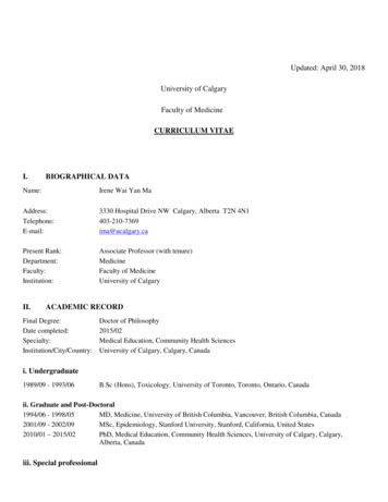
Gargani Cardiovascular Ultrasound 2011, /9/1/6pleural alterations are generally detectable, whereas inpulmonary fibrosis B-lines are often associated to an evident thickening of the pleura (Figure 5).Integrated cardio-pulmonary ultrasoundevaluationEchocardiography is an essential tool for the cardiologist, providing a huge amount of information on bothacute and chronic situations. The addition of LUS toechocardiography provides an additive insight on theeventual pulmonary involvement. The cardiopulmonarysystem is so interconnected, that an integrated approachis mandatory. Presence of multiple, diffuse, bilateral Blines associated to left ventricular systolic and/or diastolic dysfunction or valvular heart disease is highly indicative of cardiogenic pulmonary congestion (Figure 5).Moreover, for any given level of cardiac dysfunction, theresponse of the pulmonary vascular bed may be variable:LUS helps identifying those patients who, althoughasymptomatic, are going to decompensate and require amore aggressive treatment.Presence of multiple, diffuse, bilateral B-lines, associated to a normal heart, indicates a non-cardiac causeof pulmonary edema, as acute lung injury (ALI)/ARDS,interstitial pneumonia; alternatively, especially in achronic setting, it should pose the suspicion of pulmonary fibrosis (Figure 5). It is important to distinguish themultiple, diffuse, bilateral B-lines pattern from focalmultiple B-lines, that can be present in normal lungs ormay be seen around many pathologic conditions, aslobar pneumonia, pulmonary contusion, pulmonaryinfarction, pleural disease, neoplasia. This further underlines the importance to integrate LUS findings withpatients’ history, clinical presentation and other instrumental data. An overview of the main clinical applications of LUS for the cardiologist is shown in Figure 7.LUS is one of the easiest application of echography,much easier than echocardiography. Images patterns arereadily teachable, and minimal didactic and imagerecognition skill sessions are needed [51]. The learningcurve for B-lines evaluation and grading is very short[15]. The complement of LUS to echocardiographywould require only a few minutes in addition to thetime needed for a resting echocardiogram.LimitationsLUS limitations are essentially patient dependent. Obesepatients are frequently difficult to examine because ofthe thickness of their ribcage and soft tissues. The presence of subcutaneous emphysema or large thoracicdressings alters or precludes the propagation of ultrasound beams to the lung periphery.The main limitation of B-lines is the lack of specificity.As already mentioned, they are a sign of interstitialPage 7 of 9Clinical applicationsReferencesAcute dyspneaDifferential diagnosis ofcardiogenic vs nonnon-cardiogeniccardiogenicdyspnea11, 12Chronicheart failureAssessing and gradingcongestion6, 7, 9, 10, 22, 24Tailor therapy13ALI/ARDS- Early diagnosis- Differential diagnosis withcardiogenic pulmonary edema- Lung recruitment evaluation83842, 43HAPEPre-clinical detection40, 41DialysisLung fluid dynamic evaluation30, 31Acute coronarysyndromesPrognostic stratification34 3534,Stress-echoIdentification of alveolarcapillary membrane stressfailure as sign of overt heartfailure,failure22Level ofevidenceALI acute lung injury; ARDS acute respiratory distress syndrome; HAPE high altitude pulmonary edema.Green star recommendation papers, statements by scientific communitiesYellow star original papers on ISI journalsFigure 7 Overview of the main clinical applications of lungultrasound for the cardiologist.syndrome, therefore they are a very sensitive but notspecific sign of cardiogenic pulmonary edema. How todistinguish the different etiologies of B-lines has beendiscussed. However, it must be always reminded that allinstrumental data should be evaluated within the clinicalcontext and integrated with patient’s history. No singletest alone allows to establish the diagnosis.ConclusionsProviding a reliable, simple and repeatable estimation ofEVLW, B-lines assessment by LUS represents a new,helpful tool for the cardiologist, to be employed at allstages of the management of HF patients, and for thedifferential diagnosis of dyspnea. LUS can further helpin the diagnosis of other pulmonary conditions, thatmay be challenging in a Cardiology or Cardiac SurgeryDepartment. Adding LUS to echocardiography may helpto differentiate the main causes of acute dyspnea.Additional materialAdditional file 1: Sonographic pattern of the normal lung: thehorizontal hyperechoic line moving synchronously with respirationis the pleura.Additional file 2: Sonographic pattern of interstitial syndrome:multiple B-lines originate from the pleural line.AcknowledgementsThe author would like to thank dr. Eugenio Picano, who has been the firstone to introduce lung ultrasound in a Cardiology Department.Competing interestsThe author declares that they have no competing interests.
Gargani Cardiovascular Ultrasound 2011, /9/1/6Page 8 of 9Received: 27 December 2010 Accepted: 27 February 2011Published: 27 February 201121.References1. Harrison’s principles of internal medicine. New York, McGraw-Hill;, 172008.2. Lichtenstein DA: General Ultrasound in the Critically Ill. Berlin, SpringerVerlag;, II 2007.3. Ziskin MC, Thickman DI, Goldenberg NJ, Lapayowker MS, Becker JM: Thecomet tail artifact. J Ultrasound Med 1982, 1:1-7.4. Thickman DI, Ziskin MC, Goldenberg NJ, Linder BE: Clinical manifestationsof the comet tail artifact. J Ultrasound Med 1983, 2:225-30.5. Targhetta R, Chavagneux R, Balmes P, Lemerre C, Mauboussin JM,Bourgeois JM, Pourcelot L: Sonographic lung surface evaluation inpulmonary sarcoidosis: preliminary results. J Ultrasound Med 1994,13:381-8.6. Lichtenstein DA, Meziere G, Biderman P, Gepner A, Barre O: The comet-taila
lung ultrasound (LUS) may represent a useful tool for the evaluation of many pulmonary conditions in cardiovascular disease. The main application of LUS for the cardiologist is the assessment of B-lines. . from Jambrik et al, 2004 [7]). No B-lines One B-line Two B-lines Three B-lines Five B-lines Full white screen 10 B-lines Figure 3 How to .