Transcription
The RUSH Exam:Rapid Ultrasoundin SHock in theE v a l u a t i o n of th eCritically lllPhillips Perera, MD, RDMS, FACEPa,*, Thomas Mailhot, MD, RDMSb,David Riley, MD, MS, RDMSa, Diku Mandavia, MD, FACEP, FRCPCb,cKEYWORDS Rapid ultrasound in shock examination RUSH exam Shock UltrasoundCare of the patient with shock can be one of the most challenging issues in emergencymedicine. Even the most seasoned clinician, standing at the bedside of the patient inextremis, can be unclear about the cause of shock and the optimal initial therapeuticapproach. Traditional physical examination techniques can be misleading given thecomplex physiology of shock.1 Patients in shock have high mortality rates, and theserates are correlated to the amount and duration of hypotension. Therefore, diagnosisand initial care must be accurate and prompt to optimize patient outcomes.2 Failure tomake the correct diagnosis and act appropriately can lead to potentially disastrousoutcomes and high-risk situations.Ultrasound technology has been rapidly integrated into Emergency Departmentcare in the last decade. More practicing emergency physicians (EPs) are now trainedin bedside point of care or goal-directed ultrasound, and this training is now includedin all United States Accreditation Council for Graduate Medical Education EmergencyMedicine residency programs.3,4 Furthermore, the American College of EmergencyPhysicians (ACEP) has formally endorsed and embraced bedside ultrasound by theEP for multiple applications.5 This technology is ideal in the care of the critical patientin shock, and the most recent ACEP guidelines further delineate a new category ofaNew York Presbyterian Hospital, Columbia University Medical Center, Division of EmergencyMedicine, 622 West 168th Street, PH1-137, New York, NY 10032, USAbLA County1USC Medical Center, Department of Emergency Medicine, General Hospital, 1200State Street, Room 1011, Los Angeles, California 90033, USAcCedars-Sinai Medical Center, Department of Emergency Medicine, General Hospital, 1200State Street, Room 1011, Los Angeles, California 90033, USA* Corresponding author.E-mail address: pperera1@mac.com (P. Perera).Emerg Med Clin N Am 28 (2010) 29–56doi:10.1016/j.emc.2009.09.0100733-8627/09/ – see front matter ª 2010 Elsevier Inc. All rights reserved.emed.theclinics.com
30Perera et al‘‘resuscitative’’ ultrasound. Studies have demonstrated that initial integration ofbedside ultrasound into the evaluation of the patient with shock results in a more accurate initial diagnosis with an improved patient care plan.1,6,7 Instead of relying on oldertechniques, like listening for changes in sound coming from the patient’s bodysuggestive of specific pathology, bedside ultrasound now allows direct visualizationof pathology or abnormal physiological states. Thus, bedside ultrasound has becomean essential component in the evaluation of the hypotensive patient.CLASSIFICATIONS OF SHOCKMany authorities categorize shock into 4 classic subtypes.8 The first is hypovolemicshock. This condition is commonly encountered in the patient who is hemorrhagingfrom trauma, or from a nontraumatic source of brisk bleeding such as from the gastrointestinal (GI) tract or a rupturing aortic aneurysm. Hypovolemic shock may also resultfrom nonhemorrhagic conditions with extensive loss of body fluids, such as GI fluidloss from vomiting and diarrhea. The second subtype of shock is distributive shock.The classic example of this class of shock is sepsis, in which the vascular system isvasodilated to the point that the core vascular blood volume is insufficient to maintainend organ perfusion. Other examples of distributive shock include neurogenic shock,caused by a spinal cord injury, and anaphylactic shock, a severe form of allergicresponse. The third major form of shock is cardiogenic shock, resulting from pumpfailure and the inability of the heart to propel the needed oxygenated blood forwardto vital organs. Cardiogenic shock can be seen in patients with advanced cardiomyopathy, myocardial infarction, or acute valvular failure. The last type of shock isobstructive shock. This type is most commonly caused by cardiac tamponade,tension pneumothorax, or large pulmonary embolus. Many patients with obstructiveshock will need an acute intervention such as pericardiocentesis, tube thoracostomyor anticoagulation, or thrombolysis.At the bedside of a critical patient, it is often difficult to assess clinically which classification of shock best fits the patient’s current clinical status. Physical findings oftenoverlap between the subtypes. For example, patients with tamponade, cardiogenicshock and sepsis (when myocardial depression compounds this form of distributiveshock) may all present with distended neck veins and respiratory distress. Becauseof this diagnostic challenge, practitioners used to perform Swan-Ganz catheterizationin hypotensive patients, providing immediate intravascular hemodynamic data.Although the data obtained from these catheters was detailed and often helpful atthe bedside, large studies demonstrated no improvement in mortality in the patientswho received such prolonged invasive monitoring.9 Swan-Ganz catheterization hasthus declined in use, and the stage has now been set for development of a noninvasivehemodynamic assessment using point of care ultrasound.SHOCK ULTRASOUND PROTOCOL: THE RUSH EXAMGiven the advantages of early integration of bedside ultrasound into the diagnosticworkup of the patient in shock, this article outlines an easily learned and quickly performed 3-step shock ultrasound protocol. The authors term this new ultrasoundprotocol the RUSH exam (Rapid Ultrasound in SHock). This protocol involves a 3part bedside physiologic assessment simplified as:Step 1: The pumpStep 2: The tankStep 3: The pipes
The RUSH ExamThis examination is performed using standard ultrasound equipment present inmany emergency departments today. The authors recommend a phased-array transducer (3.5–5 MHz) to allow adequate thoracoabdominal intercostal scanning, anda linear array transducer (7.5–10 MHz) for the required venous examinations and forthe evaluation of pneumothorax.The first, and most crucial, step in evaluation of the patient in shock is determinationof cardiac status, termed for simplicity ‘‘the pump’’ (Table 1). Clinicians caring for thepatient in shock begin with a limited echocardiogram. The echo examination isfocused on looking for 3 main findings. First, the pericardial sac can be visualized todetermine if the patient has a pericardial effusion that may be compressing the heart,leading to a mechanical cause of obstructive shock. Second, the left ventricle can beanalyzed for global contractility. Determination of the size and contractility status ofthe left ventricle will allow for those patients with a cardiogenic cause of shock tobe rapidly identified.10,11 The third goal-directed examination of the heart focuseson determining the relative size of the left ventricle to the right ventricle. A heart thathas an increased size of the right ventricle relative to the left ventricle may be a signof acute right ventricular strain from a massive pulmonary embolus in the hypotensivepatient.12,13The second part of the RUSH shock ultrasound protocol focuses on the determination of effective intravascular volume status, which will be referred to as ‘‘the tank.’’Placement of the probe in the subxiphoid position, along both the long and shortaxis of the inferior vena cava (IVC), will allow correct determination of the size of thevessel. Looking at the respiratory dynamics of the IVC will provide an assessmentof the patient’s volume status to answer the clinical question, ‘‘how full is theTable 1Rapid Ultrasound in SHock (RUSH) protocol: ultrasonographic findings seen with classicshock statesRUSHHypovolemicEvaluation ShockCardiogenic Shock Obstructive ShockDistributive ShockHypercontractileHypercontractileheart (early sepsis)heartPericardial effusion Hypocontractileheart (late sepsis)CardiactamponadeRV strainCardiac thrombusPumpHypercontractileheartSmall chambersizeHypocontractileheartDilated heartTankFlat IVCFlat jugularveinsPeritoneal fluid(fluid loss)Pleural fluid(fluid loss)Distended IVCNormal or small IVCDistended IVC(early sepsis)Distended jugular Distended jugularveinsPeritoneal fluidveinsAbsent lung sliding(sepsis source)Lung rockets(pneumothorax) Pleural fluid (sepsis(pulmonarysource)edema)Pleural fluidPeritoneal onNormalDVTNormalAbbreviations: DVT, deep venous thrombosis; IVC, inferior vena cava; RV, right ventricle.31
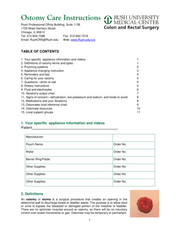
32Perera et altank?’’14,15 The clinician can also place a transducer on the internal jugular veins toview their size and changes in diameter with breathing to further assess volume.16Also included in evaluation of the tank is an assessment of the lung, pleural cavity,and abdominal cavities for pathology that could signal a compromised vascularvolume. Integration of lung ultrasound techniques can quickly allow the clinician toidentify a pneumothorax, which in the hypotensive patient may represent a tensionpneumothorax requiring immediate decompression. Tension pneumothorax presumably limits venous return into the heart due to increased pressure within the chestcavity.17,18 The lung can also be examined for ultrasonic B lines, a potential sign ofvolume overload and pulmonary edema. The clinician can further examine the thoraciccavity for a pleural effusion. Last, the clinician can perform a FAST exam (FocusedAssessment with Sonography in Trauma examination), to look for fluid in theabdomen, indicating a source for ‘‘loss of fluid from the tank.’’The third and final part of the shock ultrasound protocol is evaluation of the largearteries and veins of the body, referred to as ‘‘the pipes.’’ Clinicians should answerthe clinical question ‘‘are the pipes ruptured or obstructed’’ by first evaluating the arterial side of the vascular system to specifically examine the abdominal and thoracicaorta for an aneurysm or dissection. Next the clinician should turn to evaluation ofthe venous side of the vascular system. The femoral and popliteal veins can be examined with a high frequency linear array transducer for compressibility. Lack of fullvenous compression with direct pressure is highly suggestive of a deep venous thrombosis (DVT).19,20 Presence of a venous thrombus in the hypotensive patient may signala large pulmonary thromboembolus.RUSH Protocol: Step 1—Evaluation of the PumpFocused echocardiography is a skill that is readily learned by the EP. A smaller footprint phased-array transducer is ideal for this examination as it permits the intercostalscanning required of the heart. Imaging of the heart usually involves 4 views. The traditional views of the heart for bedside echocardiography are the parasternal long- andshort-axis views, the subxiphoid view, and the apical 4-chamber view (Fig. 1). The parasternal views are taken with the probe positioned just left of the sternum at intercostalspace 3 or 4. The subxiphoid 4-chamber view is obtained with the probe aimed uptoward the left shoulder from a position just below the subxiphoid tip of the sternum(Fig. 2). The apical 4-chamber view of the heart is best evaluated by turning the patientinto a left lateral decubitus position and placing the probe just below the nipple line atthe point of maximal impulse of the heart. It is important for the EP to know all 4 viewsFig. 1. Rapid Ultrasound in SHock (RUSH) step 1. Evaluation of the pump.
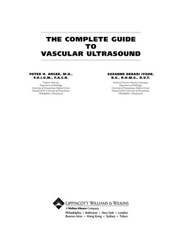
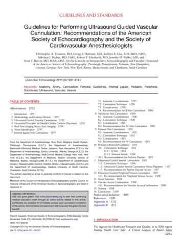
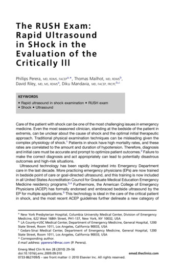
The RUSH ExamFig. 2. Subxiphoid view: cardiomyopathy with enlarged heart. LA, left atrium; LV, leftventricle; RA, right atrium; RV, right ventricle.of the heart, as some views may not be well seen in individual patients, and an alternative view may be needed to answer the clinical question at hand.‘‘Effusion around the pump’’: evaluation of the pericardiumThe first priority is to search for the presence of a pericardial effusion, which may bea cause of the patient’s hemodynamic instability. The heart should be imaged in theplanes described here, with close attention to the presence of fluid, usually appearingas a dark or anechoic area, within the pericardial space (Fig. 3). Small effusions maybe seen as a thin stripe inside the pericardial space, whereas larger effusions tend towrap circumferentially around the heart.21,22 Isolated small anterior anechoic areas onthe parasternal long-axis view often represent a pericardial fat pad, as free flowingpericardial effusions will tend to layer posteriorly and inferiorly with gravity. Fresh fluidFig. 3. Parasternal long-axis view: large pericardial effusion.33
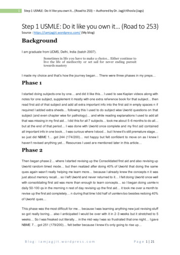
34Perera et alor blood tends to have a darker or anechoic appearance, whereas clotted blood orexudates may have a lighter or more echogenic look.Pericardial effusions can result in hemodynamic instability, due to increased pressure within the sac leading to compression of the heart. Because the pericardium isa relatively thick and fibrous structure, acute pericardial effusions may result in cardiactamponade despite only small amounts of fluid. In contrast, chronic effusions cangrow to a large volume without hemodynamic instability.23 Once a pericardial effusionis identified, the next step is to evaluate the heart for signs of tamponade. Thinking ofthe heart as a dual chamber in-line pump, the left side of the heart is under considerably more pressure, due to the high systemic pressures against which it must pump.The right side of the heart is under relatively less pressure, due to the lower pressurewithin the pulmonary vascular circuit. Thus, most echocardiographers define tamponade as compression of the right side of the heart (Fig. 4). High pressure within thepericardial sac keeps the chamber from fully expanding during the relaxation phaseof the cardiac cycle and thus is best recognized during diastole. As either chambermay be affected by the effusion, both the right atrium and right ventricle should beclosely inspected for diastolic collapse. Diastolic collapse of the right atrium or rightventricle appears as a spectrum from a subtle inward serpentine deflection of theouter wall to complete compression of a chamber.24 Whereas most pericardial effusions are free flowing in the pericardial sac, occasionally effusions may be loculated.This phenomenon is more commonly seen in patients following heart surgery, in whoma clot can form in only one area of the sac.25 In these cases, effusions can preferentially form posteriorly, and in tamponade, the left side of the heart may be compressedbefore the right side of the heart. The IVC can also be evaluated for additional confirmatory signs of tamponade.26 IVC plethora will be recognized by distention of the IVCwithout normal respiratory changes. (see later discussion on IVC in the section ‘‘Evaluation of the tank’’).Previous published studies have demonstrated that EPs, with a limited amount oftraining, can correctly and accurately identify the presence of a pericardial effusion.27Studies examining the incidence of pericardial effusions in Emergency Department orFig. 4. Subxiphoid view: cardiac tamponade.
The RUSH ExamIntensive Care patients suffering acute shortness of breath, respiratory failure, orshock have found effusions in as many as 13% of these patients.21 Another studylooked specifically at patients arriving at the Emergency Department in near-cardiacarrest states, and found a relatively large number of these cases had pericardial effusions.28 Thus, symptomatic pericardial effusions may be a cause of hemodynamicinstability in a significant number of acute patients, and EPs can quickly and accurately diagnose this condition using bedside ultrasound.As a general principle, it is easier for an EP to diagnose a pericardial effusion than toevaluate for the specific signs of tamponade.29 It is thus safer to assume tamponadephysiology in the hypotensive patient if a significant pericardial effusion is identified.Under ideal circumstances, the EP can obtain a formal echocardiogram in conjunctionwith Cardiology to specifically examine for cardiac tamponade. In the rare caseswhere there is not enough time for consultation and the patient is unstable, a pericardiocentesis under echo guidance by the EP may be life-saving. In these cases, employing bedside echocardiography also allows the EP to determine the optimalneedle insertion site for pericardiocentesis. Of note, most EPs have classically beentaught the subxiphoid approach for pericardiocentesis. However, a large reviewfrom the Mayo Clinic looked at 1127 pericardiocentesis procedures, and found thatthe optimal placement of the needle was where the distance to the effusion was theleast and the effusion size was maximal.30 The apical position at the point of maximalimpulse on the left lateral chest wall was chosen in 80% of these procedures, basedon these variables. The subxiphoid approach was only chosen in 20% of these procedures, as the investigators recognized the large distance the needle had to travelthrough the liver to enter the pericardial sac. EPs should therefore anatomically mapout the effusion before a pericardiocentesis procedure to plan the most direct andsafest route. If the apical approach is selected, the patient should optimally be rolledinto a left lateral decubitus position to bring the heart closer to the chest wall, and afterlocal anesthesia, a pericardiocentesis drainage catheter should be introduced over therib and into the pericardial sac. To maximize success and to avoid complications, thetransducer should be placed in a sterile sleeve adjacent to the needle, and the procedure performed under real-time ultrasound guidance.‘‘Squeeze of the pump’’: determination of global left ventricular functionThe next step in the RUSH protocol is to evaluate the heart for contractility of the leftventricle. This assessment will give a determination of ‘‘how strong the pump is.’’ Theexamination focuses on evaluating motion of the left ventricular endocardial walls, asjudged by a visual calculation of the percentage change from diastole to systole.Whereas in the past echocardiographers used radionuclide imaging to determineejection fraction, published studies have demonstrated that visual determination ofcontractility is roughly equivalent.31 A ventricle that has good contractility will beobserved to have a large percentage change from the 2 cycles, with the walls almostcoming together and touching during systole. As an example, a vigorously contractingventricle will almost completely obliterate the ventricular cavity during systole. Incomparison, a poorly contracting heart will have a small percentage change in themovement of the walls between diastole and systole. In these hearts, the walls willbe observed to move little during the cardiac cycle, and the heart may also be dilatedin size, especially if a long-standing cardiomyopathy with severe systolic dysfunctionis present. Motion of anterior leaflet of the mitral valve can also be used to assesscontractility. In a normal contractile state, the anterior leaflet will vigorously touchthe wall of the septum during ventricular filling when examined using the parasternallong-axis view.35
36Perera et alThe parasternal long-axis view of the heart is an excellent starting view to assessventricular contractility. Moving the probe into the parasternal short-axis orientationwill give confirmatory data on the strength of contractions. In this view, a left ventriclewith good contraction will appear as a muscular ring that squeezes down concentrically during systole. Whereas cardiologists often use the parasternal short-axis viewto evaluate for segmental wall motion abnormalities, this is a more subjectivemeasurement, and determinations may differ among different clinicians. For thatreason, it is better for the EP to initially concentrate on the overall contractility of theventricle, rather than to evaluate for segmental wall motion deficits. An easy systemof grading is to judge the strength of contractions as good, with the walls of theventricle contracting well during systole; poor, with the endocardial walls changinglittle in position from diastole to systole; and intermediate, with the walls movingwith a percentage change in between the previous 2 categories. If the parasternalviews are inadequate for these determinations, moving the patient into the left lateraldecubitus position and examining from the apical view often gives crucial data on leftventricular contractility. The subxiphoid view can be used for this determination, butthe left ventricle is farther away from the probe in this view.Published studies confirm that EPs can perform this examination and get an estimate of left ventricular contractility that compares well with that measured by a cardiologist.32 Because a large proportion of shock patients (up to 60% in one study) willhave a cardiac cause for their hypotension, this part of the examination can be veryhigh yield.10 Immediate identification of cardiogenic shock by the EP can lead tomore rapid transfer of the patient to the cardiac catheterization suite for revascularization, especially in suspected cases of cardiac ischemia.33,34 Other types of shock canbe evaluated by knowing the strength of the left ventricle during systole. Strongventricular contractility (often termed hyperdynamic, because of the strength ofcontractions of the left ventricle in addition to a rapid heart rate) is often seen in earlysepsis and in hypovolemic shock.35 In severe hypovolemic conditions, the heart isoften small in size with complete obliteration of the ventricular cavity during systole.Bedside echocardiography also allows for repeated evaluation of the patient’s heart,looking for changes in contractility over time, especially when if a patient’s status deteriorates. For example, later in the course of sepsis there may be a decrease in contractility of the left ventricle due to myocardial depression.36Knowing the strength of left ventricular contractility will give the EP a better idea ofhow much fluid ‘‘the pump’’ or heart of the patient can handle, before manifestingsigns and symptoms of fluid overload. This knowledge will serve as a critical guidefor the clinician to determine the amount of fluid that can be safely given to a patient.As an example, in a heart with poor contractility, the threshold for initiation of vasopressor agents for hemodynamic support should be lower. In contrast, sepsis patientshave been shown to benefit with aggressive early goal-directed therapy, starting withlarge amounts of fluids before use of vasopressor medications.37 Because manyEmergency Departments do not currently use the invasive catheter needed to optimally monitor the hemodynamic goals outlined for treatment of sepsis patients,bedside ultrasound gives the clinician a noninvasive means to identify and followa best management strategy.In cardiac arrest, the clinician should specifically examine for the presence orabsence of cardiac contractions. If contractions are seen, the clinician should lookfor the coordinated movements of the mitral and aortic valves. In this scenario, theabsence of coordinated opening of mitral and aortic valves will require chest compressions to maintain cardiac output. Furthermore, if after prolonged advanced cardiac life
The RUSH Examsupport resuscitation the bedside echocardiogram shows cardiac standstill, it isunlikely that the adult patient will have return of spontaneous circulation.38,39‘‘Strain of the pump’’: assessment of right ventricular strainIn the normal heart, the left ventricle is larger than the right ventricle. This aspect ispredominantly a cause of the muscular hypertrophy that takes place in the myocardium of the left ventricle after birth, with the closure of the ductus arteriosus. Theleft ventricle is under considerably more stress than the right ventricle, to meet thedemands of the higher systemic pressure, and hypertrophy is a normal compensatorymechanism. On bedside echocardiography, the normal ratio of the left to rightventricle is 1:0.6.40 The optimal cardiac views for determining this ratio of size betweenthe 2 ventricles are the parasternal long and short-axis views and the apical 4chamber view. The subxiphoid view can be used, but care must be taken to fanthrough the entire right ventricle, as it is easy to underestimate the true right ventricularsize in this view.Any condition that causes pressure to suddenly increase within the pulmonaryvascular circuit will result in acute dilation of the right heart in an effort to maintainforward flow into the pulmonary artery. The classic cause of acute right heart strainis a large central pulmonary embolus. Due to the sudden obstruction of the pulmonaryoutflow tract by a large pulmonary embolus, the right ventricle will attempt to compensate with acute dilation. This process can be seen on bedside echocardiography bya right ventricular chamber that is as large, or larger, than the left ventricle (Fig. 5).41In addition, deflection of the interventricular septum from right to left toward the leftventricle may signal higher pressures within the pulmonary artery.42 In rare cases,intracardiac thrombus may be seen floating free within the heart (Fig. 6).43 In comparison, a condition that causes a more gradual increase in pulmonary artery pressureover time, such as smaller and recurrent pulmonary emboli, cor pulmonale withpredominant right heart strain, or primary pulmonary artery hypertension, will causeboth dilation and thickening or hypertrophy of the right ventricular wall.44 These mechanisms can allow the right ventricle to compensate over time and to adapt to pumpingFig. 5. Parasternal long-axis view: right ventricular strain.37
38Perera et alFig. 6. Apical view: floating thrombus in right atrium.blood against the higher pressures in the pulmonary vascular circuit. Acute right heartstrain thus differs from chronic right heart strain in that although both conditions causedilation of the chamber, the ventricle will not have the time to hypertrophy if the timecourse is sudden.Previous published studies have looked at the sensitivity of the finding of right heartdilation in helping the clinician to diagnose a pulmonary embolus. The results showthat the sensitivity is moderate, but the specificity and positive predictive value ofthis finding are high in the correct clinical scenario, especially if hypotension ispresent.12,13,45–47 The finding of acute right heart strain due to a pulmonary emboluscorrelates with a poorer prognosis.12,48,49 This finding, in the setting of suspectedpulmonary embolus, suggests the need for immediate evaluation and treatment ofthromboembolism.50 The EP should also proceed directly to evaluation of the leg veinsfor a DVT (covered in detail later under ‘‘Evaluation of the pipes’’).The literature suggests that in general, patients with a pulmonary embolus should beimmediately started on heparin. However, a hypotensive patient with a pulmonaryembolus should be considered for thrombolysis.51,52 Bedsides, ultrasound gives thetreating clinician the clinical confidence to proceed in this more aggressive fashion.Clinical status permitting, a chest computed tomography (CT) scan using a dedicatedpulmonary embolus protocol should be obtained. If the patient is not stable enough forCT, an emergent echocardiogram in conjunction with Cardiology or bilateral duplexultrasound of the legs should be considered.RUSH Protocol Step 2: Evaluation of the Tank‘‘Fullness of the tank’’: evaluation of the inferior cava and jugular veins for size andcollapse with inspirationThe next step for the clinician using the RUSH protocol in the hypotensive patient is toevaluate the effective intravascular volume as well as to look for areas where the intravascular volume might be compromised (Fig. 7). An estimate of the intravascularvolume can be determined noninvasively by looking initially at the IVC.14,15 The ultrasound transducer should be positioned in the epigastric area in a long-axis configuration along the IVC as it runs from the abdomen into the heart. A good way of obtainingthis image is to first examine the heart in the subxiphoid 4-chamber plane and then tomove the probe into the subxiphoid 2-chamber plane, with the probe marker oriented
The RUSH ExamFig. 7. RUSH step 2. Evaluation of the tank. IVC exam, inferior vena cava; FAST views(Focused Sonography in Trauma), right upper quadrant, left upper quadrant and suprapubic; lung exam, pneumothorax and pulmonary edema.anteriorly. The aorta will often come quickly into view from this plane as a thickerwalled and deeper structure. Moving the probe to the patient’s right will bring theIVC into view, running longitudinally adjacent to the aorta. The IVC should be examined at the junction of the right atrium and the cava and followed 2 to 3 cm caudallyalong the vessel. Moving the probe into short axis to further evaluate the IVC iscomplementary as it will display the structure as an oval and allow confirmation ofsize by avoiding underestimation of the structure with the cylinder effect. As thepatient breathes, the IVC will have a normal pattern of collapse during inspiration,due to the negative pressure generated in the chest, causing increased flow fromthe abdominal to the thoracic cavity (Fig. 8). This respiratory variation can be furtheraugmented by having the patient sniff or inspire forcefully. M-mode Doppler, positioned on the IVC, can graphically document the dynamic changes in the vessel caliberduring the patient’s respiratory cycle (Fig. 9).Previous studies have demonstrated a correlation between the size and percentagechange of the IVC with respiratory variation to central venous pressure (CVP) using anindwelling catheter. A smaller caliber IVC ( 2 cm diameter) with an inspiratory collapseFig. 8. Inferior vena cava sniff test: low cardiac filling pressures.39
40Perer
SHOCK ULTRASOUND PROTOCOL: THE RUSH EXAM Given the advantages of early integration of bedside ultrasound into the diagnostic workup of the patient in shock, this article outlines an easily learned and quickly per-formed 3-step shock ultrasound protocol. The authors term this new ultrasound protocol the RUSH exam (Rapid Ultrasound in SHock).