Transcription
H. DevlinOperative Dentistry
Hugh DevlinOperativeDentistryA Practical Guideto Recent InnovationsWith 75 Figures in 102 Separate Illustrations and 5 Tables123
Dr. Hugh DevlinSchool of DentistryThe University of ManchesterHigher Cambridge StreetManchesterM15 6FHUnited KingdomLibrary of Congress Control Number: 2005939045ISBN-10 3-540-29616-6 Springer Berlin Heidelberg New YorkISBN-13 978-3-540-29616-4 Springer Berlin Heidelberg New YorkThis work is subject to copyright. All rights reserved, whether the whole or part of the material isconcerned, specifically the rights of translation, reprinting, reuse of illustrations, recitation, broadcasting, reproduction on microfilm or in any other way, and storage in data banks. Duplication of thispublication or parts thereof is permitted only under the provisions of the German Copyright Lawof September 9, 1965, in its current version, and permission for use must always be obtained fromSpringer. Violations are liable for prosecution under the German Copyright Law.Springer is a part of Springer Science Business Mediaspringer.com Springer-Verlag Berlin Heidelberg 2006Printed in GermanyThe use of general descriptive names, registered names, trademarks, etc. in this publication does notimply, even in the absence of a specific statement, that such names are exempt from the relevantprotective laws and regulations and therefore free for general use.Product liability: The publishers cannot guarantee the accuracy of any information about dosage andapplication contained in this book. In every individual case the user must check such information byconsulting the relevant literature.Editor: Gabriele Schröder, HeidelbergDesk Editor: Martina Himberger, HeidelbergCover design: Frido Steinen-Broo, eStudio Calamar, SpainTypesetting and production: LE-TEX Jelonek, Schmidt & Vöckler GbR, Leipzig, GermanyPrinted on acid-free paper 24/3100/YL - 5 4 3 2 1 0
PrefaceThis book embraces the most recent developments in modern operative dentistry, but has attempted to merge these with traditional practice. Students,colleagues, and general dental practitioners have requested an evidence-basedapproach to the practical concepts in modern restorative dentistry. One important philosophy that is emphasized in this book is that the prevention ofdental caries, restoration failure, and periodontal disease should be the basis of all operative dentistry. Recent developments in restoration design andmaterial science technology are also assessed in the light of the best availableevidence, which is referred to in the text. Innovative instrument design isdescribed and useful practical techniques are explained.The worldwide use of amalgam will continue to decline as patients demand better aesthetic restorations. For this reason, posterior resin-compositerestorations, ceramic inlay/onlay restorations, and the new high-strengthporcelain crown systems are given considerable prominence in this book.The new adhesive technologies are especially useful in the treatment of tootherosion that may have resulted from the consumption of carbonated beverages.This is a medium-sized textbook that should be used in conjunction withlarger reference texts, journal reviews, and other publications. It should complement other books in the field and will hopefully stimulate further reading.I am indebted to my friends and colleagues who generously provided illustrations. Dr. David Reekie provided the photograph in Fig. 2.15, Dr. CatherinePotter provided those in Figs. 2.4 and 2.5, Dr. Ian Pretty provided thosein Figs. 1.5–1.7, and Dr. Peter Geertsema, whose excellent standard of dental treatment is acknowledged throughout Europe, provided all of the photographs in Figs. 5.11–5.14. Their generous assistance is gratefully acknowledged.
Contents1.11.1.11.1.21.1.31.1.41.1.51.1.61.1.71New Methods of Detection of Caries . . . .The Diagnosis of Caries . . . . . . . . . . . .DIAGNOdent . . . . . . . . . . . . . . . . . .Digital Imaging Fiber-Optic TransilluminationFiber-Optic Transillumination . . . . . . . . .Quantitative Light-Induced Fluorescence . . .Radiology of Dental Caries . . . . . . . . . . .Electrical Conductance . . . . . . . . . . . . .Modern Caries Detection and Management .References . . . . . . . . . . . . . . . . . . . 2.3.22.3.3New Developments in Caries Removal and Restoration .Caries Removal . . . . . . . . . . . . . . . . . . . . . . . .Lasers . . . . . . . . . . . . . . . . . . . . . . . . . . . . .Polymer Bur . . . . . . . . . . . . . . . . . . . . . . . . .Micropreparation Burs . . . . . . . . . . . . . . . . . . . .Air Abrasion (or Kinetic Cavity Preparation) . . . . . . . .Photoactivated Disinfection . . . . . . . . . . . . . . . . .Carisolv Gel . . . . . . . . . . . . . . . . . . . . . . . . . .Atraumatic Restorative Treatment . . . . . . . . . . . . . .Caries-Detector Dyes . . . . . . . . . . . . . . . . . . . . .Restoration Following Caries Detection . . . . . . . . . . .Why Are Teeth Restored? . . . . . . . . . . . . . . . . . . .Caries as a Disease . . . . . . . . . . . . . . . . . . . . . .Preventing Dental Caries . . . . . . . . . . . . . . . . . . .When Should Caries Be Restored? . . . . . . . . . . . . . .Fissure Sealants . . . . . . . . . . . . . . . . . . . . . . . .Ozone Therapy for the Treatment of Caries . . . . . . . . .Restorative Procedures . . . . . . . . . . . . . . . . . . . .The “Tunnel” Restoration . . . . . . . . . . . . . . . . . .The Proximal “Slot” Preparation . . . . . . . . . . . . . .Traditional Cavity Preparation . . . . . . . . . . . . . . . .171718202021232324252626272830323234343435.
3.112.3.12The Repaired Amalgam Restoration . . . . . . . . . . .Cavity Preparations Involving Three or More Surfaces .Treatment of the Large Carious Lesion . . . . . . . . .The Use of Calcium Hydroxide in Direct Pulp CappingThe Foundation Restoration . . . . . . . . . . . . . . .Practical Aspects of Amalgam Retention . . . . . . . .Pins vs Bonded Restorations . . . . . . . . . . . . . . .Amalgam Bonding Procedure . . . . . . . . . . . . . .The Use of Base Materials . . . . . . . . . . . . . . . .References . . . . . . . . . . . . . . . . . . . . . . . . .103.11Posterior Resin Composite Restorations . . . . . . .Ramped Curing Lights . . . . . . . . . . . . . . . . . .Ceramic Inserts . . . . . . . . . . . . . . . . . . . . . .Nanotechnology . . . . . . . . . . . . . . . . . . . . .“Total Etch” Technique . . . . . . . . . . . . . . . . . .Fissure Sealants . . . . . . . . . . . . . . . . . . . . . .Preventive Resin Restorations . . . . . . . . . . . . . .Minimal Class II Restorations . . . . . . . . . . . . . .Posterior Composite Resin Restoration . . . . . . . . .Direct Composite Resin Restorations . . . . . . . . . .Studies of Direct Resin-Composite Restoration SurvivalReasons for Failure of Extensive Direct CompositeResin Restorations . . . . . . . . . . . . . . . . . . . .The “Sandwich” Technique . . . . . . . . . . . . . . .Packable Composite Resin Materials . . . . . . . . . .New Developments in Resin-Composite Technology . .References . . . . . . . . . . . . . . . . . . . . . . . . .5152525454555657575860.6062626464The Single Crown, Veneers, and Bleaching . . . . . .The Single Crown . . . . . . . . . . . . . . . . . . . . .Recurrent Caries and Periodontal Disease . . . . . . .The Tooth Becomes Nonvital . . . . . . . . . . . . . .The Crown Restoration Becomes Loose . . . . . . . . .Perforation of the Crown During Occlusal Adjustment .The Appearance of the Crown is Unsatisfactory . . . .Shade of the Crown . . . . . . . . . . . . . . . . . . . .Shape of the Crown . . . . . . . . . . . . . . . . . . . .Gingival Contour . . . . . . . . . . . . . . . . . . . . .Gingival Recession . . . . . . . . . . . . . . . . . . . .New Developments in Crown Provision . . . . . . . . 24.1.34.1.44.1.54.1.5.14.1.5.24.1.5.34.1.5.44.2
35.3.6.45.45.55.65.6.15.6.1.1IXVeneers . . . . . . . . . . . . . . . . . . . . .Tooth Preparation . . . . . . . . . . . . . . .Disadvantages of Veneers . . . . . . . . . . .Failure of Veneers . . . . . . . . . . . . . . . .Cementation Procedures for a Veneer . . . . .Provisional Restorations for Veneers . . . . .Resin-Bonded All-Ceramic Crowns(or “Dentin-Bonded Crown”) . . . . . . . . .Marginal Leakage . . . . . . . . . . . . . . . .Cementation Procedures for the Resin-BondedAll-Ceramic Crown . . . . . . . . . . . . . . .Bleaching of Teeth . . . . . . . . . . . . . . .Cervical Resorption . . . . . . . . . . . . . .The “Walking Bleach” Technique . . . . . . .Vital Tooth Bleaching . . . . . . . . . . . . . .In-House Tooth Bleaching . . . . . . . . . . .Microabrasion . . . . . . . . . . . . . . . . .References . . . . . . . . . . . . . . . . . . . .797981818383. . . . . . . . . . . . . . . . .8486.8687878889909092Noncarious Tooth Tissue Loss . . . . . . . . . . . . . .Noncarious Tooth Wear . . . . . . . . . . . . . . . . . .Clinical Appearance of Erosion . . . . . . . . . . . . . .Clinical Appearance of Attrition . . . . . . . . . . . . . .Clinical Appearance of Abrasion . . . . . . . . . . . . . .Prevention of Toothwear . . . . . . . . . . . . . . . . . .Recent Developments in the Treatment of Tooth Wear . .Noncarious Cervical Restorations . . . . . . . . . . . . .Clinical Procedures for Restoration of Cervical Lesions .Why Do Cervical Restorations Fail? . . . . . . . . . . . .New Developments in Direct Posterior Resin CompositesAddition of Resin Composite to Anterior Teeth . . . . . .Developments in Indirect Resin Composite Technology .Targis/Vectris Crowns . . . . . . . . . . . . . . . . . . .Sinfony . . . . . . . . . . . . . . . . . . . . . . . . . . .Belleglass HP . . . . . . . . . . . . . . . . . . . . . . . .Other Fiber Systems . . . . . . . . . . . . . . . . . . . .Ceramic Inlay and Onlay Restorations . . . . . . . . . .Inlay Restorations . . . . . . . . . . . . . . . . . . . . .Onlay Restorations . . . . . . . . . . . . . . . . . . . . .Milled Ceramic Inlays or Onlays . . . . . . . . . . . . . .Cerec 3 . . . . . . . . . . . . . . . . . . . . . . . . . . 08109111111
X5.6.1.25.6.1.35.75.7.15.7.25.85.95.10ContentsIPS Empress System . . . . . . . . . . .Fortress . . . . . . . . . . . . . . . . . .Full-Veneer Posterior Porcelain Crowns .In-Ceram . . . . . . . . . . . . . . . . .Procera AllCeram Crowns . . . . . . . .Cementation of the Restoration . . . . .Choosing the Correct Restorative SystemConclusion . . . . . . . . . . . . . . . .References . . . . . . . . . . . . . . . . .112113115115116117118119119Subject Index . . . . . . . . . . . . . . . . . . . . . . . . . . . . . . . 123
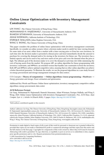
1 New Methods of Detection of Caries1.1The Diagnosis of CariesSimply looking at a tooth to determine whether caries is present is an inaccurate technique, although the exact sensitivity and specificity depends uponthe experience of the dentist (Huysmans et al. 1998). The diagnosis of cariesis one of the most difficult clinical assessments that the dentist must perform(Fig. 1.1a,b). For the best results, the teeth should be dried, and when goodillumination is used a carious occlusal lesion affecting the outer half of theenamel will appear white and opaque. The anatomy of the occlusal fissure isoften invaginated to form an expanded hidden chamber that is easily colonized by bacteria and then can become carious. However, when the walls ofthe fissure have incipient caries, the lesion is easily missed by the examiningdentist. Where the occlusal demineralization progresses to affect the outerthird of the dentin, an obvious white-spot lesion is visible without drying thesurface. Frank cavitation of the enamel surface occurs usually when the innerhalf of the dentin has undergone demineralization and is accompanied bysoftening of the outer dentin (Ekstrand et al. 1998). When cavitation of thetooth surface occurs, plaque removal by the patient becomes impossible andprogression of the lesion is inevitable. Caries progresses further by spreading along the enamel-dentin junction and undermining the overlying enamel(Fig. 1.2). Caries is a dynamic process that involves alternating periods ofdissolution of tooth mineral and its reformation, depending on the acidityof the plaque environment. A radiopaque band is often seen pulpal to thecarious lesion and results from the reprecipitation of calcium and phosphatepreviously dissolved by the carious process.Due to the reprecipitation of calcium and phosphate, the hardness ofcarious dentin increases to a maximum at a point a few millimeters awayfrom the soft surface dentin This is seen if a carious tooth is sectioned andhardness measurements are made at intervals from the carious surface to thenormal, unaffected dentin (Fig. 1.3). In the experiment shown in Fig. 1.3, the
21 New Methods of Detection of CariesFig. 1.1 (a) Caries is one of the most difficult diseases to diagnose. (b) Deep dentinalcaries beneath an intact enamel surface can often be invisible to
approach to the practical concepts in modern restorative dentistry. One im-portant philosophy that is emphasized in this book is that the prevention of dental caries, restoration failure, and periodontal disease should be the ba-sis of all operative dentistry. Recent developments in restoration design and