Transcription
SEER Program Coding and Staging Manual 2022Coding GuidelinesBladderC670–C679ReportabilityDo not report bladder cancer based on UroVysion test results alone. Report the case if there is aphysician statement of malignancy and/or the patient was treated for cancer.Not reportablePapillary urothelial neoplasms of low malignant potential (PUNLMPs)The WHO classification categorizes "PUNLMP" as borderline, 8130/1. The definition is "apapillary urothelial tumor which resembles the exophytic urothelial papilloma, but showsincreased cellular proliferation exceeding the thickness of normal urothelium." Thehistopathologic description is "the papillae of PUNLMP are discrete, slender and not fusedand are lined by multilayered urothelium with minimal to absent cytologic atypia .Mitosesare rare and have a basal location."Papilloma of bladderThe WHO classification categorizes "urothelial papilloma" as benign, 8120/0. The definitionis "composed of a delicate fibrovascular core covered by urothelium indistinguishable fromthat of normal urothelium." The histopathologic description is "characterized by discretepapillary fronds with occasional branching the epithelium lacks atypia mitoses are absentto rare and, if present, are basal in location and not abnormal. The lesions are often small andoccasionally show concomitant inverted growth pattern. Rarely, papilloma may showextensive involvement of the mucosa."Primary SiteC670 Trigone of bladderBase of bladderFloorBelow interureteric ridge* (interureteric crest or interureteric fold)C671 Dome of bladderVertexRoofVaultC672 Lateral wall of bladderRight wallLeft wallLateral to ureteral orificeSidewallC673 Anterior wall of bladderC674 Posterior wall of bladderAppendix C: Coding Guidelines1
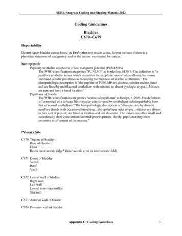
SEER Program Coding and Staging Manual 2022C675 Bladder neckVesical neckInternal urethral orificeInternal urethral/uretero orificeC676 Ureteric orificeJust above ureteric orificeC677 UrachusMid umbilical ligamentUrachal remnantC678 Overlapping lesion of bladderLateral-posterior wall (hyphen)FundusC679 Bladder, NOSLateral posterior wall (no hyphen)* The interureteric ridge is a fold of mucous membrane extending across the bladder between theureteric orifices and forms one of the boundaries for the trigone of the bladder.Bladder Anatomy and ICD-O-3Source: UICC TNM Atlas, 3rd edition, 2nd revisionAppendix C: Coding Guidelines2
SEER Program Coding and Staging Manual 2022Priority Order for Coding SubsitesUse the information from reports in the following priority order to code a subsite when the medical recordcontains conflicting information:Operative report (TURB)Pathology reportMultifocal TumorsAssign site code C679 when there are multifocal tumors all of the same behavior in more than one subsiteof the bladder and the specific subsite of origin is not known.If the TURB or pathology proves invasive tumor in one subsite and in situ tumor in all other involvedsubsites, code to the subsite involved with invasive tumor.Appendix C: Coding Guidelines3
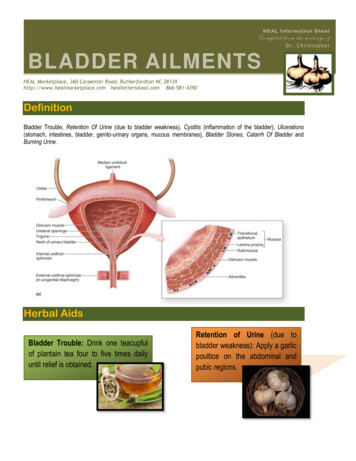
SEER Program Coding and Staging Manual 2022Bladder Wall PathologyThe bladder wall is composed of three layers. There may be “sub layers” within the major layer of thebladder.Bladder LayerMucosaSub LayerSynonymsEpithelium, transitionalepithelium, urothelium,mucosal surface,transitional mucosaBasementmembraneSubmucosaLamina propriaMuscleBladder wallSubmucous coat,lamina propria, areolarconnective tissueStagingNo blood vessels,in situ/noninvasiveNo invasion ofbasementmembrane is insituInvasion/penetration of basementmembrane onnective tissue,subepithelial tissue,stroma, muscularismucosa, transitionalepitheliumMuscularis, muscularis Invasivepropria, muscularisexterna, smooth muscleDescriptionFirst layer oninside ofbladder; Linesbladder, ureters,and urethraSingle layer ofcells that liesbeneath themucosal layerseparating theepithelial layerfrom the laminapropriaAreolarconnective tissueinterlaced withthe muscularcoat. Containsblood vessels,nerves, and insome regions,glandsTumor extends through the bladder wall (invades regional tissue) when the tumor is stated to involve oneof the following areas.Serosa (Tunica serosa): The outermost serous coat is a reflection of the peritoneum that covers thesuperior surface and the upper parts of the lateral surfaces of the urinary bladder. The serosa is partof visceral peritoneum. The serosa is reflected from these bladder surfaces onto the abdominal andpelvic walls.Perivesical fatAdventitia: Some areas of the bladder do not have a serosa. Where there is no serosa, the connectivetissue of surrounding structures merges with the connective tissue of the bladder and is calledadventitia.Appendix C: Coding Guidelines4
SEER Program Coding and Staging Manual 2022Histology 1Over 90% of bladder cancers are urothelial (transitional) cell carcinomas, derived from the uroepithelium.Other types include squamous cell carcinoma (about 2% to 7%) and adenocarcinoma (about 2%).Adenocarcinomas may be of urachal origin or nonurachal origin, with the nonurachal type generallythought to arise from metaplasia of chronically irritated transitional epithelium. Small cell carcinoma, andrarely sarcoma, can also occur. Childhood rhabdomyosarcoma, a type of sarcoma, can form in muscletissue of the bladder.Behavior CodeEspecially for bladder, the behavior code and the stage may not match. Different rules apply to assigningbehavior code compared to assigning stage. Use the rules as they apply to each data item, do not mixinstructions from one data item to another.Code the behavior as malignant (/3), not in situ (/2), when The diagnosis is high grade urothelial carcinoma AND there is no information regarding invasionOR The pathology report says the submucosa is invaded with tumorOR The only surgery performed is a transurethral resection of the bladder (TURB) documenting that depthof invasion cannot be measured because there is no muscle in the specimen ANDo There is no information regarding invasion and the physician’s TNM designation is not availableORo The pathology report does not mention whether the submucosa is free of tumor or has beeninvadedCode the behavior as in situ (/2) when The diagnosis is low grade urothelial carcinoma AND there is no information regarding invasionOR The pathology report says the submucosa is free of adder-treatment-pdq# 33Appendix C: Coding Guidelines5
SEER Program Coding and Staging Manual 2022 The only surgery performed is a transurethral resection of the bladder (TURB) documenting that depthof invasion cannot be measured because there is no muscle in the specimen ANDoThe TNM designation is Ta or Tis for TURB with no muscle in the specimenOR The pathology report includes terminology using terms such asNon-invasiveNo stromal invasion identifiedNo invasion identifiedNo lamina propria invasion identifiedExtent of invasion: AbsentFirst Course TreatmentBCGCode BCG as both surgery and immunotherapy. See the SEER manual, Appendix C, Bladder SurgeryCodes, SEER Note under code 16.Treatment Modalities (most common treatments)TURB with fulgurationTURB with fulguration followed by intravesical BCG (bacillus Calmette-Guerin) is usually used forpatients with multiple tumors or for high-risk patients.TURB with fulguration followed by intravesical chemotherapyPhotodynamic therapy (PDT) using laser light and chemotherapySegmental cystectomy (rare)Radical cystectomy in patients with extensive or refractory superficial tumorInternal irradiation (needles, seeds, wires, or catheters placed into or near the tumor) with or withoutexternal-beam irradiationChemotherapyImmunotherapy/biologic therapyAppendix C: Coding Guidelines6
SEER Program Coding and Staging Manual 2022 Appendix C: Coding Guidelines 6 The only surgery performed is a transurethral resection of the bladder (TURB) documenting that depth of invasion cannot be measured because there is no muscle in the specimen AND. o The TNM designation is Ta or Tis for TURB with no muscle in the specimen . OR