Transcription
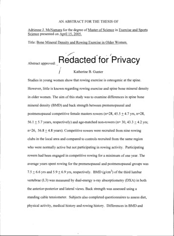
AN ABSTRACT FOR THE THESIS OFAdrienne J. McNamara for the degree of Master of Science in Exercise and SportsScience presented on April 15, 2005.Title: Bone Mineral Density and Rowing Exercise in Older Women.Abstract approved:Redacted for PrivacyKatherine B. GunterStudies in young women show that rowing exercise is osteogenic at the spine.However, little is known regarding rowing exercise and spine bone mineral densityin older women. The aim of this study was to examine differences in spine bonemineral density (BMD) and back strength between premenopausal andpostmenopausal competitive female masters rowers (n 28, 45.5 4.7 yrs, n 28,56.1 5.7 years, respectively) and age-matched non-rowers (n 30, 43.3 4.2 yrs;n 26, 56.8 4.8 years). Competitive rowers were recruited from nine rowingclubs in the local area and compared to controls recruited from the same regionwho were normally active but not participating in rowing activity. Participatingrowers had been engaged in competitive rowing for a minimum of one year. Theaverage years spent rowing for the premenopausal and postmenopausal groups was7.5 6.6 yrs and 5.9 6.9 yrs, respectively. BMD (g/cm2) of the third lumbarvertebrae (L3) was measured by dual-energy x-ray absorptiometry (DXA) in boththe anterior-posterior and lateral views. Back strength was assessed using astanding cable tensiometer. Subjects also completed questionnaires to assess diet,physical activity, medical history and rowing history. Differences in BMD and
back strength between groups were determined by analysis of covariance,controlling for lean mass. Compared to controls, postmenopausal rowers had3.2% higher BMD at the anterior-posterior spine (p .02) and 4.4% higher lateralspine BMD (p .04). Furthermore, isometric back strength was 22.6% greater inthese rowers than controls (p .0 1). In contrast, controls had higher lateral BMDthan rowers, with no differences in AP spine BMD or back strength between thepremenopausal rowers and controls. Back strength was a significant predictor ofAP spine BMD in premenopasual rowers and controls (R2 0.137, p 0.004) andof lateral spine BMD in postmenopausal rowers only (R2 0.153, pO.O4). Therewere no differences in calcium intake, age, menopausal status, weight, or leanmass between rowers and controls in either the premenopausal or postmenopausalsamples. Since both increased BMD and back strength are associated withreductions in vertebral fracture risk, our results suggest that rowing exercise maybe an important strategy to promote bone health and reduce vertebral fracture riskin postmenopausal women. However, the forces applied in rowing may not begreat enough to alter bone mass before the onset of menopause. Therefore moreresearch is needed examining rowing exercise in these older populations.
Copyright by Adrienne J. MeNamaraApril 15, 2005All Rights Reserved
Bone Mineral Density and Rowing Exercise in Older WomenbyAdrienne J. McNamaraA THESISsubmitted toOregon State Universityin partial fulfillment ofthe requirement for thedegree ofMaster of SciencePresented April 15, 2005Commencement June 2005
Master of Science thesis of Adrienne J. McNamara presented on April 15, 2005.APPROVED:Redacted for Privacyjor Professor, representing Exercise and Sports ScienceRedacted for PrivacyChair of the Detj'tiof Exercise and Sports ScienceRedacted for PrivacyDean of th'e)Graduate SchoolI understand that my thesis will become part of the permanent collection of OregonState University libraries. My signature below authorized release of my thesis toany reader upon request.Redacted for PrivacyAdrietme J. McNamara, Author
ACKNOWLEDGEMENTSI have been truly blessed these past years to have the wonderful support of somany people that have helped me get to this point. I want to express especialgratitude to the following people:To Kathy, thank you so much for all of your constant help and guidance. Frominitially teaching me about bone in class, to taking me as your student, to all of theplanning of the study and editing of drafts, you have really made this experienceenjoyable. Thank you for always pushing me to do better.To the bone lab, I truly enjoyed working and learning with all of you. This hasbeen a great group to be a part of.To Kevin, thank you for believing in me and supporting me in everything that Idecide to do. Everything in life is better and easier by having you to share it with.To my family, thank you for your support and interest in what I do.Above all, glory to God who has given me my passions and the opportunities topursue them and who has blessed my life tremendously.
CONTRIBUTION OF AUTHORSDr. Katherine B. Gunter was instrumental in the design and planning of thisresearch. In addition she provided great help in the editing of the two manuscripts.
TABLE OF CONTENTSCHAPTER ONE: INTRODUCTION.1BACKGROUND . 1PURPOSE . 19RESEARCH QUESTIONS AND HYPOTHESIS . 20CHAPTER TWO: INCREASED SPINE BONE MINERAL DENSITY INPOSTMENOPAUSAL ROWERS . 23ABSTRACT . 24INTRODUCTION . 26METHODS. 29RESULTS . 33DISCUSSION . 36REFERENCES . 41CHAPTER THREE: BONE MINERAL DENSITY AND ROWING EXERCISEIN PREMENOPAUSAL WOMEN . 46ABSTRACT . 47INTRODUCTION . 49METHODS . 52RESULTS . 56DISCUSSION . 59REFERENCES . 64CHAPTER FOUR: CONCLUSIONS . 70BIBLIOGRAPHY . 73APPENDICIES . 79
LISTS OF TABLESTable2.1Subject Characteristics by Group . 342.2Bone Mineral Density and Performance Differences3.1Subject Characteristics by Group3.2Bone Mineral Density and Performance Differences. 35. 57. 58
LIST OF APPENDICIESAppendixA. Informed Consent80B. Health History Questionnaire85C. Aerobics Center Longitudinal Physical Activity Questionnaire 90D. Food Frequency Questionnaire93E. Rowing History Questionnaire101F. Spine Loading Questionnaire104
Bone Mineral Density and Rowing Exercise in Older WomenCHAPTER ONE: INTRODUCTIONBACKGROUNDOnce regarded as a normal part of the aging process, osteoporosis is nowconsidered one of the leading causes of morbidity and mortality in Americansociety. Currently over 10 million Americans are osteoporotic, with 18 millionmore at risk due to low bone mass. Osteoporosis and related fractures significantlyimpact quality of life and carry a financial burden of over 14 billion dollarsannually (NIH consensus panel, 2001). Among the consequences of osteoporosis,vertebral fractures predominate all osteoporotic fractures, occur earlier in life thanhip fractures and may rival hip fractures in terms of morbidity and debilitation(Gold et al, 1999; Ross, Santura, Yates, 2001). Women are at the greatest risk ofthese fractures due to the accelerated bone loss of up to 5% per year in the yearsfollowing menopause resulting from the cessation of estrogen production. Thoughit is possible to supplement estrogen through hormone replacement therapy (HRT),recent evidence has shown there are significant cardiovascular and cancer risksassociated with the treatment (Humphries & Gill, 2003). Thus, fewer women arewilling to use HRT, despite its effectiveness in the prevention of bone loss.Therefore a great need exists to find viable alternatives to HRT for the preventionof bone loss and subsequent fractures.
2Resistance and Impact ExerciseExercise appears to be an effective intervention for the preservation ofbone mass, as long as the stimulus at the site of interest is sufficient to produce anoverload (Myburgh, 1993). Additionally, falls, which are a common stimulus forfracture in the elderly, can also be reduced through exercise. While impactexercises such as jumping and running seem necessary to produce changes in boneat the hip (Snow, Shaw, Winter, Witzke, 2000; Bassey, Rothwell, Littlewood, Pye,1998; Fuchs, Bauer, Snow 2001), the spine appears to adapt to non-impactresistance exercise. For example, Kohrt, Ehsani and Birge (1997) conducted an 11month intervention examining the effects of different types of exercise on bone.Thirty nine sedentary women (age 60-74), not taking HRT were assigned to one ofthree groups: a) exercises involving predominately ground reaction forces (GRF)(such as running; walking, and stairs), b) exercises consisting of predominatelyjoint reaction forces (JRF) (such as weight lifting and rowing) or c) a no exercisecontrol group. Both exercise groups performed specific supervised exercises 3-5days a week for nine months, after a two month lead-in phase involving activitiesto increase flexibility and range of motion. Participants in the GRF group walked30-45 minutes at 60-85% maximal heart rate (duration and intensity progressedthrough these ranges for the duration of the study) and were encouraged to jog asmuch as possible. Stair climbing was added after the third month. Participants inthe JRF program spent half of each session rowing (up to three 10 minutes boutson a rowing ergometer at 80-85% maximal heart rate) and the remainder of thesession weight training (2-3 sets of standing free weight exercise at an intensity
resulting in fatigue after 8-12 repetitions). Bone mineral density (BMD) of thewhole body, lumbar spine, proximal femur and distal forearm were assessed atbaseline and then in 3 month intervals throughout the study. The change at thespine was 1.5 0.7% and 1.8 0.5% in the GRF and JRF groups respectively.Changes in whole body BMD were also similar between groups. Although onlythe GRF group increased femoral neck BMD, increases in lower body musclestrength were greater in response to the JRF program than to the GRF program(15 5% vs. 9 4% respectively). Additionally, only the JRF group increased fatfree mass. Therefore, despite the absence of change in hip BMD, the outcomesspecific to the JRF group are important factors for fall reduction and subsequentfracture prevention.Numerous other studies have also used resistance training to elicit boneadaptations in adult populations (Kerr, Ackland, Maslen, Morton, Prince, 2001;Vincent & Braith, 2002; Pruitt, Jackson, Bartels, Lehnhard, 1992; Dornemann,McMurray, Renner, Anderson, 1997; Maddalozzo & Snow, 2000; Smidt, Lin,O'dwyer, Blandpied, 1991; Revel, Mayoux-Benhamjou, Rabourdin, Bagheri,Roux, 1993). Pruitt el a! (1992) examined the effects of a nine month resistancetraining intervention on 17 early postmenopausal women (1-7 years sincemenopause onset; age 54-57) with no history of resistance training. The exerciseprotocol included three, one hour weight training sessions per week, whereexercisers performed one set (10-12 repetitions) of each exercise designed to targeteither the trunk (trunk extension, hip extension, lateral flexion), lower extremities(leg press, leg ab/adduction, leg curl, leg extension), or upper extremities (biceps
curl, lat puildown, bench press, wrist roller). All exercises were performed at 60%of the subject's one repetition maximum (1-RM). The control group (n10) wasinstructed to maintain their average daily activity. Bone analysis showed that thechange in spine BMD of the weight trained group was significantly different fromthat of the control group (1.6 1.2% vs. -3.6 1.5% respectively), with nosignificant differences at any other bone site. This study shows the potential ofresistance training to maintain and even increase spine BMD at a time in awoman's lifespan when rapid loss of bone mass is expected.Similarly, in a 12-month study of 89 postmenopausal women (51-57 yearsof age), Revel et al (1993) had two groups of subjects perform exercises targetingeither 1) the psoas muscle, or 2) the deltoid muscle groups and measured theeffects on the lumbar vertebrae. The psoas exercise consisted of 60 flexions of 30 of each hip with a 5-kg sandbag on the knee whereas the deltoid exercise consistedof 60 abductions of both arms holding a 1-kg sandbag in each hand. All exerciseswere performed 2-3 times per day for the duration of 1 year. Bone loss at thelumbar spine in the control group (deltoid exercise) was greater than in thetreatment group (psoas exercise) indicating a protective effect of site-specificweight training as well as the specificity of bone response to muscles trained. Itshould be noted that compliance in this study was very low, with only 55% ofwomen completing the prescribed amount of exercise. Therefore it is likely toassume that there is potential for even greater differences in a more compliantpopulation.
5Work from our laboratory (Winters and Snow, unpublished) reinforced thesite-specific adaptations of bone in premenopausal women. We examined theaddition of upper body resistance exercises (using resistance tubing) to a jump plusresistance training protocol designed to overload the lower
(Gold et al, 1999; Ross, Santura, Yates, 2001). Women are at the greatest risk of these fractures due to the accelerated bone loss of up to 5% per year in the years following menopause resulting from the cessation of estrogen production. Though it is possible to supplement estrogen through hormone replacement therapy (HRT), recent evidence has shown there are significant cardiovascular and .
