Transcription
Combining fNIRS and EEG to improve motor cortexactivity classification during an imagined movementbased taskDarren J. Leamy, Rónán Collins and Tomas E. WardBiomedical Engineering Research Group,Department of Electronic Engineering,NUI Maynooth,Ireland.Adelaide & Meath Hospital,Tallaght,Dublin .ietomas.ward@eeng.nuim.ieAbstract.This work serves as an initial investigation into improvements to classificationaccuracy of an imagined movement-based Brain Computer Interface (BCI) bycombining the feature spaces of two unique measurement modalities: functionalnear infrared spectroscopy (fNIRS) and electroencephalography (EEG). Ourdual-modality system recorded concurrent and co-locational hemodynamic andelectrical responses in the motor cortex during an imagined movement task,participated in by two subjects. Offline analysis and classification of fNIRS andEEG data was performed using leave-one-out cross-validation (LOOCV) andlinear discriminant analysis (LDA). Classification of 2-dimensional fNIRS andEEG feature spaces was performed separately and then their feature spaceswere combined for further classification. Results of our investigation indicatethat by combining feature spaces, modest gains in classification accuracy of animagined movement-based BCI can be achieved by employing a supplementalmeasurement modality. It is felt that this technique may be particularly useful inthe design of BCI devices for the augmentation of rehabilitation therapy.IntroductionA brain-computer interface (BCI) is a system for generating computer controlsignals based on changes in monitored brain activity [1], [2]. BCIs have been used formany diverse reasons, such as for allowing tetraplegics to interact with computers [3],amputees to control prosthetic robotic limbs [4] and healthy subjects to controlcomputer interfaces through thought alone [5]. Our research interest however is in the
2Darren J. Leamy, Rónán Collins and Tomas E. Warduse of a BCI for stroke rehabilitation. We aim to use this system to circumvent thestroke affected area of a patient‟s brain by encouraging the neuroplastic process.Neuroplasticity is the process by which the human brain physically alters neuronalconnections within itself in order to adapt to sensory input. During the 20th century itwas widely believed that physical changes in the adult brain as a response to sensoryinput were impossible. Research articles challenging this consensus appeared duringthe past decade and began changing this belief about the brain. Researchers thusbegan exploring the capabilities and possibilities of a brain that can physically adaptto changing sensory inputs. Research into neuroplasticity has found that dyslexia inchildren can be treated [6], discovered that blind people use the visual cortex to helpprocess other information [7] and that a musician can improve their musical abilitiesthrough mental rehearsal [8] - each of these as a result of a physically changing brain.In cases where a stroke sufferer has lost the use of a limb, the neuroplastic processis capable of reassigning a different area of the brain to take over from the strokedamaged area [9]. In certain stroke cases, it may still be possible to record a patient‟sattempt to move a stroke-affected limb in the motor cortex. A stroke patient‟sattempts to move a stroke-affected limb may be similar to a healthy subject imagininglimb movement when the the motor cortex is still intact (as can be the case for lacunarstrokes). For this reason, this paper investigates imagined movement-related activityin the motor cortex of healthy subjects.BCIs require a method for monitoring brain activity, from the analysis of whichexternal control signals are generated. Our research is in stroke rehabilitation so oursubjects may be weak, have low mobility and may move during measurement. Wetherefore use two measurement modalities that are portable and oscopy(fNIRS)andelectroencephalography (EEG). Both of these modalities have unique advantages andthey do not interfere with each other. EEG has very high temporal resolution whereasfNIRS is not affected by electromagnetic interference and is not as susceptible tomovement artefact as EEG. By using both modalities on the same area of cortex, extrainformation about the cortical activity can be recorded. As this implements acombined electrical and hemodynamic recording of cortical activity, we are makingdirect observations of neurovascular coupling. Such information may prove to be vitalfor our research into stroke rehabilitation.fNIRSfNIRS is a measurement modality based on changing concentrations of oxyhaemoglobin (HbO) and deoxy-haemoglobin (HbR) in cortical areas of the brain.Multiple wavelengths of light in the red to near-infrared range of the electromagneticspectrum (620 nm - 1200 nm) are emitted into the scalp of a subject from the surfaceof the head from an fNIRS “source”. Light incident on the head disperses through thebiological tissue, a portion of which exits the head again after passing through corticalareas of the brain, where the chromophores HbO and HbR are present. For a givenentry and exit point on the scalp, the photons are known to have followed a roughlybanana-shaped path through the head, known as the “photon path” [10]. The meandepth of the photon path is related to the physical distance between the points of entry
Combining fNIRS and EEG to improve motor cortex activity classification during animagined movement-based task3and exit on the scalp. The intensity of the wavelengths of light transmitted through thehead is measured with a fNIRS “detector”, which is then used to infer time-changingconcentrations of HbO and HbR along the photon path. This is done using themodified Beer-Lambert law, which describes optical attenuation in a highly-scatteringmedium [11]:(1)where OD is the optical densities, I0 is the incident light intensity, I is thetransmitted intensity, α is the absorption coefficient of the chromophore, c is theconcentration of the chromophore, B is the differential pathlength factor, L is thedistance between the source and detector and G is a term to account for scatteringloss. If measurements are only made of the changes in light attenuation then B, L andG remain constant and changes in HbO and HbR concentration can be derived fromthe expression:(2)A typical hemodynamic activation response is an initial decrease in HbO andincrease in HbR followed by a large increase in HbO and a decrease in HbR while thecortex is active. When the cortex is at rest, HbO and HbR concentrations return tobaseline levels. These changes in HbO and HbR concentration are used to determinehemodynamically whether an area of cortex is active or not.EEGNon-invasive EEG is the measurement of the spatially integrated dendritic activityof similarly oriented neurons near the surface of the brain. EEG features a spectralstructure which changes locally in response to neuronal activity. Spectral powerchanges in the EEG which occur in temporal relation to subject engagement with atask are known as Event Related Synchronisation (ERS) and Event RelatedDesynchronisation (ERD). The particular ERS/ERD responses in the motor cortex tomotor tasks have been detailed elsewhere [12]. Immediately before a subject engageswith a motor task, the motor cortex EEG exhibits a suppression of power in the µfrequency range (8-12 Hz), known as Pre Movement Mu Desynchronisation (PMMD)[13]. Similarly, when a subject rests from motor activity, an increase of power in the βfrequency range (12-30 Hz) is observed shortly after, known as Post Movement BetaSynchronisation (PMBS) [14]. These changes in spectral power are used to determineelectrical changes in motor cortex activity associated with movement and imaginedmovement.
4Darren J. Leamy, Rónán Collins and Tomas E. WardMethodologySubjectsTwo healthy individuals participated in the study. Subject A was 38 years old andleft-handed. Subject B was 26 years old and right-handed. Both participants hadnormal or corrected vision. Neither participant had consumed any stimulant prior tothe experiment. Each participant gave informed oral consent. The experiment wasapproved by the ethics board of the National University of Ireland Maynooth.Experimental procedureThe subjects were seated in a comfortable chair facing a computer monitor withtheir feet flat on the floor and their arms resting on armrests. Instructions weredelivered visually via a computer monitor diagonally measuring 43 cm and positioned80 cm from the subject‟s eyes, centred at eye-level. Instruction presentation andtrigger signal generation were carried out with custom software written using C# andthe .NET framework. Trigger signals were recorded simultaneously by both thefNIRS and EEG systems.Each subject completed 40 experimental trials, during which instructions werepresented to the subject. Before instruction periods began there was a 10 second waitduring which the computer screen was blank. Two types of instruction period wereused - an activity period during which the screen displayed the message “ImagineMovement” and a rest period during which the screen displayed the message “Relax”.Instruction periods lasted 10 seconds, facilitating a total experimental time of 410seconds.Prior to the commencement of the experiment, the subject was handed a fist-sizedsoft ball and asked to practice squeezing the ball with their dominant hand for oneminute. The subject was instructed to imagine squeezing the ball during an activityinstruction period. Subjects were told to not make any actual movements. Subjectswere instructed to stop imagining the movement during a rest instruction period.Signal acquisitionMultichannel fNIRS and EEG systems were implemented concurrently to recordboth hemodynamic and electrical responses in the motor cortex of the subjects duringexperiments. Three fNIRS sources, three fNIRS detectors and seven EEG electrodeswere arranged in a unique montage on a custom-made head mount. The head mountwas made of low-density polythene backed by polyurethane foam. fNIRS sources anddetectors were positioned with 3 cm spacing, with the EEG electrodes positioned atthe mid-way point of each pair of adjacent fNIRS sources and detectors. fNIRSacquisition was performed with a TechEn CW6 system (TechEn Inc., MA, USA)using wavelengths of 690 nm and 830 nm. fNIRS data was digitally sampled at 25
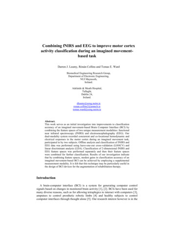
Combining fNIRS and EEG to improve motor cortex activity classification during animagined movement-based task5Hz. EEG acquisition was performed with a BioSemi Active Two system (BioSemiInc., The Netherlands). DC-coupled EEG data was digitally sampled at 2048 Hz.Figure 1 shows the layout of the fNIRS-EEG montage. fNIRS source positions arelabelled S1-S3, fNIRS detector positions are labelled D1-D3 and EEG electrodepositions are labelled E1-E7. Seven channels of fNIRS-EEG were recorded. ThefNIRS sources and detectors and EEG electrodes used for each channel are displayedin Table 1. During experiments, the fNIRS-EEG montage was centred over thesubject‟s dominant-side motor cortex (C4 of the 20/20 system for left-handed SubjectA and C3 for right-handed Subject B). Figure 2 shows the orientation of the montagein place on Subject A‟s D1D2D1D2D2D3D3E1E2E3E4E5E6E7Table 1. Channel fNIRS and EEG designationsFig. 1. fNIRS-EEG montageFig. 2. Dual fNIRS-EEG module over subject‟s motor cortex
6Darren J. Leamy, Rónán Collins and Tomas E. WardSignal processing and feature extractionfNIRSThe fNIRS system records raw light intensity signals. These are first converted tochanges in optical density (ΔOD) and then, using the modified Beer-Lambert law, tochanges in concentration (ΔHbO and ΔHbR).The raw intensity signals were initially low-pass filtered with a cut-off frequencyof 0.5 Hz to remove high-frequency components, such as those associated with thecardiac cycle. Next, the intensity signals are normalised using their mean amplitudeover the entire recording. The normalised intensity signals are then high-pass filteredwith a cut-off frequency of 0.01 Hz to remove baseline drift and Mayer waveinterference. ΔOD signals are obtained by taking the negative logarithm of thisfiltered signal. 4th order Butterworth filters were used for all filtering steps.The modified Beer-Lambert law is then used to find the ΔHbO and ΔHbR signalsfrom the ΔOD signals for each channel. Once the source-detector separation for thechannel, the extinction coefficients for both the 690 nm and 830 nm light in HbO andHbR and the differential pathlength factor are known, the operation involves a simplematrix inversion and multiplication to obtain the ΔHbO and ΔHbR signals. Adifferential pathlength factor of 5.93 was used (in accordance with the literature [15]).The ΔHbO and ΔHbR data was then separated into individual 10 second trials.During each trial, the change in average amplitude of the ΔHbO and ΔHbR signalsfrom the initial 5 seconds to the subsequent 5 seconds was used as the feature to trainand test the classifier.EEGThe EEG data was first high-pass filtered with a cut-off frequency of 1 Hz toremove DC and near-DC components. The EEG data was then analysed to identifythe frequencies at which PMMD and PMBS occurred for the different events. Theevents were “imagined movement onset” and “imagined movement offset”, whichcoincide with a change in on-screen instruction to the subject. The frequencies atwhich ERD and ERS occurred were identified by visual inspection of the averageFFT plots for the reference and activity periods for both events.The reference period was chosen to be between 4.5 and 3.5 seconds before bothtypes of event. For an imagined movement onset event, the activity window waschosen to be from between 0 and 1 second after the event. For an imagined movementoffset event, the activity window was chosen to be from between 0.5 and 1.5 secondsafter the event. The activity windows were chosen because of the expected timings ofPMMD and PMBS. The raw EEG is filtered for the identified ranges of PMMD andPMBS. These µ-range and β-range signals are then squared to get power signals.The change in µ-range power and β-range power from reference window toactivity window were used as features of EEG activity for classification.
Combining fNIRS and EEG to improve motor cortex activity classification during animagined movement-based task7ClassificationThe goal of classification is to decode the subject's current state based on thefeatures extracted from the fNIRS and EEG responses. The classifier attempted toclassify features into one of two classes: „active‟ and „rest‟. We employed the LinearDiscriminant Analysis (LDA) classifier and calculated classification accuracy viaLeave-One-Out Cross Validation (LOOCV), as we had employed previously [16]. Inparticular, for a total of N trials of data, N-1 trials were used for training the classifierand the remaining trial was used for testing. This was repeated N times with each trialused for testing once. Accuracy was measured as the amount of correct classificationsover N trials. Following classification of the fNIRS and EEG 2-dimensional featurespaces, the individual feature spaces were combined into an all-encompassing 4dimensional feature space. Every trial of data thus had 4 features available forclassification - change in HbO over the trial, change in HbR over the trial, change inµ-range power at the start of the trial and change in β-range power at the start of thetrial. This combined 4-dimensional feature space was also classified using LDA %69%61%56%56%59%Subject 62%66%fNIRS64%51%61%64%41%74%15%53%Subject 49%58%Table 2. LDA classification results for fNIRS, EEG and combined featuresThe classification results are presented in Table 2. Shown are the classificationaccuracies of the classifier when operating on fNIRS features alone, EEG featuresalone and combined fNIRS/EEG features.DiscussionOur results demonstrate that through combining fNIRS and EEG features into asingle fNIRS-EEG feature space, an increase in classification accuracy of imaginedmovement can be obtained. The two experimental subjects had very differentclassification accuracies of the cortical activity. For Subject A, all but one channel
8Darren J. Leamy, Rónán Collins and Tomas E. Wardmade gains in classification accuracy by combining feature spaces. For Subject B,however, combining feature spaces often results in a classification accuracy resultintermediate to those of fNIRS and EEG alone.From these results, it appears that when fNIRS and EEG classification isreasonably accurate, the combined classification result tends to be higher than both.When either fNIRS or EEG classification accuracy is good and the other is not,combining feature spaces seems to result in intermediate classification accuracy. It isalso worth pointing out that when both fNIRS and EEG classification was poor,combining feature spaces resulted in better performance.These results show that combining fNIRS and EEG feature spaces can results inhigher classification accuracy. This is of more importance to improving an fNIRSsystem than an EEG system. Improving EEG classification accuracy can beaccomplished by increasing the density of electrodes over a cortical area. However,fNIRS has a limit to the proximity of source-detector pairs. An fNIRS detector cansuffer from “source-blinding” if sources are placed too close, even if that detector isnot intentioned to record light from that source. Therefore, for the sole purposes ofimproving classification accuracy, supplementing an fNIRS system with EEG data ismore useful than adding fNIRS to an EEG system.An advantage of a dual-modality system such as this one is that for the samemeasurement space on the head, more information about the underlying neurovascularrelationship is being recorded. An EEG of fNIRS alone system can only record theelectrical or hemodynamic response in an area of cortex. Our system records fNIRSand EEG but also records information about the relationship between them, even ifwe do not yet fully understand that relationship.We expect a similar improvement in classification accuracy when using dry EEGelectrodes instead of the standard wet electrodes used here. Dry electrodes have muchlower signal-to-noise ratio (SNR), so combining a dry EEG set-up with fNIRS couldhelp offset the poor SNR. We expect a completely dry fNIRS-EEG system wouldsignificantly reduce set-up time, reduce subject discomfort and have similarclassification performance to a wet electrode EEG system.ConclusionInvestigation into dual-modality measurement is of importance to BCI researchersdue to the potential gains in classification accuracy while utilising the same area ofcortex. This work has demonstrated that improvements to imagined-movement basedBCIs are possible by implementing multi-modal measurements. We believe thisresearch will lead to more accurate BCIs and smaller measurement devices.AcknowledgementThe authors gratefully acknowledge the contribution of Science Foundation Ireland:Research Frontiers Program 2009, Grant No. 09/RFP/ECE2376.
Combining fNIRS and EEG to improve motor cortex activity classification during animagined movement-based task9References[1] E. Angelakis et al, Brain-computer interface: a reciprocal self-regulatedneuromodulation., Acta Neurochir Suppl, vol. 97, 2007, pp 555-559.[2] J. R. Wolpaw et al, Brain-computer interfaces for communication and control,Clinical Neurophysiology, vol. 113, 6, 2002, pp 767-791.[3] J. Conradi et al, Brain-Computer Interfacing in Tetraplegic Patients with HighSpinal Cord Injury, International journal of bioelectromagnetism, vol. 11, 2, 2009, pp65-68.[4] L. R. Hochberg et al, Neuronal ensemble control of prosthetic devices by a humanwith tetraplegia, Nature, vol. 442, 2006, pp 164-171.[5] R. Scherer et al, The Self-Paced Graz Brain-Computer Interface: Methods andApplications, Comput. Intell. Neurosci., 2007, 79826.[6] A. Meyler et al, Modifying the brain activation of poor readers during sentencecomprehension with extended remedial instruction: A longitudinal study ofneuroplasticity, Neuropsychologia, vol. 46, 10, 2008, pp 2580-2592.[7] H. Burton, Visual Cortex Activity in Early and Late Blind People, The Journal ofNeuroscience, vol. 23, 10, 2003, pp 4005-4011.[8] A. Pascual-Leone et al, Modulation of muscle responses evoked by transcranialmagnetic stimulation during the acquisition of new fine motor skills, J Neurophysiol.,vol 74, 3, 2005, pp 1037-1045.[9] B. B. Johansson, Brain Plasticity and Stroke Rehabilitation : The Willis Lecture,Stroke, vol. 31, 2000, pp 223-230.[10] E. Okada et al, Theoretical and experimental investigation of near-infrared lightpropagation in a model of the adult head, Appl. Opt., vol. 36, 1, 1997, pp 21-31.[11] A. Sassaroli and S. Fantini, Comment on the modified Beer–Lambert law forscattering media, Phys. Med. Biol., vol. 49, 14, 2004, N255.[12] G. Pfurtscheller and F. H. Lopes da Silva, Event-related EEG/MEGsynchronization and desynchronization: basic principles., Clinical Neurophysiology,vol. 110, 1999, pp 1842-1857.[13] G. Pfurtscheller and A. Aranibar, Evaluation of event-related desynchronization(ERD) preceding and following voluntary self-paced movements., Electroenceph.Clin. Neurophysiol., vol. 46, 1979, pp 138–146.[14] G. Pfurtscheller and A. Stanc ak, Jr. and C. Neuper, Post-movement betasynchronization. A correlate of an idling motor area?, Electroenceph. Clin.Neurophysiol., vol. 98, 1996, pp 281–293.[15] P. van der Zee et al, Experimentally measured optical pathlengths for the adulthead, calf and forearm and the head of the newborn infant as a function of interoptode spacing, Adv. Exp. Med. Biol., vol. 316, 1992, pp 143-153.[16] D. J. Leamy and T. E. Ward, A novel and concurrent fNIRS/EEG measurementsystem: design and initial results., Engineering in Medicine and Biology Society(EMBC), 2010 Annual International Conference of the IEEE, 2010, pp 4230-4233.
combining the feature spaces of two unique measurement modalities: functional near infrared spectroscopy (fNIRS) and electroencephalography (EEG). Our dual-modality system recorded concurrent and co-locational hemodynamic and electrical responses in the motor cortex during an imagined movement task, participated in by two subjects.