Transcription
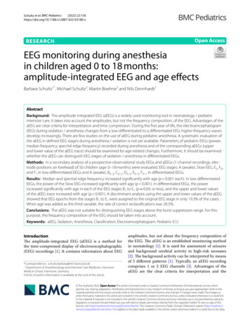
(2022) 22:156Schultz et al. BMC -xOpen AccessRESEARCHEEG monitoring during anesthesiain children aged 0 to 18 months:amplitude‑integrated EEG and age effectsBarbara Schultz1*, Michael Schultz2, Martin Boehne3 and Nils Dennhardt1AbstractBackground: The amplitude-integrated EEG (aEEG) is a widely used monitoring tool in neonatology / pediatricintensive care. It takes into account the amplitudes, but not the frequency composition, of the EEG. Advantages of theaEEG are clear criteria for interpretation and time compression. During the first year of life, the electroencephalogram(EEG) during sedation / anesthesia changes from a low-differentiated to a differentiated EEG; higher-frequency wavesdevelop increasingly. There are few studies on the use of aEEG during pediatric anesthesia. A systematic evaluation ofthe aEEG in defined EEG stages during anesthesia / sedation is not yet available. Parameters of pediatric EEGs (power,median frequency, spectral edge frequency) recorded during anesthesia and of the corresponding aEEGs (upperand lower value of the aEEG trace) should be examined for age-related changes. Furthermore, it should be examinedwhether the aEEG can distinguish EEG stages of sedation / anesthesia in differentiated EEGs.Methods: In a secondary analysis of a prospective observational study EEGs and aEEGs (1-channel recordings, electrode positions on forehead) of 50 children (age: 0–18 months) were evaluated. EEG stages: A (awake), Slow EEG, E2, F0,and F1 in low-differentiated EEGs and A (awake), B0–2, C0–2, D0–2, E0–2, F0–1 in differentiated EEGs.Results: Median and spectral edge frequency increased significantly with age (p 0.001 each). In low-differentiatedEEGs, the power of the Slow EEG increased significantly with age (p 0.001). In differentiated EEGs, the powerincreased significantly with age in each of the EEG stages B1 to E1 (p 0.04, or less), and the upper and lower valuesof the aEEG trace increased with age (p 0.001). A discriminant analysis using the upper and lower values of the aEEGshowed that EEG epochs from the stages B1 to E1 were assigned to the original EEG stage in only 19.3% of the cases.When age was added as the third variable, the rate of correct reclassifications was 28.5%.Conclusions: The aEEG was not suitable for distinguishing EEG stages above the burst suppression range. For thispurpose, the frequency composition of the EEG should be taken into account.Keywords: aEEG, Sedation, Anesthesia, Classification, Electroencephalogram, Pediatric ICUIntroductionThe amplitude-integrated EEG (aEEG) is a method forthe time-compressed display of electroencephalographic(EEG) recordings [1]. It contains information about EEG*Correspondence: schultz.barbara@mh-hannover.de1Department of Anesthesiology and Intensive Care Medicine, HannoverMedical School, Hannover, GermanyFull list of author information is available at the end of the articleamplitudes, but not about the frequency composition ofthe EEG. The aEEG is an established monitoring methodin neonatology [1]. It is used for assessment of seizuresand background cerebral activity in high-risk neonates[2]. The background activity can be interpreted by meansof 5 different patterns [1]. Typically, an aEEG recordingcomprises 1 or 2 EEG channels [3]. Advantages of theaEEG are the clear criteria for interpretation and the The Author(s) 2022. Open Access This article is licensed under a Creative Commons Attribution 4.0 International License, whichpermits use, sharing, adaptation, distribution and reproduction in any medium or format, as long as you give appropriate credit to theoriginal author(s) and the source, provide a link to the Creative Commons licence, and indicate if changes were made. The images orother third party material in this article are included in the article’s Creative Commons licence, unless indicated otherwise in a credit lineto the material. If material is not included in the article’s Creative Commons licence and your intended use is not permitted by statutoryregulation or exceeds the permitted use, you will need to obtain permission directly from the copyright holder. To view a copy of thislicence, visit http:// creat iveco mmons. org/ licen ses/ by/4. 0/. The Creative Commons Public Domain Dedication waiver (http:// creat iveco mmons. org/ publi cdoma in/ zero/1. 0/) applies to the data made available in this article, unless otherwise stated in a credit line to the data.
Schultz et al. BMC Pediatrics(2022) 22:156time compression. Compared to conventional multichannel EEG recordings, establishing and maintaining such arecording requires less effort. In addition to being usedfor newborns, aEEG devices are also used in pediatricintensive care patients beyond the neonatal age [4]. In arecent survey, main indications were neurological complications or disease and altered mental state [4].The background activity of the EEG can be changedin pediatric patients due to illness. Another factor thataffects background activity is sedation. Increasing dosesof sedative substances, administered intravenously orby inhalation, lead to typical changes in the EEG. Thesechanges, which, e.g., occur with propofol and with inhalation anesthetics such as sevoflurane, consist in a progressive slowing up to the occurrence of intermittentsuppression phases and finally to a continuous suppression of the EEG [5, 6]. The EEG changes can be describedusing a stage classification, and they can be used to assessthe depth of sedation and anesthesia [7, 8].Changes in the EEG caused by brain maturation mustbe taken into account when assessing sedation effectsin the child’s EEG. An age effect that can be observedin the pediatric EEG during anesthesia is an increase inthe power of the EEG with increasing age. This effectwas described for example in a study in children aged0 to 17 months [9] and in another study comparing twogroups of children aged 0–3 months and 4–6 months,respectively [10]. During the first few months of life, thefrequency composition of the EEG during anesthesiachanges. While negligible frontal alpha power (8–12 Hz)was found in children aged 0–3 months, increased alphapower was present in children aged 4–6 months [10]. Furthermore, it was reported that theta (4–8 Hz) and alpha(8–12 Hz) oscillations emerge by 4 months, that alphaoscillations increased in power from 4 to 10 months, andthat frontal alpha-oscillation predominance emerged at 6 months [11].In a study by de Heer et al. [12], correlations betweenend-tidal sevoflurane concentrations and the relative power in EEG frequency bands were not found inchildren up to 6 months, while such correlations wereobserved in children from 6 to 12 months and 1–6 years.In another study, obvious concentration-dependentchanges in EEG parameters, including the spectral edgefrequency, were observed in children aged 6 months to2 years during sevoflurane anesthesia, while there werefew changes in children younger than 3 months [13].Several studies have examined EEG changes duringemergence from anesthesia [14–16]. In some of the studies, little or no changes in power [14, 15] were found inyounger children, while changes were seen in older children, with the upper age limit of the younger children inthese studies being 3 or 6 months. On the other hand,Page 2 of 12Cornelissen et al. [16] described that, in children with anage of less than 3 months, frontal EEG frequency bandsshifted in power with decreasing end-tidal sevoflurane.There are few studies on the use of aEEG during anesthesia in children [14, 15, 17–19]. A systematic evaluation of the aEEG with regard to defined EEG stagesduring anesthesia is not yet available.In most children, at the age of 6 months, the EEG is sofar developed that during sedation and anesthesia a rangeof EEG stages, that is analogous to EEG stages of olderchildren and adults, can be distinguished [20]. The termslow-differentiated EEG and differentiated EEG wereintroduced to describe if in an EEG, due to its developmental state, the full range of these EEG stages cannot orcan be distinguished [20, 21].The aEEG is used in many pediatric intensive careunits [4]. For its use in children with differentiatedEEGs knowledge on how sedative drugs affect the aEEGis important. In addition to its use in the intensive careunit, the aEEG might be a valuable addition to the monitoring of pediatric patients in the operating room.In this analysis, EEGs recorded during administration of sevoflurane in children between the ages of 0 and18 months were examined in order to gain informationabout age-related changes in the EEG and the aEEG. Furthermore, it was assessed whether aEEG in children withdifferentiated EEG is suitable to distinguish between different EEG stages of sedation and anesthesia.Patients and methodsData basisAs data basis for the analysis, EEG and aEEG recordingsfrom a previous, single-center, prospective, observationaltrial published by Dennhardt et al. were used [21]. Thestudy had been approved by the responsible ethics committee (Ethics Committee of Hannover Medical School,Germany, Chairperson Prof. Dr. H. D. Troeger, No. 3259–2016 dated June 22, 2016). Because of the observationaldesign without change of the anesthetic management,written consent was not necessary according to the Ethics Committee. Children between 0 and 24 months oldundergoing elective surgery were eligible for the study.Excluded were children who had a neurological disorder or a severe developmental delay, or who were preoperatively given medication that altered the EEG, with theexception of midazolam as premedication. The preoperative physical status was scored according to the AmericanSociety of Anesthesiologists (ASA) physical status scale.Sevoflurane and remifentanil were administered duringthe steady state of anesthesia after induction with sevoflurane, remifentanil and atracurium. Anesthetic management followed the usual practice.
Schultz et al. BMC Pediatrics(2022) 22:156aEEGThe aEEG contains information about the amplitudes of anEEG [1]. The calculation steps of the aEEG include filtering, rectification, smoothing, and time compression of theEEG. Over time, a band-shaped pattern results, the uppermargin of which represents the highest amplitudes and thelower margin of which represents the lowest amplitudes ofsuccessive EEG epochs. The aEEG is represented using aso-called semilogarithmic scale, i.e., the scale is linear upto 10 μV, and logarithmic above [1, 22–24]. For the use ofthe aEEG in neonatology, there are suggestions for distinguishing different patterns based on the position of theupper and lower margin of the aEEG trace [25]. There are5 patterns in the classification by Hellström-Westas et al.[22]. Figure 1 shows examples of these patterns from theown data set.EEG recording, stage classification, spectral parameters,and aEEG parametersThe EEGs were recorded using the Narcotrend-CompactM (MT MonitorTechnik, Bad Bramstedt, Germany) as1-channel registrations, for which the required threeelectrodes were attached to the patient’s forehead. TheNarcotrend-Compact M checks the anesthetic EEG inchildren in the first year of life to determine whether thePage 3 of 12EEG is either low-differentiated or differentiated. In thecase of low-differentiated EEGs, the stages A (awake),Slow EEG, E 2, F0, and F1 are used. The Slow EEG is characterized by continuous activity without suppressionperiods. Suppression periods begin to occur in stage E2, and the length of suppression periods increases instages E2, F0, and F 1. In the case of differentiated EEGs,the Narcotrend-Compact M distinguishes the stages A(awake), B0–2, C0–2, D0–2, E0–2, F0–1, and it calculates theNarcotrend Index between 100 and 0 [7, 8]. Figure 2Aand B show examples of different EEG stages in a lowdifferentiated and in a differentiated EEG recorded during anesthesia in a 1-month-old and in an 11-month-oldchild. The associated power spectra show the power offrequency components of the EEG. Regardless of thedegree of differentiation, the Narcotrend-Compact Mcalculates the aEEG, the density spectral array (DSA),and other parameters.As basis for this evaluation, the original recordingswere classified with the Narcotrend software (Version3.4). 50 EEG recordings with aEEG courses were reanalyzed, 11 recordings were not available due to technicalreasons.The following EEG parameters were considered:the EEG power (μV) (calculated as root of the signalFig. 1 aEEG background patterns (C: Continuous, DC: Discontinuous, BS: Burst Suppression, LV: Low Voltage, FT: Flat Trace). Definitions accordingto Hellström-Westas et al. [22]. According to Schettler, for LV the lower margin is around or below 5 μV and the upper margin is no more than 10 μV[3]. The assignment of an aEEG section to an aEEG pattern is based on the upper and lower margins of the aEEG. The visual determination of theposition of the upper or lower margin for an aEEG section can be carried out in such a way that 50% of the associated values are above and 50%below [26]
Schultz et al. BMC Pediatrics(2022) 22:156Page 4 of 12Fig. 2 Raw EEGs and power spectra recorded during anesthesia. The power spectra were calculated for 20s epochs. A Low-differentiated EEG,1-month-old child. Examples of the Slow EEG, E2, and F 0 stages. B Differentiated EEG, 11-month-old child. Examples of stages B, C, D, E, and Fpower (μV2)), the median frequency (50% quantile ofthe power spectrum), and the spectral edge frequency(SEF95, 95% quantile of the power spectrum). Furthermore, the two aEEG parameters upper and lower valueof the aEEG trace, which are calculated by the Narcotrend-Compact M, were evaluated. All parameters,the Narcotrend stage, and the Narcotrend Index werecalculated for consecutive 20 s epochs. For each of the50 patients, the entire EEG during the steady state (cutto suture) was evaluated. To characterize the coursesin the steady state, a mean value was calculated fromthe 20 s values for each patient and for the parameters median frequency, SEF95, power, and NarcotrendIndex.Tinning and Acworth (2007) developed formulas forcalculating the mean body weight in children of different ages [27]. The children’s documented body weightand the calculated weight according to Tinning andAcworth [27] were used in analyses.
Schultz et al. BMC Pediatrics(2022) 22:156StatisticsThe calculations were carried out with the statistical software SAS (version 9.3). Fisher’s exact test wasused to compare nominal data. The t-test was used formean value comparisons for two groups. Linear regression models were used to investigate the relationshipbetween age and the parameters EEG power, and theupper and the lower value of the aEEG trace. Spearman’s rank correlation was used to examine correlations between EEG spectral parameters (median andSEF95), patients’ age and patients’ weight.A multiple correlation coefficient between the Narcotrend Index and the upper and the lower values ofthe aEEG trace was calculated.By way of a linear discriminant analysis, it was evaluated whether the aEEG parameters upper and lowervalue of the aEEG trace are suitable for distinguishingthe stages B 1 to E 1 of the differentiated EEG; an additional linear discriminant analysis was carried out withthe patient’s age as third parameter. The sample sizewas n 10,153.As part of the discriminant analyses, reclassificationsinto the stages B1 to E1 were carried out.For the discriminant analyses of the differentiatedEEGs, stage B0 was not considered due to the smallnumber of cases.The awake EEG was not included in the analyses; furthermore, EEGs characterized by intermittent or continuous suppression periods were not included as forsuch EEGs defined patterns exist in the aEEG classification by Hellström-Westas et al. [22].The significance threshold was assumed to be p 0.05.ResultsOf the 50 patients, 21 had a low-differentiated EEGaccording to the Narcotrend evaluation and 29 patientshad a differentiated EEG.Demographic dataThe included 50 patients and the not included 11patients did not differ significantly with regard to age(p 0.96), documented weight (p 0.96), and sex(p 1.00). There was no evidence that the 11 patientsnot included in the analysis could skew the results ofthe study.The patients with low-differentiated EEGs wereyounger and lighter than the children with differentiatedEEGs (age: 2.5 1.2 vs. 9.7 4.3 months, weight: 4.9 1.2vs. 8.5 2.0 kg; each p 0.001). All patients were under19 months of age. Regarding the ASA status (ASA: American Society of Anesthesiologists), i.e., the preoperativePage 5 of 12physical condition, there was no difference between thetwo patient groups (p 0.84).The documented weight was significantly lowerthan the calculated weight according to Tinning andAcworth [27], both in children with a low-differentiatedEEG (p 0.007) and children with a differentiated EEG(p 0.04). The difference between documented and calculated weight was 11.9% (mean standard deviation:0.6 0.9 kg) in the group with low-differentiated EEGsand 8.4% (mean standard deviation: 0.7 1.7 kg) in thegroup with differentiated EEGs.EEG spectral parameters, patients’ age, and patients’weightFor the course of each anesthesia, the means of medianfrequency and spectral edge frequency (SEF95) werecalculated as parameters from the frequency range. Themedian frequency and SEF95 increased significantly withage (p 0.001 each) (Fig. 3). The median frequency andthe spectral edge frequency also increased significantlywith the patients’ documented body weight and with thecalculated weight according to Tinning and Acworth [27](p 0.001 each).In children with low-differentiated EEGs, 89.9% of theclassifications for the steady state of anesthesia were SlowEEG, 7.5% were E2, 2.6% were F0, and 0.1% were F1. Therewas a significant increase in power of the Slow EEG withage (p 0.001) (Fig. 4).For each of the differentiated EEGs, the median Narcotrend Index was calculated as a measure to describethe mean depth of hypnosis at steady-state anesthesia.The mean of all 29 medians was 53.6 (IQR (48.5; 62)),which can be assigned to stage D1.For the children with differentiated EEGs, a relationbetween age and EEG power was examined for the stages B1 to E1. First, the mean EEG power was calculated foreach child and for each stage. In each of the stages, thepower increased significantly with age (B1: p 0.04, B2:p 0.004, C0: p 0.001, C1: p 0.001, C2: p 0.001, D0:p 0.04, D1: p 0.003, D2: p 0.007, E0: p 0.004, E1:p 0.005). Figure 4 shows, as an example, the increase inpower with age for stage D1.aEEG parameters and patients’ ageAccompanying the described increase in power in thestages, the upper and lower values of the aEEG increasedsignificantly with age in each of the stages B 1 to E1 (ineach of the stages B 1 to E 1 upper values p 0.001, lowervalues p 0.001).aEEG parameters and EEG stagesIn order to analyze whether there is a correlation betweenEEG stages B 1 to E 1 and the aEEG in the differentiated
Schultz et al. BMC Pediatrics(2022) 22:156Page 6 of 12Fig. 3 Change in spectral edge frequency (SEF95) and median frequency with ageFig. 4 EEG power, dependent on age. Slow EEG and Stage D1EEGs, a multiple correlation coefficient between theNarcotrend Index and the upper and the lower valuesof the aEEG trace was calculated. The correlation coefficient was r 0.09. In Fig. 5A and B, no relationship withthe Narcotrend Index can be seen. As an example, Fig. 6shows the course of the EEG stages or EEG index values(cerebrogram) and the aEEG from one course of anesthesia in an 11 month-old child. EEG stages from the E to theB range occurred between 09:50 and 12:00 h. In spite ofthe different EEG stages, there are no significant changesin the aEEG during this period. (The raw EEG segmentsshown in Fig. 2B were taken from the course of anesthesia shown in Fig. 6 at the marked times.)By way of a linear discriminant analysis, it was evaluated whether the upper and the lower values of the aEEGtrace are suitable for distinguishing the EEG stages B1 to E1.For each of the EEG stages from B 1 to E 1, it was checkedwhich of these stages an EEG epoch was, based on the two
Schultz et al. BMC Pediatrics(2022) 22:156Page 7 of 12Fig. 5 Values of the aEEG in the stages B 0 to E 1. A Upper values of the aEEG trace. B Lower values of the aEEG traceassociated aEEG parameters, assigned to. A correct classification was made in 19.3% of the cases. In 80.7% of thecases, an incorrect stage was chosen. When age was addedas the third variable, the rate of correct reclassifications was28.5%.Table 1A and B show the results (as percentages) ofthe classification into the substages B 1-E1 by meansof discriminant analysis without (Table 1A) and with(Table 1B) patient age as additional variable. Correctclassifications are indicated by boldface. The total number of epochs per substage is indicated in an extra column on the left side of each table. The total number ofall epochs is 10,153.DiscussionThe current analysis focused on age-related changes inthe EEG and the aEEG and on the ability of aEEG-derivedparameters to distinguish between different EEG stagesof sedation and anesthesia. It was shown for the first timethat the increase in power with age occurs across all EEGsub-stages from very light anesthesia / sedation (B1) tovery deep anesthesia / sedation ( E1). In the differentiatedEEGs, EEG stages from the range B1 to E1 could neitherbe distinguished on the basis of the upper and lower values of the aEEG trace, nor on the basis of the two aEEGparameters and the patient’s age. This result supportsthe view that, in EEG stages above the burst suppression
Schultz et al. BMC Pediatrics(2022) 22:156Page 8 of 12Fig. 6 Course of anesthesia in an 11-month-old child. A EEG stages and index. B aEEG. The shift to lower μV values that occurred at the end of theaEEG course is due to very low EEG amplitudes in stage B1. The raw EEGs shown in Fig. 2B originate from this course of anesthesia. The markingsshow the times at which the raw EEG sections were drawn
Schultz et al. BMC Pediatrics(2022) 22:156Page 9 of 12Table 1 Results of the classification based on A) upper and lower value of the aEEG, B) upper and lower value of the aEEG plus patientage(A)Classification based on aEEG %0%10%26%Classification accord- 332ing to Narcotrend322Compact M447489(B)Classification based on aEEG parameters and ageB1B2C0C1C2D0D1D2E0E1Classification accord- 332ing to Narcotrend322Compact 6%42E17%7%5%2%12%2%5%17%24%19%range, the aEEG is of limited value for depth of sedationand anesthesia monitoring in children aged 0–18 months.Growth and maturation of the brain are particularly rapidin the first year of life [28]. The observed increase in medianfrequency and SEF95 in both low-differentiated and differentiated EEGs can be explained on the one hand by thedevelopment of higher frequencies in the EEG with advancing age. On the other hand, the median frequency andSEF95 depend on the intraoperative anesthetic EEG stage.The increase in power with age is consistent with observations made by other authors [9, 10, 15]. Sankupellay et al.,who observed a rise of spectral power in different stages ofsleep within the first two years of life, speculated that thesechanges of the spectral power represented a sleep stateindependent aspect of EEG development and were relatedto structural brain development [29].In the present analysis, based on the upper and lowervalues of the aEEG trace, EEG stages in the range fromstage B1 to E1 were not reliably distinguished in children with differentiated EEGs, even when additionallyconsidering the children’s age. When calculating theaEEG, one of the calculation steps is filtering the EEG.This is done in order to reduce the effect of artifacts[30]. An asymmetrical bandpass filter is used, whichstrongly suppresses activity below 2 Hz and above15 Hz [22–24]. The primary feature of the B stages ofthe anesthetic EEG are waves in the beta frequencyrange, i.e., above 12.5 Hz. The anesthetic EEG stagesfrom D0 to E1 are characterized by increasingly pronounced delta activity (0.5–3.5 Hz). Due to the filtering,part of the waves from the frequency ranges, which arecharacteristic for the B range and for the D / E range,are therefore not represented in the aEEG. This couldhave contributed to the fact that in our investigation itwas not possible to distinguish between stages in the B 1to E1 range using the aEEG. The frequency compositionof the EEG, which is the essential characteristic for distinguishing the anesthetic EEG stages, is not taken intoaccount in the aEEG. Figure 2B illustrates that the rawEEGs differ significantly in their frequency composition. A distinction between stages C and D based on theamplitudes is not possible. However, in the examples inFig. 2B, there are higher amplitudes in stage E, while instage B there is a tendency towards lower amplitudes.In B stages, the EEG amplitude can be very small, sothat the upper and lower values of the aEEG trace canconsequently assume small values (see also Fig. 5A andB). Such portions of the aEEG originating from EEGs
Schultz et al. BMC Pediatrics(2022) 22:156with high-frequency waves and low amplitude may fulfil the criteria of the aEEG pattern Low Voltage (LV).A Low Voltage pattern in the aEEG is associated withan unfavorable prognosis after perinatal asphyxia [31,32] and is classified as severely abnormal [32]. A LowVoltage pattern in the B stage of the anesthetic EEG is anormal finding.Figure 5A shows that the range of values for the uppervalues of the aEEG is similar across stages B 1 to E 1,with a tendency towards smaller values in the B and Estages. The same applies to the lower values of the aEEG(Fig. 5B). Figure 5A and B show that the upper and thelower values of the aEEG provide little EEG stage-specific information. The reclassification results obtainedby means of the linear discriminant analysis (Table 1Aand B) confirm this visual assessment. Apart from lineardiscriminant analysis, other methods, e.g., neuronal networks, are available for classification tasks [33]. However,such alternative methods would also be limited by theinformation content of the available variables.Although the aEEG is a widely used monitoring tool inneonatology and pediatric intensive care [4], so far useof the aEEG for sedation monitoring was only reportedby some authors [34, 35]. Reports on the use of the aEEGduring anesthesia differ in their estimation of the valueof the aEEG for depth of anesthesia monitoring [17–19].In children in the first two years of life, absorption, distribution, and excretion of medication change due tomaturation. There are also pharmacodynamic maturationprocesses [36]. This makes individually adequate dosing difficult. Even though aEEG is not suitable for reliably distinguishing between stages in the B1 to E 1 range,it can support the detection of burst suppression EEGsas well as complete EEG suppression (stages F0 and F1).For these EEG patterns, which can occur at higher dosesof hypnotically acting substances, the classes according to Hellström-Westas et al. [22] include the classesBurst Suppression (BS) and Flat Trace (FT). In a guideline on postoperative delirium, the European Society ofAnaesthesiology recommends avoiding burst suppressionpatterns in adults during anesthesia [37]. A consensusstatement on the role of neuromonitoring on perioperative outcomes also recommends avoiding burst suppression EEGs [38]. It should, however, be borne in mindthat, in preterm infants, an EEG pattern with intermittent, very flat sections can be a normal finding due todevelopment. A pattern with alternating phases of higheramplitude and flatter amplitude (tracé alternant) can stillbe observed in healthy deep sleep in cerebrally healthychildren up to 44–46 weeks of postconceptional age [39].In neonatology, the aEEG is a standard procedure forpatient monitoring [1]. The aEEG is also used for intensive medical care for preterm infants [1, 40] and forPage 10 of 12patients beyond the age of newborns [41]. When sedatingas part of neonatal intensive care, the aEEG can help toidentify and avoid very deep stages of sedation [35]. If theaEEG is used in older children in the pediatric intensivecare unit or in the operating room, then the relationshipsdescribed in this work should be taken into account. Ifone wants to use the aEEG for older children in the context of sedation or anesthesia, then the inclusion of information about the proportion of higher frequency wavesin the EEG helps to distinguish between EEG stages thatfall within the range of the Continuous pattern according to Hellström-Westas et al. [22]. In the case of the LowVoltage pattern according to Hellström-Westas et al. [22],the content of higher-frequency waves can be used todecide whether the EEG stage is one of very light or verydeep sedation or anesthesia.A limitation of the analysis is the fact that a power calculation was not provided as the analysis was performedon a pre-existing data base.Interesting future research topics arise with regard tothe clinical application of the aEEG and to possible methodological modifications and improvements of the aEEG:The criteria by Hellström-Westas et al. f
EEG monitoring during anesthesia in children aged 0 to 18 months: amplitude-integrated EEG and age eects Barbara Schultz1*, Michael Schultz2, Martin Boehne3 and Nils Dennhardt1 Abstract Background: The amplitude-integrated EEG (aEEG) is a widely used monitoring tool in neonatology / pediatric intensive care.