Transcription
Taylor et al. Cell Communication and Signaling 2010, 8:11http://www.biosignaling.com/content/8/1/11Open AccessREVIEWSmall molecule screening in zebrafish: an in vivoapproach to identifying new chemical tools anddrug leadsReviewKerrie L Taylor, Nicola J Grant, Nicholas D Temperley and E Elizabeth Patton*AbstractIn the past two decades, zebrafish genetic screens have identified a wealth of mutations that have been essential tothe understanding of development and disease biology. More recently, chemical screens in zebrafish have identifiedsmall molecules that can modulate specific developmental and behavioural processes. Zebrafish are a uniquevertebrate system in which to study chemical genetic systems, identify drug leads, and explore new applications forknown drugs. Here, we discuss some of the advantages of using zebrafish in chemical biology, and describe someimportant and creative examples of small molecule screening, drug discovery and target identification.Genetic and chemical screens in zebrafishZebrafish (Danio rerio) have a unique status in experimental biology. As vertebrates used for forward andreverse genetics, they have provided novel insight intodevelopment and disease genetics. More recently,zebrafish research has pushed forward the exploration ofchemical biology in a whole animal system. The advantages of using zebrafish as an experimental system forchemical biology mirror those already well established fortheir use in genetics. Only a few centimetres long asadults, thousands of zebrafish can be housed in a laboratory with relatively low husbandry costs. Breeding pairscan produce over 200 embryos each week that are fertilized outside of the mother and can be easily collectedfrom the breeding tank. Embryonic development from asingle cell, and the rapid formation of discrete tissues andorgans with physiological similarity to their human counterparts, can be viewed in real time under a light microscope [1]. Organ progenitors can be observed by 36 hpf(hours post-fertilization), hatching occurs at 48-72 hpfand independent feeding by 5 dpf (days post-fertilization). Whole-mount in situ hybridization and antibodystaining allows for detection of specific RNA or proteinexpression (or modification) in the developing whole ani* Correspondence: e.patton@hgu.mrc.ac.uk1MRC Human Genetics Unit and the Division of Cancer Research, Institute ofGenetics and Molecular Medicine, The University of Edinburgh, Crewe RoadSouth, Edinburgh, EH4 2XR, UKmal, and transgenic technology provides the tools to follow the expression of a specific gene (or series of genes) inthe living fish [2]. The zebrafish genome is sequenced,and genetic mutants affect a wide range of biological processes including development, behaviour, metabolism,vision, immunity and cancer.Genetic screens in zebrafish proceed in two mainapproaches: forward and reverse genetics [3]. Forwardgenetics, characterized as 'phenotype to genotype', firstinvolves the identification and characterization of a specific phenotype, followed by the identification of theunderlying genetic mutation. The zebrafish system hasbeen especially powerful in the identification of developmental phenotypes, caused by N-ethyl N-nitrosourea(ENU) and insertional mutagenesis, and many of theunderlying genetic mutations have been identified [4-8].Reverse genetics, 'genotype to phenotype', takes advantage of molecular biology techniques. In these cases, agene of interest is selected and targeted by morpholinooligonucleotide (MO) knockdown, TILLING (TargetingInduced Local Lesions IN Genomes), or zinc-fingernucleases to discover the function of the genetic mutation within the fish [9-14].Chemical genetics complements traditional geneticapproaches [3]. First, many small molecule libraries aremade up of compounds with known biological function,allowing rapid elucidation of biological pathways withinthe organism. Second, chemical treatment can occur atFull list of author information is available at the end of the article 2010 Taylor et al; licensee BioMed Central Ltd. This is an Open Access article distributed under the terms of the Creative CommonsAttribution License (http://creativecommons.org/licenses/by/2.0), which permits unrestricted use, distribution, and reproduction inany medium, provided the original work is properly cited.
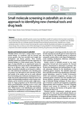
Taylor et al. Cell Communication and Signaling 2010, 8:11http://www.biosignaling.com/content/8/1/11any point during development or in the adult organism,allowing for the study of latent effects of genes duringdevelopment. Third, chemical dosage can be controlled,which can be advantageous when studying essential functions, or tissue specificity. Small molecule screening identifies relevant targets within the physiological context ofthe organism, biasing the screen for compounds that aremore likely to be cell permeable, less toxic, effective andwith an aceptable pharmacokinetic and pharmacodynamic profile [15]. Like genetics, chemical genetics canachieve both forward and reverse approaches [3,15]. Inforward chemical genetics, 'phenotype based small molecule discovery', a library of inhibitors can be screened fora specific phenotype in the animal, and from this the target(s) can be identified (Figure 1). Here, we describe someof the phenotype based chemical screens in zebrafish thatPage 2 of 14are pushing forward our knowledge of stem cells, behavior, development and disease treatment (Figure 2A, B, D,E). Conversely, reverse chemical screening entails testingchemical inhibitors with known molecular targets forspecific phenotypes in the zebrafish. Examples of thisapplication of the zebrafish system include identifyingand optimizing new bioactive compounds, applying acancer drug to developmental disease, and revealing theteratogenic mechanisms that hinder valuable clinicaldrugs (Figure 2C, F).Considerations of chemical libraries andmethodologyIn genetic screens, the type of mutagen (e.g. ENU, x-ray,insertional mutagen) will determine the range of possiblegenetic mutations and phenotypes, as well as the ease ofFigure 1 Phenotype based chemical screening in zebrafish. Male and female pairs are bred to produce hundreds of single cell embryos that arefertilized ex vivo. For high throughput screening, groups of males and females can be bred within in a larger tank (group breeding), producing highnumbers of embryos for screening. Breeding is synchronized by the light/dark cycles, and the fish tend to breed within the first two hours of light inthe morning. In this example, embryos are arrayed in 96-well plates, each with a different chemical compound, and observed for a specific phenotype.The chemical in well A2 causes a loss of melanocytes, like MoTP [18]. MoTP can specifically kill differentiated melanocytes, and has become a valuablechemical tool to explore melanocyte stem cell biology [42-44]. Other small molecule screens in zebrafish have also identified pigmentation phenotypes [85,86,96]. In another example of a small molecule screen, compounds are screened for inhibition of tail fin regeneration. The tail fin is clippedand grows back within a few days. A zebrafish embryo treated with the compound from well B3, a glucocorticoid, does not correctly regenerate itstail fin [97].
Taylor et al. Cell Communication and Signaling 2010, e 3 of 14DBEFC
Taylor et al. Cell Communication and Signaling 2010, 8:11http://www.biosignaling.com/content/8/1/11Page 4 of 14Figure 2 Creative examples of chemical biology in zebrafish. The zebrafish system can be used in a wide range of chemical biology experimentsand screens. A. Zebrafish as young as four dpf have active and sleep-like states. Continuous tracking of movement behaviours of the embryos duringrest and wake states, established by light and dark cycles, can be recorded by a camera and computer. High throughput screening for behaviouralchanges has identified new uses for poorly characterized compounds [80,82]. B. Genetic polymorphisms may underlie differences in sensitivity to poornutrition. In this example, ENU mutagenized zebrafish (parental (P) generation) were screened for genetic mutations that showed sensitivity tosub-optimal copper nutrient conditions. Zebrafish embryos fertilized with UV-inactivated sperm can live as haploid embryos until about 3 dpf [2].Haploid embryos of the heterozygous mother (F1 generation) were screened for loss of pigmentation, but only in the presence of the small moleculecopper chelator, neocuproine [41]. C. Intensive efforts by the pharmaceutical industry to develop drugs that target the MAPK pathway to treat cancerpatients may also be useful for the management of developmental diseases caused by mutations in the MAPK pathway. In the zebrafish model, expression of BRAF or MEK cardio-facio-cutaneous (CFC) mutant alleles interferes with early development. A one-hour treatment within a specific developmental time window with a MEK inhibitor is sufficient to allow normal development for a CFC zebrafish embryo [75]. D. The cardio-vasculaturesystem is conserved in fish, mice and humans. A small molecule screen for changes in hematopoietic stem cells (HSC) development identified theprostaglandin pathway as critical for HSC establishment [59]. A long-acting compound, called dmPGE2, can stimulate HSC development in the embryo and adult zebrafish, as well as in the mouse. dmPGE2 can be safely administered to people, and a clinical trial is underway to see if dmPGE2 treatment of umbilical cord blood prior to transplant can benefit transplant patients (L.I. Zon, personal communication). E. The acute myelogenous leukemiaoncogenic fusion AML1-ETO (AE) promotes a change from an erythrocytic fate to a granulocytic cell fate. Erythrocytes express the gata1 gene in theposterior blood island of the developing zebrafish (red dotted line). Heat-shock inducible expression of AE causes a cell fate change that can be visualized by loss of gata1 expression. A chemical screen identified that COX-2 inhibitors can suppress the AE cell fate change, and the embryos maintaingata1 expression in the presence of AE [72]. F. Zebrafish can play a valuable role in testing for drug toxicity and teratogenisity, as well as for testingdirect chemical targets in vivo. Thalidomide is a valuable drug for multiple myeloma and leprosy, but caused severe developmental birth defects whentaken by pregnant mothers in the late 1950s and early 1960s. Zebrafish are also sensitive to thalidomide, and treatment in early development preventsthe proper development of embryonic fins [88]. Thalidomide binds CRBN, and knockdown of crbn in the developing zebrafish also causes a loss of finphenotype. This suggests that the binding of thalidomide to CRBN in vivo may underlie its teratogenisty.identifying the mutation [2,10]. For example, ENU mutagenesis can generate hypomorphic, temperature sensitive, and gain- and loss-of-function mutations thatrequire genetic mapping to identify the genetic lesion.Insertional mutagenesis is more likely to cause gene disruptions, and allows for the rapid identification of theinsertion in the genome. Likewise, in chemical biology,the choice of chemical library directs the type of molecular processes that are disrupted, as well the approach oftarget pathway identification [16]. Traditionally, the discovery of new drugs has been achieved through the isolation of natural products from plants and microbes [17].Screening a library of natural products will be rich inchemical diversity and may identify novel active compounds, but identifying the active compound(s) and subsequent targets may require extended fractionationprocedures and biochemical techniques. Conversely,screening a library designed to target specific classes ofenzymes, such as kinases, allows for rapid identificationand direct testing of the chemical target in vivo, butrepresents a much smaller range of chemical diversity.Similarly, screening a library of compounds of pharmacologically active molecules of known bioactivity, whileagain covering a reduced chemical space, may allow for arapid transition to pre-clinical mammalian models.As in genetic screens, an important feature of chemicalscreening is consideration for the screening assay, that is,the phenotypic feature that is the basis for the screen. Thezebrafish system allows for diverse screening assaysincluding development and function of internal tissuesand organs in living animals that can be illuminated byflorescent reporter genes or molecules, or disrupted bygenetic mutations. Whole-mount RNA in situ hybridiza-tion and antibody staining provide details of cellularchanges in fixed embryos. No other vertebrate is as wellpositioned for high throughput phenotyping as thezebrafish embryo [3]. For high-throughput screening,computerized detectors can rapidly screen thousands oftreated embryos in a small treatment volume (e.g. in a 384well or 96 well plate), while lower throughput screeningcan often involve a single investigator screening multiplecharacteristics in larger treatment volumes (e.g. in a 24well plate).In the first chemical screen in zebrafish, Peterson andcolleagues (2000) screened 1,100 compounds selectedfrom the DIVERSet E Library (Chembridge) in 96-wellplates for small molecules that caused developmentalphenotypes during the first three days of development[18]. This was an important proof-of-concept studyshowing that small molecule screening in zebrafish couldidentify chemicals that, like genetic mutations, disruptspecific developmental processes. In addition, the identified chemicals could be used at different doses and at different developmental intervals to identify the timing ofthe chemical action during development. Recently, athorough review of the zebrafish chemical screens performed and libraries used has been published [16]. Here,we describe selected screens and chemical-genetic analyses performed in zebrafish to highlight the range ofexperimental screen designs and outcomes (Figures 1 and2).Chemical screening to rescue disease phenotypes:aortic coarctationMany genetic mutations identified in zebrafish are analogous to disease genotypes in humans, or cause pheno-
Taylor et al. Cell Communication and Signaling 2010, s that share clinical features with human diseases[19,20]. The zebrafish gridlock mutant suffers from a malformed aorta that prevents circulation to the trunk andthe tail, similar to coarctation of the aorta in humans. Thegridlock phenotype results from a mutation in the hey2gene, a transcriptional repressor that determines angioblast differentiation [21,22]. To screen for chemical suppressors of aortic coarctation, Peterson and colleaguestreated gridlock mutant embryos with small moleculesfrom the DIVERSet E library (Chembridge) from earlygastrulating embryos until 48 hpf [23]. From over 5000chemicals screened, two structurally related compoundssuppressed the gridlock phenotype. Altering the treatment time during development revealed a critical chemical treatment window for correcting the aorta phenotypeto be between 12 and 24 hpf. Importantly, the correctionmechanism was identified to be via upregulation of vascular endothelial growth factor (VEGF), and the subsequent promotion of blood vessel development. Smallmolecule inhibitors of the vascular endothelial growthfactor receptor (VEGFR) can inhibit blood vessel formation during zebrafish development, and tail fin regenerative angiogenesis in the adult [24,25]. The chemicallyinduced upregulation of VEGF was also effective at stimulating endothelial cell tubule formation [23]. Thus, phenotypic-based screening can identify suppressors of agenetic disease phenotype that have relevant biologicalactivity in mammalian cells. Importantly, in this example,the target compounds did not affect the disease gene orprotein directly. Rather, a pathway critical to the rescue ofthe phenotype was successfully identified, and can nowbe considered for new therapeutic approaches.Chemical screening to reveal new insight intoretinal blood vasculatureA highly metabolic tissue, the retina is fed oxygen andnutrients by an intricate vascular network. In humans,disruption of the retinal vasculature network, throughdamage to the existing network or inappropriate neovascularization, can lead to loss of vision and severe forms ofblindness. Understanding the basic biology of retinal vasculature is critical to understand the aetiology andpathology of retinal disease. In the zebrafish embryo, thishas been done through the identification and detailedcharacterization of genetic mutants which have a disruption of the retinal vasculature network [26]. These studiesare facilitated by the generation of transgenic zebrafishlines, expressing green florescent protein (GFP) under thepromoter of the endothelial vessel specific fli1 gene[26,27]. As a complementary approach to genetics, and tolearn about retinal vascularization, the fli1-GFP transgenic line was used in a screen for small molecules thatcould modulate the retinal vascular network during days3-6 of development [28]. Approximately 2000 small mole-Page 5 of 14cules from the bioactive Spectrum library were added tothe developing embryos at the pectoral fin stage (approximately 60 hpf ), and embryos were then embedded inmethyl cellulose and visualized under an inverted microscope. Of the 2000 compounds screened, five displayed areproducible effect on retinal vessel morphology: twoaffected vessel diameter without affecting vessel number,one affected vessel diameter and number, and two causedsevere collapse and loss of over 80% of the vessels.Extending these studies, Kennedy and colleaguesscreened a panel of known small molecule regulators ofangiogenesis, and found that LY294002, an inhibitor ofPI3 kinase signalling, can prevent the ocular angiogenesisin wild type fish and partially correct the extraneousangiogenesis in the out of bounds zebrafish mutant [29].Treatment with LY294002 within specific developmentaltime-windows, showed that the chemical could affect thedevelopment of new vessels, without affecting existingintraocular vessels or retinal function. Importantly, directand localized delivery of the drug was effective at inhibiting intraocular angiogenesis without additional systemeffects or altering visual function. Additional studies haveshown that chemical modulation in the adult zebrafishcan reduce hypoxia induced neovascularization [30].Studies such as these provide a groundwork for identifying chemicals and chemical methodologies that can specifically target aberrant retinal vascularization, whichaffects the sight of millions of people worldwide.Parallel in vitro and in vivo chemical screening:identifying new and known cell cycle regulatorsMany small molecules that affect the cell cycle have beenidentified through screening for compounds that alter thecell cycles of cancer cells in culture. Such high-throughput screens allow for large numbers of compounds to bescreened on multiple cell lines in a rapid and efficientmanner [31]. In the 1960s, cell based screening of naturalproducts by the National Cancer Institute identified taxolas a potent mitotic inhibitor [32]. Derived from thePacific yew tree, taxol is now considered one of the mostimportant chemotherapeutics to treat breast, lung, andovarian cancer [33]. Many mammalian cell cycle genesare conserved in zebrafish [34], and the majority of cellcycle inhibitors used in mammalian cells share sufficientconservation with the drug targets to be effective inzebrafish embryos or cell lines [35]. The zebrafish crash &burn (crb) mutant for the bmyb gene, is a MYB familytranscription factor involved in cell cycle progression andcancer. The crb mutant was identified in a genetic screenfor changes to the cell cycle, as revealed by an increasednumber of mitotic cells marked by a phospho-histone H3antibody [36]. crb mutants also reveal abnormal spindleand centrosome formation, and polyploidy. Notably,adult zebrafish heterozygous for crb have an increased
Taylor et al. Cell Communication and Signaling 2010, dence of carcinogen-induced cancer [36]. Using theDIVERSet E library (Chembridge) of 16320 compounds,Zon and colleagues screened for small molecules thatcould specifically rescue the crb phospho-histone H3mitotic defect [37]. One compound, called persynthamide, was identified that suppressed the mutant phenotype. In wild type embryos persynthamide transientlydelays S-phase via an ATR dependent checkpoint. Comparison with other known S-phase inhibitors, such ashydroxyurea and amphidicolin, showed that it was thechemically induced S-phase delay that was sufficient torescue the crb defect. In human cells, loss of B-MYB leadsto reduced levels of cyclin B1 [38], and expression ofcyclin B1 is sufficient to rescue crb mutant zebrafishembryos [36]. The persynthamide-induced S-phasecheckpoint also causes an increase in cyclin B1 expressionand thereby rescues the crb mutant. This is anotherexample of phenotypic based chemical screening that rescues a genetic phenotype by targeting the altered pathway, and not the mutated protein product directly.A common question in chemical screening is whetherthe identified compounds are specific to zebrafish, andhow the results of zebrafish chemical screening comparewith mammalian cell line based chemical screens. In thesmall molecule screen for suppressors of crb, additionalcompounds were identified that disrupted mitosis in wildtype zebrafish [35]. While the chemical library had beenpreviously screened extensively for cell cycle inhibitors inmammalian cells, 14 novel compounds that affected thecell cycle were identified via screening on zebrafishembryos. Six of the 14 hits were effective in mammaliancell lines, confirming the conservation of the affected target pathways across vertebrate species. Of the remainingseven hits, three were serum-inactivated which accountsfor their lack of activity in cell culture systems. Four hitswere only active in the zebrafish embryo, but not celllines, possibly due to activation by yolk sac proteins. Onehit was active in zebrafish embryos and zebrafish cell culture, but not in mammalian cell culture, suggesting thetarget of the compound was specific to the zebrafish andnot conserved in mammalian cells. Thus, in vivo and invitro chemical screens are complementary approaches,that when used together constitute a powerful approachto identifying a more complete set of chemically bioactivetools.Using chemicals to identify gene-nutrientinteractionsThe ability to limit the timing and regulate the dose ofsmall molecules has allowed for unique insight into thetissue-specific requirements for copper in the developingembryo. Copper is an essential nutrient, and sub-optimalcopper nutrient conditions in humans can lead to severeclinical symptoms including disorders of the nervous sys-Page 6 of 14tem, hair, and skin [39]. While copper is essential for allcells, some cells and tissues have a specialized copperrequirement. Gitlin and colleagues treated developingzebrafish embryos with the small molecule copper chelator, neocuproine, and found that loss of cuprodependentenzyme activity specifically affected some tissues, including melanocytes, the notochord, and the developinghindbrain [40]. Supported by a genetic mutant in the copper transporter atp7a (that was cloned in part by virtue ofphenotypic features indentical to the effect of the copperchelator neocuproine), Gitlin and colleagues revealed thetime, dose and tissue specific requirements for copperdependent enzymes during embryogenesis [40].Humans with mutations in ATP7A develop Menke'sdisease, a rare X-linked disorder of copper metabolism,characterized by neurodegeneration, hypotonia, andhypopigmentation. Less well understood are othergenetic conditions that lead to sensitivity to sub-optimalcopper nutrient conditions in otherwise healthy individuals. To address this problem, the Gitlin group used lowdoses of neocuproine and screened for genetic mutationsthat revealed a copper deficiency phenotype under mildcopper deficiency conditions (Figure 2B). Two geneticmutants were found: a hypomorphic allele of atp7a andmutation of the vacuolar atpase atp6. Vacuolar Atp6 isrequired for proton transport in the secretory pathway,an important process in intracellular copper transport[41]. Thus, genetic polymorphisms that otherwise havenormal developmental features can be combined withchemicals to reveal selected sensitivities to sub-optimalnutrient conditions.Chemical-genetic and chemical-chemical screens toprobe melanocyte regeneration pathwaysMoTP (4-(4-morpholinobutylthio)phenol), identified inthe original small molecule screen by Peterson and colleagues (2000), caused a dramatic loss of melanocytes bycell death [18]. Tyrosinase is a copper dependent rate limiting enzyme for melanin pigmentation in melanocytes,and MoTP exerts melanocyte-specific cell death via itsconversion to a cytotoxic form in cells that express highlevels of tyrosinase [42]. With a melanocytotoxic compound in hand, the Johnson group has been able to useMoTP to gain significant understanding of melanocyteregeneration in zebrafish. Many tissues and cells, including melanocytes, can regenerate in zebrafish, providingimportant insight into tissue-specific stem cells and progenitor development. Using MoTP to ablate embryonicmelanocytes, ENU mutagenized zebrafish were screenedfor mutants that fail to regenerate melanocytes aftertreatment [43]. Two mutants were identified, eartha andjulie, that only regenerated about 10% of the normal complement of melanocytes, affecting late stages of melanocyte differentiation in the regeneration process, and
Taylor et al. Cell Communication and Signaling 2010, iferation of melanoblasts during early melanocyteregeneration respectively. Thus, by using a chemical toprecisely control a specific cell type and process, geneticmutations necessary for distinct aspects of melanocyteregeneration can be identified.In a further small molecule screen for compounds thatspecifically blocked melanocyte regeneration followingMoTP-induced melanocyte ablation, Johnson and colleagues identified ICI-118,551, a chemical that specifically blocks melanocyte regeneration, while having noeffect on ontogenetic melanocyte development and otherdevelopmental processes [44,45]. One target of ICI118,551 is the β2-adrenergic receptor, but it is not clear ifthis is the mechanism by which it blocks melanocyteregeneration. Nonetheless, it is still a useful tool to probethe origin of melanocytes. For example, transgeniczebrafish expressing kit ligand have an increased numberof ontogenetic melanocytes, that are suppressed by ICI118,551 treatment, indicating that kit ligand exerts itseffects on the stem cell lineage of the developing zebrafish[44,45].The genetic mutant, picasso develops normal larvalmelanocytes (ontogenetic melanocytes), but shows deficits in forming new melanocytes at the onset of metamorphosis. Positional cloning identified the mutationcausing the picasso phenotype to be in the erbb3b gene,an epidermal growth factor receptor (EGFR)-like tyrosinekinase [45]. Sequential death and regeneration cycles bysuccessive MoTP treatments in picasso embryos showedthat they had a defect in their capacity to regenerate melanocytes [44]. Treatment of wildtype embryos with acommercially available Erbb inhibitor mimics the picassophenotype of abnormal adult melanocyte developmentand melanocyte regeneration after MoTP ablation.Thus, through a combination of chemical screens andgenetics, Johnson and colleagues have identified twoexisting populations of melanocytes that contribute tozebrafish pigmentation: ontogenetic erbb3b-independent melanocytes that develop within the first 72 hpf, andan erbb3b-dependent melanocyte progenitor populationthat is laid down before the first 48 hours of developmentand provides the adult melanocytes, as well as the adultand larval regenerating melanocytes following ablation.In humans, melanocyte stem cells maintain hair color,and cancer stem cells may support melanoma development and chemoresistance. A series of chemical toolsthat control distinct aspects of melanocyte biology inzebrafish will be valuable tools to test in zebrafish andmammalian models of melanoma [46-48].Gene-drug interactions that modulatemechanosensory hair cell deathHearing loss caused by the death of inner ear sensory hairis a common medical problem. Over a third of ageingPage 7 of 14people have significant hearing loss, and younger peoplecan also suffer irreversible hearing loss after antibiotic orchemotherapeutic treatments. In zebrafish, the lateralline helps the fish to detect changes in water pressure andwater movement, and shares structural and functionalsimilarities with mammalian inner ear hair cells [49]. Thedeveloping lateral line system in zebrafish consists ofmechanosensory hair cells supported by rosette-likestructures of neuromasts [50,51]. Like mammalian earhair cells, treatment with aminoglycoside antibiotics,such as neomycin and gentamicin, can cause mechanosensory hair cell death [52,53]. Using vital dyes thatallowed for visualization of the living neuromasts, Raibleand colleagues screened over 10,000 small moleculesfrom the DIVERSet E Library (Chembridge) for compounds that protected the mechanosensory hair cellsfrom neomycin toxicity [52]. Two compounds, PROTO-1and PROTO-2 (benzothiophene carboxamides) wereidentified, that showed significant protection from theototoxic effects of neomycin. Importantly, when combined with neomycin in microbiological testing, PROTO1 and PROTO-2 did not interfere with neomycin's antibacterial activity. Aminoglycoside drug uptake dependson ion channel activity which can be significantly blockedby altering extracellular calcium concentrations [
posterior blood island of the develo ping zebrafish (red dotted line). Heat-shock inducible expression of AE causes a cell fate change that can be visu-alized by loss of gata1 expression. A chemical screen identified that COX-2 inhibitors can suppress the AE cell fate change, and the embryos maintain gata1 expression in the presence of AE [72]. F.