Transcription
FonorceDegradation Comparative StudyBiosimilar Adalimumab and HumiraEstudio comparativo de la degradación de la fuerza en biosimilar Adalimumab y Humira496CinnoRA has been developed by CinnaGen Co., as abiosimilar adalimumab of originatHUMIRA . Biosimilarsare defined as “similar” or “highly similar” to the reference medicinal prod ucts (originator products) which arelarge molecules with an inherent physicochemical complexity. Such complex molecules with a variety of functional groups are susceptible to instability through different degradation pathways, it is a matter of great concernas it affects the quality, safety and efficacy of the drugproduct. So force degradation or stress stability studies areexpected to be part of the demonstration of similarity ofa biosimilar to reference product as well as orthogonalanalytical techniques to characterize the physicochemicaland functional properties of biosimilar in comparison withoriginator. A force degradation protocol designed to expose sample to oxidative, acid and base hydrolysis, thermal, photolysis and mechanical stress conditions. Sampleswere tested in defined time points by validated analytical method to detect aggregated forms by size exclusionchromatography, acidic and basic charged variants by ionexchange chromatography and TNF-α neutralizing activityof adalimumab by invitro bioassay. The results were analyzed by Minitab statistical software and the results demonstrate the comparable degradation pathway betweenbiosimilar and reference product in all conditions.Keywords: degradation product(s), biosimilar(s), monoclonal antibody(s), stability, protein, protein aggregation.AbstractResumenAida Badamchi Shabestari1, Seyed Mojtaba Mostafavi2, Hanieh Malekzadeh31Quality Control department, CinnaGen Company, Tehran, Iran2College of Sciences and Engineering, University of Tasmania, Australia3Quality Assurance, Analytical Method Validation Department, CinnaGen Company, Tehran, IranCinnoRA ha sido desarrollado por CinnaGen Co., comoun adalimumab biosimilar del originador HUMIRA . Losbiosimilares se definen como “similares” o “muy similares” a los productos medicinales de referencia (productosoriginadores) que son moléculas grandes con una complejidad fisicoquímica inherente. Tales moléculas complejascon una variedad de grupos funcionales son susceptibles ala inestabilidad a través de diferentes vías de degradación,es un motivo de gran preocupación ya que afecta la calidad, la seguridad y la eficacia del producto farmacéutico.Por lo tanto, se espera que los estudios de degradación dela fuerza o de estabilidad del estrés formen parte de la demostración de la similitud de un producto biosimilar al dereferencia, así como de las técnicas analíticas ortogonalespara caracterizar las propiedades fisicoquímicas y funcionales del biosimilar en comparación con el originador. Unprotocolo de degradación de fuerza diseñado para exponer la muestra a condiciones de estrés por oxidación, ácido y base de hidrólisis, térmica, fotólisis y estrés mecánico.Las muestras se analizaron en puntos de tiempo definidos mediante un método analítico validado para detectarformas agregadas mediante cromatografía de exclusiónpor tamaño, variantes cargadas ácidas y básicas mediantecromatografía de intercambio iónico y actividad neutralizadora de TNF-α de adalimumab mediante bioensayo deinvitro. Los resultados fueron analizados por el softwareestadístico Minitab y los resultados demuestran la vía dedegradación comparable entre biosimilares y productosde referencia en todas las condiciones.Palabras clave: producto (s) de degradación, biosimilar(es), anticuerpo (s) monoclonal (es), estabilidad, proteína,agregación de proteínas.
Introductionwww.revhipertension.comAbbreviation: International Conference on Harmonization (ICH), United States Food and Drug AdministrationU.S FDA, European Medicines Agency (EMA), WorldHealth Organization (WHO)., olium-5-carboxanilide (XTT), Nmethyl dibenzopyrazine Methyl Sulfate (PMS).Introduction: ICH the international conference on harmonization, U.S FDA United States food and drug administration, EMA European medicines agency, WHO, TheWorld Health Organization. HOS high order structure,XTT zolium-5-carboxanilide, PMS N-methyl dibenzopyrazinemethyl sulfate,Introduction: Adalimumab is a mono clonal antibody which blocks tumor necrosis factor (TNF)-alpha andis indicated in a number of inflammatory conditions fortreatment of Rheumatoid Arthritis (RA), Psoriatic Arthritis,Ankylosing Spondylitis, Crohn’s Disease and Plaque Psoriasis1,2. With the increasing use of monoclonal antibodies,especially in the areas of cancer and autoimmune disease,there has been a great demand for the development ofbiosimilar therapeutic monoclonal antibodies. Biosimilarshaving a similar level of quality, efficacy and safety compared to that of the originator products provide additional advantages to patients in terms of affordability andlow cost of therapy, while expanding patient access totherapies. Force degradation or stress stability studies areexpected to be part of the demonstration of similarity toestablish degradation profiles and this study provides direct comparison of the proposed biosimilar product (CinnoRA manufactured by CinnaGen Co.) with the reference product (HUMIRA manufactured by Abbvie)3,4. On theother hand, these experiments play an important role inthe drug development process, drug formulation design,selection of storage conditions and packaging, better understanding of potential liabilities of the drug olecule chemistry, and solving of stability-related problems5. Also theneed of force degradation studies is described in variousinternational guidelines such as ICH6-11, EMA12-14, U.SFDA15 and WHO guidelines16-18.To provide an orthogonal assessment of degradationtrends, typically the exposure of drug substance or drugproduct to the relevant stress conditions of light, heat,humidity, acid/base hydrolysis, and oxidation is involved.The chemical and physical instabilities could disturb tertiary and frequently secondary structure of proteins; formation and/or breakage of covalent bonds in the first order structure of biopharmaceuticals, this conformationaland chemical alteration could impair biological activity ofbiopharmaceuticals and could induce irreversible proteinaggregation, oxidation, hydrolysis adsorption, deamidation, unfolding, denaturation, elimination, racemizationand disulfide exchange19-21,36,37.Oxidation is one of the major degradation routes for proteins, potentially all 20 natural amino acids can be oxidized,however, methionine (Met), cysteine (Cys), histidine (His),Revista Latinoamericana de Hipertensión. Vol. 13 - Nº 6, 2018phenylalanine (Phe), tryptophan (Trp) and tyrosine (Tyr) aregenerally most prone to oxidation, due to the high reactivity of sulfur atoms and aromatic rings towards variousreactive oxygen species22-25. Oxidation of Met residues,has been proposed as a marker for protein degradationby cellular machinery26,34. There are three major oxidativepathways important to consider for drug degradation: (i)radical- initiated oxidation (also known as autoxidation);(ii) peroxide-mediated oxidation; (iii) electron transfer mediated oxidation27,39. H2O2 is an excellent starting point,generally mainly yielding methionine sulfoxide28. Oxidative modifications can lead to HOS changes with aggregateformation resulting in potentially reduced biological activity22,23,42. Baertschi et al. employed hydrogen peroxidein force degradation at strengths from 0.3% to 30%27;moreover, Bhagyashree et al. used H2O2 30% to reach thesufficient degradation of mefenamic acid47. In addition,some studies were conducted at elevated temperaturesby Alsante et al. and Srivastava for oxidative stressing with3% Hydrogen Peroxides in up to 60 C for 5 days to result 5-20% degradation of sample32,33, and Timm et al.reached to the methionine residue oxidation of rat/mousehybrid monoclonal antibody by H2O2 0.05% at 37 C48.Hydrolysis is a common degradation problem hydrolysisreactions are typically acid or base catalyzed. Acidic andbasic conditions should therefore be employed in orderto induce all potential hydrolytic reactions27. For acid andbase degradation recommended condition by Baertschi etal. and Huynh-Ba is 1-13 pH range during a 7-day study27,5. More so, Iram recommends pH 2,4,6,8 in 40 and60 C in 5 days to achieve degradation in active pharmaceutical ingredients37. Hoitinc evaluated the stability ofgonadotropin releasing hormone in the pH range of 5–6resulted in hydrolysis at the N-terminal side of the serineresidue49. Hydrochloric acid or sulfuric acids (0.1–1 M) foracid hydrolysis whereas sodium hydroxide or potassiumhydroxide (0.1–1M) for base hydrolysis are suggested assuitable reagents for hydrolysis30-32,37,29.Photostability testing is accepted as an integral part ofstress testing, especially when the drug substance or drugproduct are photosensitive. It needs to be confirmed thatwhen a drug product or substance experiences an exposure to light this does not result in a not acceptable change18. The method that has been used in different literatureis according to Q1B8.Thermal analysis can help in development of degradationprofile of proteins which are sensitive to temperature, liquid drug substance and drug products can be exposed toheat without humidity.30 In thermal degradation studiesthe temperature should be equal or greater than accelerated stability study are normally conducted at 40 C to80 C18, Sharma recommends 60 C and 80 C in 5 daysto reach 5-20% degradation in drug substance and drugproduct38, for proteins Tamizi and her colleague indicate40 C to result degradation in rhGH and mAbs42.Mechanical stress condition (agitation) is the most important factor in influencing the particle size and Shaking-497
In force degradation study it should be carefully considered that conditions selected on a case-by-case basis theconditions have varied greatly from compound to compound and from investigator to investigator7.All oxidation, pH, heat and agitation could lead aggregated forms in proteins, aggregation is the formation ofcomplexes between macromolecules. They can be dimers,trimers, and heavier multimers. The complexes may be covalently bonded or just associated through hydrophobicinteractions. The formation of aggregates can cause changes in protein binding and activity (potency), and havebeen implicated in immunogenic reactions in the patient.Control of aggregate formation during process and formulation development is, therefore, very important5, theaggregated forms were analyzed by Liu et al. by SE-HPLCsystem which was equipped with an LS detector and AUCsedimentation velocity (AUC-SV)40.498Charged isoforms in drug product samples of adalimumab were separated on a CEX-HPLCPro Pac WCX-10 and assessing biological activity wereindicated by TNFa-mediated apoptosis inhibition by Liu etal. and Moo-Young Song et al.40,41.Materials and methodsThe main the present work was to design a complete force degradation study which is included all stress to conditions to illustrate degradation profile of biosimilar adalimumab in comparative way with reference product whichhas not studied before.ll reagents were of analytical grade,all aqueous solutions were preparedusing purified water from milli-q water purification system merck [Germany], 0.2 M phosphate buffer (pH 6.2 0.1) containing 27.2g di-potassiumhydrogen phosphate merck [Germany] and 18.65g potassium chloride merck [Germany] made up to 1000ml withmilli-q water, 20 mM MES (morpholinoethane sulfonicacid) himedia buffer [India] (pH 6.0 0.1) containing 8.52gMES made up to volume 2L with milli-q water and pH wasadjusted with sodium hydroxide 10N merck [Germany].Agilent HPLCsSystem 1290 [Santa Clara, CA, USA] - LCcolumn: weak cation exchange, dionex pro pac WCX-I0thermo fisher scientific [Waltham, MA, USA] 4mm ID x250mm L. for Ion exchange chromatography, the systemequipped with UV detector and refrigerated auto sampler.LC Column: Tosoh [Tokyo, Japan] 7.8mm ID x 30cm L, 5µl,250A was used for Size exclusion chromatography. Mu-rine L929 cells ATCC (American Type Culture Collection),[Manassas, VA, USA], RPMI-1640 medium [ATCC, Manasas, VA, USA], XTT tetrazolium salt [ATCC, Manassas, VA,USA], PMS [ATCC, Manassas, VA, USA]Methodsstressed protein aggregates were made by shaking samples side to side.hotolysis study is performed same as Q1B8.3 batches of cinnora Drug product in industrial scale were entered in the study incomparison with 3 batches of HUMIRA . The force degradation study designed (table 1) according to physicochemical and structural properties of adalimumab by considering mostly used conditions in the different scientificliterature29,33-38.Table 1: Force degradation condition for drug productStress typeConditionsTime(day)Oxidative3.0% H2O2/25 2 CDP Control (without H2O2)1, 3, 7Acid hydrolysisHCL 0.1M /25 2 C/ pH 2DP Control (without acid)1, 3, 7IECNaOH 0.1M /25 2 C/ pH 81, 3, 7DP Control (without base)SECBase hydrolysisThermalhydrolysis40 2 C1, 3, 7Photolytic1.2 million lux hours/25 2 CDP wrapped in aluminumfoil as control1, 3, 8Mechanical study512 rpm/ 25 2 C2,4TestsBiologicalactivityIn order to determine aggregated forms, the sample wasdiluted in the SEC running buffer (0.2 M phosphate buffer, pH:6.2 0.1) to obtain the concentration of 1mg/ml.20µl of the prepared sample was injected into a SE column operated with a flow rate of 0.5 ml/min for 35 minand aggregated forms were detected by UV spectrophotometer at 280 nm.To detect various charged species variants of adalimumabAgilent HPLC system 1290 was used Prior to injection thesample to HPLC, dionex pro pac WCX-I0 column was saturated with 100% V/V 20 mM MES buffer (pH 6.0 0.1) with the flow rate of 0.7mL/minute. 1mg/ml Concentration of adalimumab incubated Differently chargedspecies variants of adalimumab were separated out ofthe column in 115 minutes and detected by UV spectrophotometer at 280 nm.To assess the TNF-αneutralizing activity of adalimumab.L929 cells were cultured on T-flask. Prior to addition tothe cell plate, a series of diluted originator humira andcinnora samples were prepared by RPMI 1640 and added
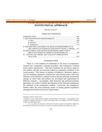
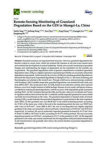
Revista Latinoamericana de Hipertensión. Vol. 13 - Nº 6, 2018www.revhipertension.comPLA of TNF neutralizing assay on L929 cells. All result obtained in the study were statistically analyzed by Minitab18 in ANOVA interval plot to check comparability of resultsbetween biosimilar and reference product statistically.Resultsto 96 well cell plate in the presence of optimized amountsof TNF-α to allow neutralization. Cell plates were furtherincubated at 37 C with 5% CO2 and 95-98% humidityfor 46 2 hours. At the end of incubation, TNF-neutralizing activity was estimated by Adding a mixture of XTTand PMS to well plates, incubation at 37 C with 5% CO2and 95-98% humidity for 2h 30 min and reduction assay.Results obtained as a percent of cell viability (absorbanceat 570 nm) were plotted against different concentrations(ng/mL) of samples applied in a four-parameter sigmoidaldose-response curve by using graphpad prism software.The TNF-α neutralizing activity of cinnora was expressedas a relative potency with respect to the originator Humira Photolytic:The results of detected parameters in photolytic condition as representative data shown in (table 2). The resultsanalyzed by minitab ANOVA interval plot in Figure1,2 and3 which illustrate no significant difference of bioassay, aggregated forms and acidic/basic charged variants betweenCinnora and Humira in all 1,3 and 8 days.499Table 2: The results of bioassay, total aggregated forms and acidic/basic charged variants in the photolytic stress testing are presented. The results are given in each time point (day 1,3,8) beside control samples of cinnora and Humira .ConditionsParametersSamplesControlDay 1ControlDay 3ControlDay 8CinnoRA 09CinnoRA 10CinnoRA 11HUMIRA 01HUMIRA 02HUMIRA 03CinnoRA 09CinnoRA 10CinnoRA 11HUMIRA 01HUMIRA 02HUMIRA 03CinnoRA 09CinnoRA 10CinnoRA 7HUMIRA 01HUMIRA 02HUMIRA 03181618161718171617171718181617171918CinnoRA 09CinnoRA 10CinnoRA 111191210111299131112159101510914HUMIRA 01HUMIRA 02HUMIRA 03131110131412141210121513141212121513 Bio assayPhotolyticTotal aggregated formsAcidic charged variantsBasic charged variantsFigure 1: Comparison of acidic and basic charged variants by Ion exchange chromatography in photolysis condition (day 1) between Humira and Cinnora . Representative Ion exchange chromatography profile is presented for 3 batches of Cinnora (09,10,11) and 3 batchesof Humira (01,02,03). This degradation pattern is followed in both day 3 and day 7.
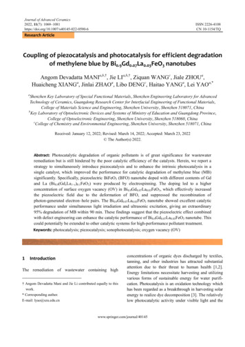
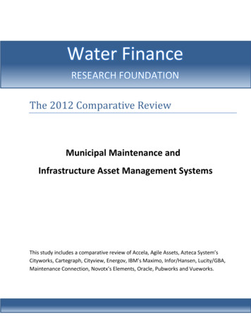
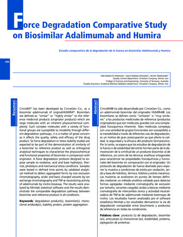
Figure 2: Comparison of total aggregated forms by size exclusion chromatography in photolytic condition (day 1) between Humira andCinnora . Representative Ion exchange chromatography profile is presented for 3 batches of Cinnora (09,10,11) and 3 batches of Humira (01,02,03). This degradation pattern is followed in both day 3 and day 7.500Figure 3: Interval plot of the photolytic condition of Cinnora in comparison with Humira Basic hydrolysis:Basic pH used for degradation of Humira and Cinnora batches the results of aggregated forms, acidic and basic charged variants and bioassay are presented (table 3).Based on ANOVA statistical analysis in figure 4,5 and 6the degradation profile of biosimilar and the referenceproduct is similar. In addition, a significant difference isobserved between control and degraded sample (table 3).
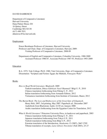
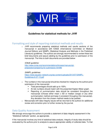
Revista Latinoamericana de Hipertensión. Vol. 13 - Nº 6, 2018www.revhipertension.comTable 3: The results of bioassay, total aggregated forms and acidic/basic charged variants in basic, acidic and oxidative stress testingare presented. The results are given in each time point (day 1,3,8) beside control samples of Cinnora and Humira .ControlBasic HydrolysisAcidic HydrolysisOxidativeControlBasic HydrolysisAcidic HydrolysisOxidativeControlBasic HydrolysisAcidic HydrolysisOxidativeBio assayTotal aggregated formsAcidic charged variantsDay 7936076694570599748056CinnoRA 101015746097480559525047CinnoRA 111026325998540489246043HUMIRA 01HUMIRA 02HUMIRA 0304238000485550CinnoRA 09267052670527865CinnoRA 10257252779627876 Basic charged variantsDay 3CinnoRA 09SamplesParametersDay 1 CinnoRA 11267822783527856HUMIRA 01HUMIRA 02HUMIRA noRA 0915179111520891526159CinnoRA 10151681215209815261513CinnoRA 11161791316201181627149HUMIRA 01HUMIRA 02HUMIRA 625262516151610125CinnoRA 09883166984272985275CinnoRA 10107336510842681085175CinnoRA 11118306410741641175175HUMIRA 01HUMIRA 02HUMIRA 757559 Figure 4: Comparison of acidic and basic charged variants by Ion exchange chromatography in basic hydrolysis condition (day 1) betweenHumira and Cinnora . Representative Ion exchange chromatography profile is presented for 3 batches of Cinnora (09,10,11) and 3batches of Humira (01,02,03). This degradation pattern is followed for day 3 and 7. *mAU:milli area under curve501
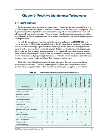
Figure 5: Comparison of total aggregated forms by size exclusion chromatography in basic hydrolysis condition (day 1) between Humira and Cinnora are given. Representative Ion exchange chromatography profile is presented for 3 batches of Cinnora (09,10,11) and 3batches of Humira (01,02,03). This degradation pattern is followed for day 3 and 7.502Figure 6: Interval plot of the basic hydrolysis of Cinnora in comparison with Humira Figure 7: Comparison of acidic and basic charged variants by Ion exchange chromatography in acidic hydrolysis condition (day 1) between Humira and Cinnora is presented. Representative Ion exchange chromatography profile is presented for 3 batches of Cinnora (09,10,11) and 3 batches of Humira (01,02,03). This degradation pattern is followed for day 3 and 7.
www.revhipertension.comRevista Latinoamericana de Hipertensión. Vol. 13 - Nº 6, 2018Acidic hydrolysis:Acidic hydrolysis was implemented by exposing the sample to HCL, and the degaradation is considrable in table3. Besides, according to figure 7,8 and 9 all changes arecomparable between Cinnora and Humira .Thermal stress:Thermolytic degradation is implemented by 25 2ºC. Theresults of all batches of Cinnora and Humira are given intable 4 also figures 13,14 and 15 illustrate the comparability of all parameters in Cinnora and Humira .Oxidative stress:As result of H2O2 exposure to adalimumab exerts its negative impact on the biological activity by increasing aggregate formation which is considerable in table 3. Significant difference between control and sample and similarityin degradation pattern of Cinnora and Humira was observed in figures 10,11 and 12.Mechanical stress:Slightly aggregates increasing was detected in mechanical stress, yet there is no significant difference betweenday 2 and day 4 (table 5). The result of Cinnora and Humira are completely similar and comparable (figure 16,17 and 18).503Figure 8: Comparison of total aggregated forms by size exclusion chromatography in acidic hydrolysis condition (day 1) between Humira and Cinnora is demonstrated. Representative Ion exchange chromatography profile is presented for 3 batches of Cinnora (09,10,11)and 3 batches of Humira (01,02,03). This degradation pattern is followed for day 3 and 7.Figure 9: Interval plot of the acidic hydrolysis of Cinnora in comparison with Humira
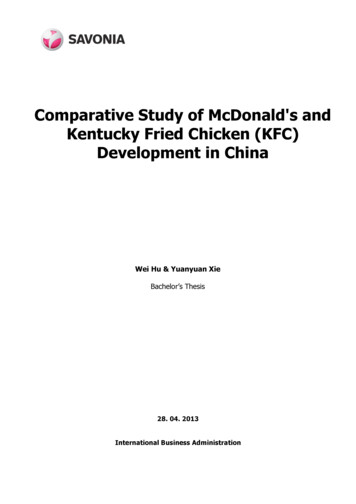
Figure 10: Comparison of acidic and basic charged variants by Ion exchange chromatography in oxidative stress condition (day 1) betweenHumira and Cinnora is given. Representative Ion exchange chromatography profile is presented for 3 batches of Cinnora (09,10,11)and 3 batches of Humira (01,02,03). This degradation pattern is followed for day 3 and 7.504Figure 11: Comparison of total aggregated forms by size exclusion chromatography in oxidative stress condition (day 1) between Humira and Cinnora is demostarated. Representative Ion exchange chromatography profile is presented for 3 batches of Cinnora (09,10,11) and 3 batches of Humira (01,02,03). This degradation pattern is followed for day 3 and 7.Figure 12: Interval plot of the oxidative stress of Cinnora in comparison with Humira
Revista Latinoamericana de Hipertensión. Vol. 13 - Nº 6, 2018www.revhipertension.comTable 4: The results of bioassay, total aggregated forms and acidic/basic charged variants of Cinnora and Humira in the thermal stress condition.ConditionsParametersBio assayTotal aggregated formsThermalAcidic charged variantsBasic charged variantsSamplesCinnoRA 09CinnoRA 10CinnoRA 11HUMIRA 01HUMIRA 02HUMIRA 03CinnoRA 09CinnoRA 10CinnoRA 11HUMIRA 01HUMIRA 02HUMIRA 03CinnoRA 09CinnoRA 10CinnoRA 11HUMIRA 01HUMIRA 02HUMIRA 03CinnoRA 09CinnoRA 10CinnoRA 11HUMIRA 01HUMIRA 02HUMIRA 03Day 19591909710296445433212121201920798576Day 3919294911009356553421232121212291010688Day 782818384898556554425262524232311111181011Figure 13: Comparison of acidic and basic charged variants by Ion exchange chromatography in thermal stress condition (day 1) betweenHumira and Cinnora is demonstrated. Representative Ion exchange chromatography profile is presented for 3 batches of Cinnora (09,10,11) and 3 batches of Humira (01,02,03). This degradation pattern is followed for day 3 and 7.Figure 14: Comparison of total aggregated forms by size exclusion chromatography in thermal stress condition (day 1) between Humira and Cinnora is given. Representative Ion exchange chromatography profile is presented for 3 batches of Cinnora (09,10,11) and 3 batches of Humira (01,02,03). This degradation pattern is followed for day 3 and 7.505
Figure 15: Interval plot of the thermal stress of Cinnora in comparison with Humira 506Table 5: The results of bioassay, total aggregated forms and acidic/basic charged variants of Cinnora and Humira in the mechanical stress conditionConditionsParametersBio assayTotal aggregated formsMechanicalAcidic charged variantsBasic charged variantsSamplesCinnoRA 09CinnoRA 10CinnoRA 11HUMIRA 01HUMIRA 02HUMIRA 03CinnoRA 09CinnoRA 10CinnoRA 11HUMIRA 01HUMIRA 02HUMIRA 03CinnoRA 09CinnoRA 10CinnoRA 11HUMIRA 01HUMIRA 02HUMIRA 03CinnoRA 09CinnoRA 10CinnoRA 11HUMIRA 01HUMIRA 02HUMIRA 03Day 210195989910095535243212019181718899141310Day 491899094929053635422222119182010910141412Figure 16: Comparison of acidic and basic charged variants by Ion exchange chromatography in mechanical stress condition (day 1) between Humira and Cinnora is demonstrated. Representative Ion exchange chromatography profile is presented for 3 batches of Cinnora (09,10,11) and 3 batches of Humira (01,02,03). This degradation pattern is followed for day 3 and 7.
www.revhipertension.comRevista Latinoamericana de Hipertensión. Vol. 13 - Nº 6, 2018Figure 16: Comparison of acidic and basic charged variants by Ion exchange chromatography in mechanical stress condition (day 1) between Humira and Cinnora is demonstrated. Representative Ion exchange chromatography profile is presented for 3 batches of Cinnora (09,10,11) and 3 batches of Humira (01,02,03). This degradation pattern is followed for day 3 and 7.507DiscussionFigure 17: Comparison of total aggregated forms by size exclusion chromatography in mechanical stress condition (day 1) between Humira and Cinnora is given. Representative Ion exchange chromatography profile is presented for 3 batches of Cinnora (09,10,11) and 3batches of Humira (01,02,03). This degradation pattern is followed for day 3 and 7.Photolysis:Ion exchange and size exclusion chromatography andbioassay confirmed that acidic/basic charged variant, dimers and biological activity are identical and meet theacceptable range with similarity in all batches. Thus, theproducts are photo resistant in examined conditions.Basic hydrolysis:Changing pH significantly altered the mechanism of aggregate growth in adalimumab which leads to a decrease in antibody activity and could elicit an immunologicalresponse43-45; in addition, acidic charged variants increase in basic solution and protein get acidic properties. Nosignificant differences in charged variants, high molecularweights and bioassays between Humira and Cinnora batches were observed (Figure 4, 5, 6).Acidic hydrolysis:Acidic hydrolysis caused the generation of a vast amount of aggregated forms which leads to precipitation andcomplete degradation of protein with no biological activity (table 3). Besides, basic charged variants increase inacidic condition. In all parameters, significant differencewas observed between control and sample groups in table 3 which demonstrates the degratative effecs of HCLtreatment on protein degradation.Oxidation:The oxidation of methionine to methionine sulfoxide presumably affects protein structure and stability by increasing basic charged variants reducing side chain hydrophobicity and increasing the capacity for hydrogen bonding[46]. As result of H2O2 exposure to adalimumab exerts its
negative impact on the biological activity by increasing aggregate formation which is considerable in table 3.Thermal stress:Thermolytic degradation is usually thought of as degradation caused by exposure to temperatures high enough toinduce covalent bond breakage, that is, pyrolysis. Raisingthe temperature above accelerated condition (25 2 C)caused aggregated formation and biological activity decreases (table 4). Whereas, it should be considered theresults in bioassay maintain still is an acceptable range aswell as charged variants which demonstrate relative thermolytic stability in Cinnora and Humira .Mechanical stress:A slight increase in aggregated forms is noticeable otherparameters meet the acceptable limits, so Cinnora andHumira are relatively resistant to mechanical stresses.508ConclusionsTotally the information provided by this study shows significant similarity in drug product characteristics, and basedon their similarity in orthogonal characterization studiesthe comparability in degradation pattern can be considered. However, to get information of structural changes inorder to develop a higher resistant medicine in exposurewith different stress conditions some type of structural determinations, such as N-terminal amino acid analysis andglycosylation sites in drug product could be implemented.comprehensive force degradationstrategy implemented to assess thesimilarity between biosimilar and innovator in protein degradation profile. Acidic and basiccharged variants, aggregates, biological activities wereevaluated and the results support the conclusion thatCinnora is analytically similar to Humira under differentstress conditions.Acknowledgement and Funding: This work was supported by CinnaGen Co., In addition the authors wouldlike to thank to all quality control team of CinnaGen Co.for their great efforts in conducting the tests.References1.HUMIRA (adalimumab) lable-FDA. Available at: https://www.accessdata.fda.gov2.Therapeutic monocolonal antibodies. Available at: https://www.uptodate.com3.Bandyopadhyay S, Mahajan M, Mehta T, Singh A.K, Parikh A, GuptaA. K, Kalita P, Patel M, Mendiratta S K.Physicochemical and functionalcharacterization of a biosimilar adalimumab ZRC-3197.Biosimilars2015;5:1–18.4.Magnenata L, Palmeseb A, Fremauxc C, D’Amicid F, TerlizzesedM, Rossib M , Chevalete L. Demonstration of physicochemical andfunctional similarity between the proposed biosimilar adalimumabMSB11022 and Humira. MABS 2017;9(1):127–39.5.Huynh-Ba K. Handbook of Stability Testing in Pharmaceutical D
profile of proteins which are sensitive to temperature, li-quid drug substance and drug products can be exposed to heat without humidity.30 In thermal degradation studies the temperature should be equal or greater than accele-rated stability study are normally conducted at 40 C to 80 C18, Sharma recommends 60 C and 80 C in 5 days