Transcription
original article 19Nightstick Fractures, Outcomesof Operative and Non-Operative TreatmentMohammed Ali*, D. I. Clark, Amole TambeABSTRACTIntroduction: A nightstick fracture is an isolated fracture of the ulnar shaft. Although operative and non-operative treatments have beencommonly decided by the degree of displacement of the fracture, still there is a controversy specially in those moderately displaced. Hereinwe report our experience with nightstick fractures.Objective: To evaluate operative and non-operative treatment of nightstick fracture.Materials and methods: We retrospectively reviewed the clinical notes, physiotherapy letters and radiographs of 52 patients with isolatedulnar shaft fractures. Outcome Measurements included radiographic healing, post-operative range of motion and complications.Results: The study included 13 females and 39 males, with a mean age of 26 years [range, 18–93 years]. The mean Follow-up period was32 months ranged from 12 to 54 months. Ten patients were treated non-operatively; forty-two patients had open reduction and internalfixation including six open fractures. The average wait for surgery was 2.5 days. Mobilisation was commenced immediately after thesurgeries non-load bearing. 40 patients had no complications post-operatively with good outcome and average of four visits follow-up. Inthe non-operative group, five out ten failed and had a mean follow-up of nine visits.Conclusion: Satisfactory outcome is to be expected with open reduction and internal fixation. Fractures with less than 50% displacementshould be treated on individual bases, considering; age, pre-morbid functional status, co-morbidities, compliance and associated injuries.KEYWORDSnightstick fracture; ulnar shaft fracture; non-operative management; non-unionA U T H O R A F F I L I AT I O N SRoyal Derby Hospital, Derby, United Kingdom* Corresponding author: Trauma and Orthopaedics Department, South Tyneside District Hospital, Harton Ln, South Shields NE34 0PL,United Kingdom; e-mail: mohammedkhider84@hotmail.comReceived: 14 January 2018Accepted: 8 November 2018Published online: 1 April 2019Acta Medica (Hradec Králové) 2019; 62(1): 19–23https://doi.org/10.14712/18059694.2019.41 2019 The Authors. This is an open-access article distributed under the terms of the Creative Commons Attribution ), which permits unrestricted use, distribution, and reproduction in any medium,provided the original author and source are credited.
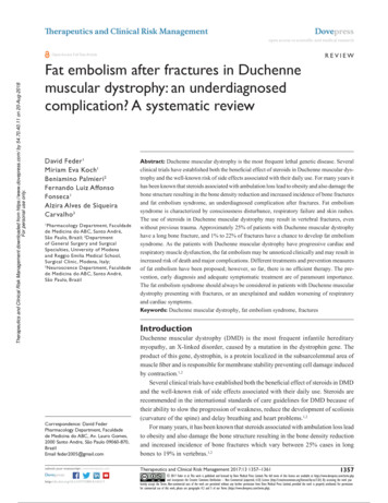
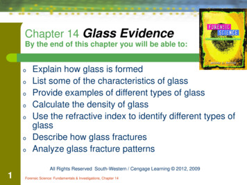
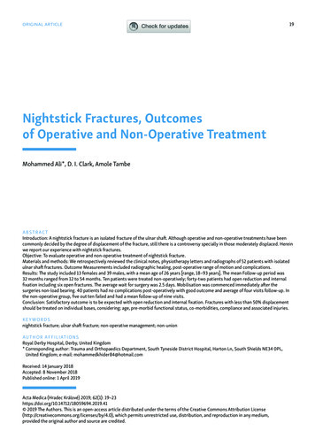
20 INTRODUCTIONMohammed Ali et al. Acta Medica (Hradec Králové)A nightstick fracture is an isolated fracture of the ulnarshaft (IUSF) associated with a direct blow usually as a result of the forearm being held in protection across the face(1). It can also occur with excessive supination or pronation. In these fractures, the integrity of the periosteumand the interosseous membrane determines the stabilityof the fracture and this is normally indicated by the initialdisplacement. Although typically closed fractures, theyhave a higher rate of delayed union or non-union. The aimof management is to prevent the complications of malunion and non-union and restore the best possible function of the limb. Majority of nightstick fractures used tobe treated non-operatively (1) and numerous methods ofimmobilisation have been adopted by surgeons. However,the treatment of isolated ulnar fractures remains controversial, with different authors advocating both surgicaland non- surgical management. Herein we report our experience with nightstick fractures.they either discharged with an open appointment or givenfurther follow-up appointment.In the non-operative group; immobilisation was obtained through an above elbow back-slab in a mid-proneposition with the elbow in 90 degrees flexion and the wristin a neutral position. After one week, patients had a repeatradiograph to check the position of the fracture. The backslab was then either completed or converted to a full cast atone week if the fracture position still acceptable. Patientshad x-rays at two and four weeks in view of late slipping.Patients also had x-rays at 8 weeks and when there wasgood evidence of healing, cast was removed they were referred to physiotherapy. At 12 weeks, they will be reviewedagain with plain radiographs to assess the healing of thefracture specially if patients are symptomatic.Upon review, fracture union was considered whenthere is a bridging callus with no tenderness or movementat the fracture site. Non-union was declared when therewas evidence of hypertrophic callus without bridging andpersistent pain at the fracture site.METHODSRESULTSRetrospectively, we reviewed 96 consecutive patientswith ulnar shaft fractures admitted to our hospital fromSeptember 2010 to December 2015. The study comprisedreview of patients’ clinical notes and radiographs. Ulnarshaft fractures with ipsilateral radial, humeral, or wristfractures were excluded. Add to that, Monteggia fractureswere excluded, as were fractures of the olecranon or coronoid or styloid processes. These fractures resulted fromassaults with a stick like weapon, road accidents or falls.52 patients met the inclusion criteria. The method of treatment was decided by the treating surgeon. Part of thesefractures was treated with open reduction and internalfixation using plates and the other part were treated closedwith an above elbow arm cast.We also reviewed the site of these fractures (proximal,middle, or distal shaft) and the degree of displacement.Surgeries were performed either under general anaesthesia with local anaesthetic infiltration or regional block.Patients were placed in a supine position with the armplaced on an upholstered arm-board. Pneumatic tourniquet was used in all cases. An ulnar approach to the ulnarshaft was performed in all operative cases.For antimicrobial prophylaxis, Cefuroxime was usedintravenously, with 1.5 g injected intra-operatively and0.75 g injected after 8 and 16 hours. The operated arm of allpatients had a wool and crepe dressing and put in a broadarm sling post-operatively.Patients started gentle wrist and shoulder movementnon-load bearing immediately after surgery. Patients thenseen after two weeks in outpatient clinics, where they hada wound check and started gentle elbow exercise including active pronation/ supination movement. All patientshad post-operative physiotherapy referral initiated at twoweeks as per our hospital protocol. Reduction and fixationwere checked with plane radiographs at two weeks, sixand 12 weeks post-operatively. Based on the progress withthe physiotherapy and the bony union on the radiographs52 cases were found to be isolated ulnar shaft fractures.This included 13 females and 39 males, with a mean age of26 years [range, 18–93 years]. The mean Follow-up periodwas 32 months ranged from 12 months to 54 months. Onepatient had proximal third shaft fracture, 12 patients haddistal shaft fractures and 39 patients demonstrated midshaft fractures. 16 fractures were comminuted, 22 obliquedisplaced fractures, 13 transverse displaced fractures, oneun-displaced transverse fracture. 6 patients had Openfractures.THE OPERATIVE GROUP42 patients had open reduction and internal fixation usingplate and screw fixation, this group included six open fractures. 38 fractures were fixed using a dynamic compression plate (DCP) and four with a limited contact dynamiccompression plate (LC-DCP) (Figure 1). The mean waitingtime for surgery was 2.5 days, ranging from 1 to 7 days.None developed wound infection or wound breakdownpost-operatively. There were no recorded instances ofnerve damage.Adequate union was achieved in 40 (95%) cases. Twopatients (5%) after DCP fixation, developed non-unionduring the follow-up (20 and 24 weeks) period and required a revision surgery (Figure 2). Fixation in both cases was done after anatomical reduction. Both patients haduneventful early post-operative course however one patient was reported to be a smoker and the other patientwe could not find any mechanical or biological reason forthe non-union.In the 42 surgical cases; Anatomical reduction fromtime of surgery was maintained in all patients duringfollow-up. All patients were reported to have full supination, pronation and mean flexion arc of 10 to 130 degrees( / 10) at 12 Week. Good callus formation was noted in40 patients at 12 weeks. No reported cases of mal-union or
Nightstick Fractures, Operative vs Non-Operative Treatment 21Fig. 1 x-rays show anatomical reduction of the fracture and fixationusing plate and screws.Fig. 2 X-rays show revision of a non-united fracture.metal work failure. Only one patient was reported to develop metal work irritation from a DCP plate and requireda metal work removal after 6 months. The average numberof hospital visits was 4 (ranged from 4 to 6 visits).surgical route. At two weeks, four patients had furtherdisplacement beyond the acceptable limits. Three of themhad a surgical fixation after two weeks and one patient wasdeemed not to be fit for surgery and went into non-union.The average number of hospital visits was 9 (ranged from7 to 10 visits).The other five patients had above elbow plaster cast ina mid-prone position. Cast was reduced to below elbow at4 weeks and removed at eight weeks. Patients were thenreferred to physiotherapy. At 12 weeks, they were reviewedagain with plain radiographs and fractures deemed to beTHE NON-OPERATIVE GROUPTen patients were treated non-operatively (Table 1). Fivepatients were declared to fail non-operative treatment.One patient developed a mal-union however; he was happy with the function and did not want to go down theTab. 1 Patients who received non-operative plications73/M4IHD, OPNoMid-shaft disp-obliqueAbove elbow castingNon-union49/M1NoneNoMid-shaft comminutedAbove elbow castingNone89/M4OP, IHD, CKDNoMid-shaft comminutedAbove elbow castingNone35/F1NoneNoDistal third disp-obliqueAbove elbow castingNoneFurther displacementbeyond acceptable limit21/M1NoneYesDistal third disp-obliqueAbove elbow casting/ ORIF73/M2PVD, OPNoMid-shaft disp-transAbove elbow castingNone59/F3CKD, OPNoMid-shaft disp-transAbove elbow castingNoneFurther displacementbeyond acceptable limit51/F1NoneNoDistal third disp-transAbove elbow casting/ ORIF32/M1NoneNoMid-shaft disp-transMUA / Above elbowcastingMal-union18/M1NoneNoMid-shaft undisp-transAbove elbow casting/ ORIFFurther displacementbeyond acceptable limit
22 united based on radiographs and clinical examination. Patients were reported to have ( 5 degrees) from full supination and pronation and mean flexion arc of 10 to 120 degrees ( / 10). At 12 weeks, all were discharged to the careof the physiotherapy and left with an open appointmentshould they have any problems in the future.DISCUSSIONNon-operative treatment has been embraced by manyauthors and traditionally, benign neglect or non-operative management was reported to give satisfactory results with a prompt return to function and good healingrates. Traditionally, non-operative treatment was mainlyrecommended for non-displaced fractures or fractureswith less than 50% displacement (2–4). The 1984 paper byDymond et al. (5) which was authored based on a cadaveric study, concluded that more than 50% displacementinvolves considerable disruption of the periosteum andof the interosseous membrane. These displaced fractureswere deemed to be unstable and necessitate above-elbowimmobilisation for stability. In a minimally displaced ulnar shaft fracture, these structures, together with the intact radius, provide a strong stabilizing effect, which mayexplain why some authors (6, 7) were able to achieve satisfactory outcomes by treating low-energy ulnar fractureswith minimum immobilization (8, 9). Furthermore, twostudies have concluded that minimally displaced IUSFsin the middle and distal third of the ulna are stable andcan be mobilised at earlier stages (10, 11). On the otherhand, some authors suggested that proximal IUSF are besttreated by ORIF, believing that the soft-tissue forces tendto destabilize fractures in this region. Also, it is possiblethat some of these are occult Monteggia fractures thathave spontaneously reduced (9, 10). Hopper and Sarmiento although they advised non-operative treatment for diaphysial fractures in the distal two thirds, they excludedthose in which the bone ends are displaced by 5 mm ormore and advised to be treated with open reduction andinternal fixation, particularly if the mechanism of injury was high energy (2, 11). Riska and Nottage, shared thesame views however, they excluded patients with head injury, spinal cord injury, or poly-trauma patient in whominternal fixation of distal two-thirds ulnar fractures mayfacilitate acute care or rehabilitation (12, 13). Szabo et al.(14) retrospectively reviewed the treatment and outcomeof 46 isolated fractures of the ulnar shaft. 18 fractures hadinternal fixation and 28 were treated closed. One openfracture became infected following fixation and failed tounite. Seven failed the non-operative treatment and ended up with non-union. They suggested prognostic factorsfor non-union in non-operatively treated fractures whichinclde; proximal third fractures and those with displacement 5 mm or more.Immobilisation positions have been discussed as having a great role in maintaining fracture reduction andthe eventual acceptable alignment of the healed fracture.Traditionally, recommendations for immobilizing theforearm in neutral, supination, or pronation positionshave been based on theory, anecdotal experience, andMohammed Ali et al. Acta Medica (Hradec Králové)tradition (15–18). A few clinical studies have shown success with the forearm immobilized in either pronation orsupination (16, 18). Altner et al. (19) in their series, theyimmobilised patients in a mid-prone position and reported good results. Add to that Boyer et al. (20) evaluated theeffect of forearm position on the healing outcomes following non-surgical treatment using above elbow cast. Theyconcluded that residual fracture angulation at the time ofunion was not significantly affected by forearm position.A review by Mackay (21) et al. in 2000, included 33 series involving 1876 patients. The outcomes of the non-surgical treatment of minimally displaced ulnar fractureswith a stable configuration were consistently satisfactory.Below elbow plaster cast, functional brace and early mobilisation all achieved similar results. Above elbow cast wasdeemed to be unnecessarily restrictive. Mackay also concluded that open reduction and internal fixation is betterused in widely displaced or unstable fractures to preservethe forearm rotation.Moed et al. (22) Reported the outcomes of immediateinternal plate fixation of an open diaphyseal fracture ofthe forearm in fifty patients. Although they had two cases of deep infection and non-union in six, the functionalresults were excellent or good in 85 per cent of the series.They related these results to the severity of the initialsoft-tissue injury and the surgical technique and recommended autogenous cancellous bone-grafting in comminuted fractures. On the other hand, Wright et al. (23)studied 198 forearm fractures to determine the union ratewhere acute bone grafting was recommended but not performed. The overall union rate in comminuted, non-grafted forearm fractures (open and closed) was 98%. Anotherstudy by Wei et al. (24) concluded that acute bone graftingof diaphyseal forearm fractures did not affect the unionrate or the time to union.Recently, Cai and his fellow researchers (1) reviewed allpublished randomised controlled trials and observationalstudies that have assessed the outcome of these fracturesfollowing above- or below-elbow immobilisation, bracingand early mobilisation. They included 27 studies comprising 1629 fractures. They found that early mobilisationproduced the shortest radiological union time and thelowest mean rate of non-union. They advised early mobilisation, with a removable forearm support for the treatment of nondisplaced or partially displaced nightstickfractures.Coulibaly et al. in 2015 conducted a retrospectivecase-control analysis on patients diagnosed with IUSF tocompare surgical and nonsurgical outcomes (25). Theymeasured complications and functional ability. They foundthat nonsurgical treatment of IUSF is prone to complications and is associated with mal-union and non-unionwhile Surgical treatment with rigid plate fixation andearly range of motion resulted in a shorter period of castimmobilization and an earlier return to weight bearing,and led to reduced patient morbidity.In our study, although the numbers are not comparable, complication rates in the non-operatively treatedgroup were significantly higher than those reported inthe operated group. In our study five fractures, Failed thenon-operative treatment. Another notable correlate in
Nightstick Fractures, Operative vs Non-Operative Treatment our series was location in the middle third of the shaft,an experience not shared in previous papers. Also, patients who developed these complications either werevery young and might not be very compliant or elderlywith multiple co-morbidities and this was also noted byCoulibaly.The limitations of our study include its retrospectivenature, the small number of patients and the relativelyshort duration of follow-up. There are other elements thatwe did not measure and that could have contributed to ourconclusion.CONCLUSIONBased on our study and the published literature we believethat IUSFs with more than 5 mm displacement should betreated operatively. Satisfactory outcome is to be expectedwith open reduction and internal fixation with rigid plateas it allows early mobilisation and enables earlier return tofunction with very low risk of wound problems. Althoughour study did not reveal good results with non-operativetreatment, we believe those with less than 5 mm displacement should be treated on individual bases, considering;age, pre-morbid functional status, co-morbidities, compliance and associated injuries.REFERENCES1. Cai XZ, Yan SG, Giddins G. A systematic review of the non-operativetreatment of nightstick fractures of the ulna. Bone Joint J 2013 Jul;95-B(7): 952–9.2. Hooper G. Isolated fractures of the shaft of the ulna. Injury 1974;6(2): 180–4.3. Du Toit FP, Grabe RP. Isolated fractures of the shaft of the ulna. S AfrMed I 1979; 56(1): 21–5.4. Corea JR, Brakenbury PH, Blakemore ME. The treatment of isolatedfractures of the ulnar shaft in adults. Injury 1981; 12(5): 365–70.5. Dymond IW. The treatment of isolated fractures of the distal ulna.J Bone Joint Surg (Br) 1984; 66(3): 408–10.6. Pollock FH, Pankovich AM, Prieto JJ, Lorenz M. The isolated fracture23of the ulnar shaft. Treatment without immobilization. J Bone JointSurg Am 1983; 65(3): 339–42.7. Sarmiento A. Isolated ulnar shaft fractures treated with functionalbraces. J Orthop Trauma 1998; 12(6): 420–3.8. Ekelund AL, Nilsson OS. Early mobilization of isolated ulnar shaftfractures. Acta Orthop Scand 1989; 60(3): 261–2.9. de Jong T, de Jong PC. Ulnar shaft fracture needs no treatment. A pilotstudy of 10 cases. Acta Orthop Scand 1989; 60(3): 263–4.10. Zych GA, Latta LL, Zagorski JB. Treatment of isolated ulnar shaftfractures with prefabricated functional fracture braces. Clin Orthop1987; (219): 194–200.11. Sarmiento A. Treatment of ulnar fractures by functional bracing.J Bone Joint Surg Am 1976; 58(8): 1104–7.12. Nottage WM. A review of long bone fractures in patients with spinalcord injuries. Clin Orthop 1981; (155): 65–70.13. Riska EB, von Bonsdorff H, Hakkinen S, Jaroma H, Kiviluoto O,Paavilainen T. Primary operative fixation of long bone fractures inpatients with multiple injuries. J Trauma 1977; 17(2): 111–21.14. Szabo RM, Skinner M. Isolated ulnar shaft fractures. Retrospectivestudy of 46 cases. Acta Orthop Scand 1990 Aug; 61(4): 350–2.15. Davis DR, Green DP. Forearm fractures in children. Clin Orthop 1976;120: 172–84.16. Evans EM. Rotational deformity in the treatment of fractures of bothbones of the forearm. J Bone Joint Surg 1945; 27: 373–9.17. Gibbons CLMH, Woods DA, Pailthorpe C, et al. The management ofisolated distal radius fractures in children. J Pediatr Orthop 1994; 14:207–10.18. Gupta RP, Danielsson LG. Dorsally angulated solitary metaphysealgreenstick fractures in the distal radius: Results after immobilizationin pronated, neutral, and supinated position. J Pediatr Orthop 1990;10: 90–219. Altner, PC, Ted Hartman, J. Isolated Fractures of the Ulnar Shaft inthe Adult. Surg Clin North Am 1972; 52(1): 155–70.20. Boyer BA, Overton B, Schrader W, Riley P, Fleissner P. Position of Immobilization for Pediatric Forearm Fractures. J Pediatr Orthop 2002;22(2): 185–7.21. Mackay D, Wood L, Rangan A. The treatment of isolated ulnar fractures in adults: a systematic review. Injury 2000; 31(8): 565–70.22. Moed BR, Kellam JF, Foster RJ, Tile M, Hansen ST Jr. Immediate internal fixation of open fractures of the diaphysis of the forearm. J BoneJoint Surg Am 1986 Sep; 68(7): 1008–17.23. Wright RR, Schmeling GI, Schwab JP. The necessity of acute bonegrafting in diaphyseal forearm fractures: a retrospective review.J Orthop Trauma 1997 May; 11(4): 288–94.24. Wei SY, Born CT, Abene A, Ong A, Hayda R, DeLong WG Jr. Diaphyseal forearm fractures treated with and without bone graft. J Trauma1999; 6: 1045–825. Coulibaly MO, Jones CB, Sietsema DL, Schildhauer TA. Results of70 consecutive ulnar nightstick fractures. Injury 2015 Jul; 46(7):1359–66.
uneventful early post-operative course however one pa-tient was reported to be a smoker and the other patient we could not find any mechanical or biological reason for the non-union. In the 42 surgical cases; Anatomical reduction from time of surgery was maintained in all patients during follow-up. All patients were reported to have full supina-