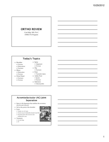
Transcription
10/29/2012Curt Stilp, MS, PA-COHSU PA Program Shoulder DislocationOveruseWrist/Hand Spine Hip Knee Ankle/Foot FracturesOveruse FracturesTraumatic injurySprainFractureInfections AlignmentOveruse Elbow OveruseDislocationsFracturesOsteomyelitisInjury to the ligaments that stabilize the acromion,clavicle and coracoid Fall on the point of the shoulder Exam Tender over AC joint Clavicle may be ballotable (Grade II and III) Increased pain with crossover andpulling down arm Treatment Ice and sling PT1
10/29/2012 Continuum of Impingement – Bursitis – Strain –Partial Tear – Complete Tear History Symptoms Repetitive overhead motion Diffuse anterior pain Pain worse in arc of 60-120⁰ Exam X-ray MRI Impingement testsShape of AcromionNeer TestSupraspinatus TestHawkinsTest2
10/29/2012Treatment Rest Sub-acromialinjection Rehab Surgical repairAnterior – most commonABduction/Ext Rot 98% of all dislocations Sulcus sign PosteriorADduction/Int RotSeizures, electrocutions Must get a lateral X-ray Treatment Reduction - modified Kocher method Surgery Must do a pre and post-reduction neuro exam3
10/29/2012Easy to managebecause of large ROMof shoulderHanging traction vs.IM RodTry to preventstiffness of the elbowInjury to the radialnerve Anatomy Wrist Extensor attachmentCauses Repetitive wrist extension Repetitive grippingSigns and Symptoms Pain with wrist extension Shaking hands Extension of middle finger againstresistance Tender over lateral epicondyleTreatment Rest PT Compression wraps4
10/29/2012TreatmentReduction by extension, supination, thenflexion (can put pressure over radial headto feel for “clunk”) Child feels relief almost immediately andstarts using the arm Educate parents about lifting child fromunder the arms Carpal Tunnel Syndrome(CTS)5
10/29/2012Repetitive wrist/finger movementForceful loading of tendons in carpal tunnelExtreme wrist flexion/extensionVibrationNon-occupational factors DiabetesConnective Tissue DzPregnancyMal-union of a wrist fractureAnatomically small carpal tunnelSigns Positive Phalens test (flexion) Positive Tinels test (tap) Positive compression test 6mm 2-point discrimination Bilateral symptoms thinksystemic diseaseConservative Approach SymptomsNumbness (worse in AM)Grip Thumb-finger opposition Thenar atrophy Activity ModificationNSAIDSSplintingInjectionSurgical Decompression OpenEndoscopic Technique6
10/29/2012 Anatomy – First Dorsal Compartment of the Wrist AbPL EPBCauses Repetitive gripping or extension of the ThumbSigns and Symptoms Tenderness Positive Finkelstein test Must differentiate from Scaphoid (navicular)tenderness and CMC arthritisTreatment Splint Ice PT NSAID’s Cortisone injection Surgical decompression7
10/29/2012 Colles’ and Smith’s fractureFall on the outstretched handObvious deformity on exam Re-establish proper alignment Dinner fork deformity – Colles’Short arm cast (4-6 weeks) vs. closed reductionvs. External fixator vs. ORIFROM of fingers and elbowConsider PT/OT8
10/29/2012 Fracture of the 5th MC neck(not base)Can accept 40⁰ of dorsalangulationTreatment Ulnar Gutter SplintMCP flexed at 60⁰4-6 weeksHistory Fall on outstretched hand Exam X-ray Complications Snuffbox tendernessMay be negative at firstAVN9
10/29/2012 Repeat X-ray in 7-10 days May get CT or MRI Treatment Thumb spica cast or splint for 8-10 weeks 10 curve as measured by Cobb angle3-D deformity that includes:Curvature in the coronal planeKyphosis or lordosis in the sagittal plane Rotation in the axial plane Adolescent Idiopathic Scoliosis (AIS)Most common type9-10 y/o female Found on school screening Change in appearance Uneven shoulders, hips, skin folds Asymptomatic and painless10
10/29/2012 Adams forward bend testLook for a rib or muscle prominence Looking at rotation only Full column PA and lateral standing films,shoes removed, on single long cassetteDetermine type of curveGrade severity Evaluate skeletal maturity Cobb angleRisser sign11
10/29/2012 Options are observation, bracing and surgeryTreatment generally depends on the age of thepatient and the severity and risk forprogression of the curveThe following table serves as a guideline fortreatment based on the spinal curvature:Curve DegreeTreatment Options 20 degreesObservation20-40 degreesBracing 40 degreesSurgery Low back pain when ambulating orextending the lower back (walkingdownhill)Relieved when lying, sitting or squatting orbending forwardBilateral radicular leg painPositive “shopping cart” signDifferent that vascular claudication pain Symptoms with walking a predictable distanceand relieved by stopping and standingNSAIDS, Lumbar corsetPhysical Therapy- lumbar flexion programEpidural steroid injections (may have up to3 per year)12
10/29/2012Low back pain increased with prolongedsitting and activityPain relieved with extensionRadicular leg painPhysical Therapy - lumbar extensionprogram History of lifting something heavy /- back painUnilateral radicular leg painNumbness/tingling Treatment 80% get better on their ownLaminotomy/Discectomy13
10/29/2012 Presentation Types Lying on the gurney with a externally rotated andslightly shortened legSubcapital, Femoral Neck, Basilar Neck, Intertrochanteric, Sub-trochantericTreatment Pinning vs Hemi-arthroplastyMechanisms of Injury Valgus InjuriesPivoting InjuriesDiagnosis Physical Examination Lachman, Anterior Drawer Acute (1-2 hours) Bloody Effusion MRITreatment Non-surgical vs. SurgicalAssociated with medial meniscal tear and MCLrupture – “Bloody Triad”14
10/29/2012AnteriorDrawerLachman’sDiagnosis History Twisting injury Effusion over 1-2 days Exam Joint line tenderness McMurray’s or Apley Grind Test MRITreatment Arthroscopic repair vs. menisectomyInversion – most commonEversion15
10/29/2012PE Findings Drawer test Talar tilt testDiagnosis Grades I-III based on nSwellingAble to walkUnable torunIIPartial TearPainSwellingBruisingPain withwalkingIIITearPainSwellingBruisingUnable towalkTreatment - Early Mobilization- Use RICE treatment acutely- Ankle Rehab Flexibility Strength Proprioception16
10/29/2012- AP, Lateral, Mortise view- Follow Ottawa Ankle Rules (OAR) Get an X-ray for: Malleolar pain and tenderness Inability to bear weight for 4 steps- Swelling is NOT a reliable guide to thepresence of a fracture Stress fractureTransverse fracture through the base of the 5thMT1-2 cm from the proximal tipShort-leg cast vs. ORIFHigh incidence of nonunion17
10/29/2012 Bacterial infection of the bonePathogensStaph aureus – MOST COMMONSalmonella in children with sickle celldisease Pseudomonas after puncture wounds(animal bites) Symptoms – Vary by ageLocalized bone pain Fussiness and fever Swelling, warmth and erythema of theoverlying tissue Decreased movement of the affectedextremity (pseudo paralysis in infants,refusal to walk or limp in older children) 18
10/29/2012Radiographic findings: Remember: Radiographic findings lag behind infection by 1-2weeks Bone scan is most sensitive for detecting earlyosteomyelitis Plain film findings: Blurring or obliteration of soft tissue fat planes (theearliest sign) Elevation of the periosteum (pus) with periostealreaction Cortical destruction and endosteum scalloping Eventually a sequestrum and involucrum maydevelop AC joint dislocation Rotator Cuff Radial N. injuryTennis Elbow Pain with overheadactivityHumerus Fx Fall on point ofshoulderPain with wristextension DeQuervain’s Colles’ Fx Boxers Fx Scaphoid Fx Scoliosis shopping cart signUlnar gutter splintSnuff box tendernessUnilateral leg painShortened externallyrotated legACL Acute bloodyeffusionMeniscusNon-bloody effusionover 1-2 days Ankle Sprain Ankle Fx Jones Fx Osteomyelitis Hip Fx FOOSH, dinner fork HNP Cobb angleLSSS FinkelsteinsPicture Phalens, Tinels, thenaratrophy Nursemaids CTS Inversion, ATFLMortise wideningNon-unionStaph A.19
10/29/2012 1 Curt Stilp, MS, PA-C OHSU PA Program Shoulder Overuse Dislocations Fractures Elbow Dislocation Overuse Wrist/Hand Fractures Overuse Spine Alignment Overuse Hip Fractures Knee Traumatic injury Ankle/Foot Sprain Fracture Infections Osteomyelitis Injury to the ligaments that stabilize the acromion, .