Transcription
TECHNOLOGY STATUS EVALUATION REPORTEndoscopic mucosal resectionPrepared by: ASGE TECHNOLOGY COMMITTEEJoo Ha Hwang, MD, PhD, FASGE, Vani Konda, MD, Barham K. Abu Dayyeh, MD, MPH,Shailendra S. Chauhan, MD, FASGE, Brintha K. Enestvedt, MD, MBA, Larissa L. Fujii-Lau, MD,Sri Komanduri, MD, John T. Maple, DO, FASGE, Faris M. Murad, MD, Rahul Pannala, MD, MPH,Nirav C. Thosani, MD, Subhas Banerjee, MD, FASGE, ChairThis document was reviewed and approved by the Governing Board of the American Society for GastrointestinalEndoscopy.The ASGE Technology Committee provides reviews ofexisting, new, or emerging endoscopic technologies thathave an impact on the practice of GI endoscopy.Evidence-based methodology is used, by using a MEDLINEliterature search to identify pertinent clinical studies onthe topic and a MAUDE (U.S. Food and Drug Administration Center for Devices and Radiological Health) database search to identify the reported adverse events of agiven technology. Both are supplemented by accessingthe “related articles” feature of PubMed and by scrutinizing pertinent references cited by the identified studies.Controlled clinical trials are emphasized, but in manycases, data from randomized, controlled trials are lacking. In such cases, large case series, preliminary clinicalstudies, and expert opinions are used. Technical data aregathered from traditional and Web-based publications,proprietary publications, and informal communicationswith pertinent vendors. For this review, the MEDLINEdatabase was searched for publications in Englishthrough September 2014 by using the keywords “endoscopic lesion removal,” “endoscopic resection,” “endoscopic mucosal resection,” and “EMR.”Technology Status Evaluation Reports are drafted by 1or 2 members of the ASGE Technology Committee, reviewed and edited by the committee as a whole, andapproved by the Governing Board of the ASGE. Whenfinancial guidance is indicated, the most recent codingdata and list prices at the time of publication are provided. Technology Status Evaluation Reports are scientificreviews provided solely for educational and informational purposes. Technology Status Evaluation Reportsare not rules and should not be construed as establishinga legal standard of care or as encouraging, advocating,requiring, or discouraging any particular treatment orpayment for such treatment.BACKGROUNDEMR was developed for minimally invasive, organ-sparingendoscopic removal of benign and early malignant lesions inthe GI tract. This report focuses on instruments, injectionsolutions, and techniques currently used for EMR. Thisreport is an update of a previous Technology Status Evaluation Report titled “Endoscopic Mucosal Resection and Endoscopic Submucosal Dissection.”1 The topic of endoscopicsubmucosal dissection (ESD) is now discussed in aseparate Technology Status Report.2TECHNOLOGY UNDER REVIEW: EMREMR is an endoscopic technique developed for theremoval of sessile or flat neoplasms confined to the superficial layers (mucosa and submucosa) of the GI tract. Thecommonly used techniques can be categorized as injection-, cap-, and ligation-assisted EMR. Underwater EMR isa newer technique that is useful, particularly for salvageEMR.Proper patient and lesion selection for EMR with endoscopic and/or endosonographic evaluations is essential.Before the start of any EMR procedure, close visual inspection to delineate the margins, particularly of flat lesions, isimperative because manipulation of the lesion mayobscure landmarks. It may be helpful to mark the marginsof the targeted lesion with superficial cautery marks withthe tip of a snare or with argon plasma coagulation(APC). Electrosurgical unit settings for polypectomy andEMR are discussed in a previous Technology committeedocument.3 A retrieval device may then be used toretrieve EMR specimens.Injection-assisted EMRCopyright ª 2015 by the American Society for Gastrointestinal Endoscopy0016-5107/ jection-assisted EMR is also often called saline solutionlift–assisted polypectomy. This technique was introducedin 1955 for rigid sigmoidoscopy and then in 1973 for flexible colonoscopy.4,5 The procedure starts with injection ofwww.giejournal.orgVolume 82, No. 2 : 2015 GASTROINTESTINAL ENDOSCOPY 215
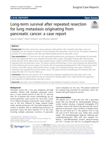
Endoscopic mucosal resectiona solution into the submucosal space under the lesioncreating a safety cushion. The cushion lifts the lesion, facilitating capture and removal by using a snare while minimizing mechanical or electrocautery damage to thedeeper layers of the GI wall. The lesion may be removedin a single resection or a piecemeal fashion.Cap-assisted EMRCap-assisted EMR also uses submucosal injection to liftthe target mucosal lesion. Dedicated mucosectomy deviceshave been developed that use a cap affixed to the tip of theendoscope (EMR Kit; Olympus America Inc, Center Valley,Pa) (Table 1).6 These single-use devices come equippedwith a specially designed crescent-shaped electrocauterysnare that must be opened and positioned on the internalcircumferential ridge at the tip of the cap (Fig. 1). Theendoscope is then immediately positioned over the targetlesion, and suction is used to retract the mucosa into thecap after which the snare is closed to capture the lesion.The lesion is then resected with standard snare excisiontechnique by using electrocautery. The available capassisted mucosectomy devices differ primarily in the characteristics of the cap. Caps are composed of clear plastic,which may be soft or hard. The caps are cylindrical and available with a flat circular (straight)– or oval (oblique)–shapedtip, both with outer diameters ranging from 12.9 to 18 mm.Ligation-assisted EMRIn ligation-assisted EMR, a band ligation device (DuetteMulti-Band Mucosectomy device, Cook Medical Inc.,Winston-Salem, NC) is attached to the endoscope, andthe banding cap is positioned over the target lesion withor without previous submucosal injection. Suction isapplied to retract the lesion into the banding cap, and aband is deployed to capture the lesion, thereby creatinga pseudopolyp. An electrocautery snare is then used toresect the pseudopolyp above or below the band.7,8 Thehandle of the EMR band ligator allows insertion andadvancement of a snare device through the endoscopeworking channel without requiring removal of the bandingapparatus. The kit also includes a 1.5 2.5-cm hexagonalbraided electrocautery snare available with a 5F (for diagnostic endoscopes) or 7F (for therapeutic endoscopes)insertion sheath. In addition, the band ligation device incorporates 6 bands, allowing potential resection at asmany as 6 mucosal sites without the need to change thedevice. Two sizes of ligating caps are available to fit endoscopes with outer diameters of 9.5 to 13 mm and 11 to14 mm.away from the deeper muscularis propria layer and allowsEMR without requiring submucosal injection.9 Thistechnique has the theoretical advantages of eliminatingany risk of tracking neoplastic cells into deeper layers ofthe GI tract wall by the injection needle and makingcapture of flat lesions easier. This method has also beenreported to be effective in managing recurrences afterprevious EMR, as well as patients with previous partialresections and biopsies of lesions10 because theseinterventions may result in submucosal fibrosis, makinglifting of the lesion with submucosal injection difficult.11-18Adjunctive techniquesAdditional ablative techniques are used in an organspecific manner in addition to EMR for the ablation ofresidual tissue. In the esophagus, radiofrequency or cryoablation is frequently used to ablate additional Barrett’sesophagus after EMR of the dysplastic lesions. Duringresection of flat adenomas in the GI tract, APC or theuse of hot biopsy forceps (also known as the hot avulsiontechnique) may be used to ablate residual adenomatoustissue at the base and edges of the resection site.13,19-22However, the application of APC to ablate residual adenomatous tissue was associated with a higher risk of adenomarecurrence.23 Use of the snare tip with soft coagulation forresidual tissue that cannot be removed by snare resectionin the colon is currently being evaluated in a randomized,controlled trial.Specimen handlingBecause EMR specimens are larger than biopsy samples,it is helpful for pathologic interpretation to orient andmount the specimen before submerging it in fixative.The specimen is often pinned onto a paraffin wax blockand fully submerged in fixative before transporting thespecimen to pathology. A paraffin wax block is beneficialbecause it will not float in fixative.Submucosal injection solutionsIn the underwater EMR (UEMR) technique, luminal air issuctioned, and water is instilled to fill the GI lumen andimmerse the target lesion. Water immersion allows lesionvisualization without over distention of the GI tract wall.It is postulated to “float” the mucosa and submucosaAlthough submucosal injection is not essential for allEMR procedures, it is an integral part of injection-assistedEMR. Various solutions have been used for submucosal injection (Table 2). The ideal agent should be inexpensive,readily available, nontoxic, easy to inject, and provide along-lasting submucosal cushion.24,25 Normal saline solutionis widely available and often used for injection-assisted EMR.However, a cushion made with normal saline solution oftendissipates within minutes. Multiple studies have demonstrated longer lasting cushions made with various agentsincluding hyaluronic acid (HA), hydroxypropyl methylcellulose (HPMC), succinylated gelatin, glycerol, and a fibrinogensolution.26-32 Currently, there are no injection solutions thatare specifically approved for EMR by the U.S. Food andDrug Administration; therefore, all solutions mentioned inthis document would be considered off-label. A 0.4% solution of HA is approved as an injection solution for216 GASTROINTESTINAL ENDOSCOPY Volume 82, No. 2 : 2015www.giejournal.orgUnderwater EMR
Endoscopic mucosal resectionTABLE 1. Commercially available devices for EMRMucosectomy devicesEndoscopediameter, mmEMR kits (include cap, needle,and 25-mm crescent snare)Hard straight 13.9-mm capManufacturerCost, US *Minimum working channelsize required, mmOlympus America Inc,Center Valley, PA347 each2.0Cook Medical Inc,Winston-Salem, NC315 each9.3-10Hard straight 14.9-mm cap10-11Hard wide oblique 16.1-mm cap9.3-10Hard wide oblique 16.1-mm cap10-11Hard straight 12.9-mm cap8.6-9.2Hard wide oblique 16.1-mm cap8.6-9.2Soft oblique 18-mm cap8.6-9Soft oblique 18-mm cap9.1–9.8Soft oblique 18-mm cap9.8-10.4Soft oblique 18-mm cap10.3-11.3Soft oblique 18-mm cap11.2-11.8Duette Multi-Band Mucosectomy *Manufacturer list price is provided as a reference. The actual price may vary depending on specific contracts.submucosal injection in Japan (MucoUp; Johnson & Johnson, Tokyo, Japan) and has been demonstrated to sustainmucosal lifting longer than saline solution, reducing the volume of injection solution necessary to complete an endoscopic resection.28Randomized studies evaluating HA solutions (0.4%, 0.2%,and 0.13%) used for submucosal injections in endoscopicresection demonstrate that HA solutions are more effectivein maintaining mucosal elevation than saline solution.28,33,34An inexpensive, over-the-counter preparation of HA (0.15%concentration) is available in the United States (Blink Contacts; AMO, Santa Ana, Calif) and was reported in a retrospective case series to be an effective agent for submucosal liftingfor EMR.35 HPMC has also been demonstrated to be effectivein mucosal lifting for EMR.29,30,36 HPMC preparations areavailable in the United States as ophthalmic lubricants andare typically viscous solutions and must be diluted to 0.3%to 0.8% to facilitate injection. The use of succinylated gelatinas a submucosal injection solution for colonic EMR has beendemonstrated to facilitate removal of large colonic lesions( 20 mm) in fewer pieces for piecemeal EMR in a randomized, double-blind trial compared with normal saline solution.37 This study also demonstrated that the use ofsuccinylated gelatin resulted in fewer resections per lesion,fewer injections per lesion, lower injection volumes, andshorter procedure duration. Glycerol (10% solution) and asolution containing fibrinogen have also been reported aseffective in sustaining mucosal lifting after submucosalinjection; however, the use of these agents has not beenreported in the United States.31,32 Tissue damage, delayedhealing, and local inflammatory reaction have been reportedat the injection sites when using HPMC, hypertonic sodiumchloride (3.75%), and hypertonic dextrose ( 20%) in animalmodels; however, the clinical significance of these findingshas not been studied.38,39The volume of submucosal injection solution used during EMR varies depending on the size of the lesion and thesolution used for injection. Repeated injections may berequired if the cushion dissipates before complete removalof the lesion.Dilute epinephrine (1:100,000–1:200,000) is often addedto the submucosal injection fluid because of the theoreticalbenefits of decreased bleeding and a sustained submucosalcushion (due to delayed absorption of fluid resulting fromdecreased vascular flow) and is generally considered to besafe.40 Submucosal injection of epinephrine potentiallycan result in systemic effects such as severe hypertension,ventricular tachycardia, and intestinal ischemia; however,case reports regarding these complications are rare andresult from procedures in which the goal of therapy washemostasis rather than EMR and during which higherconcentrations of epinephrine (1:10,000) were used.41-43Staining dye (ie, diluted indigo carmine or methyleneblue) is frequently added to the injection solution to facilitate identification of the lateral and deep margins of thetarget lesion before and during the resection process. Thestaining dye may also improve recognition of muscularispropria injury and intraprocedural perforation.44,45www.giejournal.orgVolume 82, No. 2 : 2015 GASTROINTESTINAL ENDOSCOPY 217
Endoscopic mucosal resectionFigure 1. A, A clear plastic EMR cap attached to the end of an endoscope. B, The snare is opened and positioned on the internal circumferential ridge atthe tip of the cap before suctioning the lesion into the cap. (Photographs courtesy of Olympus America, Center Valley, Pa.)CLINICAL APPLICATIONSEMR may be used for definitive therapy of premalignantand early-stage (T1N0) malignant lesions of the digestive tractif there is limited submucosal invasion. EUS is often used forlocoregional staging before endoscopic resection. EMR canalso be used to obtain larger histologic specimens (comparedwith the standard endoscopic tissue sampling techniques)and can provide an accurate histological T stage for these superficial malignancies.46 These techniques can also be usedto obtain a histologic diagnosis of subepithelial lesions inthe GI tract located in the muscularis mucosa or superficialsubmucosa.47,48 In general, EMR should not be attempted ifinvasion into the deep submucosa or beyond is suspected.Nonlifting of the lesion after submucosal injection is a predictor of deep invasion and indicates that the lesion is notamenable to endoscopic removal.49,50 However, EMR maybe attempted if the nonlifting is thought to be a consequenceof submucosal fibrosis related to previous manipulation (previous biopsy or attempted/incomplete EMR) of the lesion.10In addition, it has been shown that deep submucosalinvasive cancer can be predicted with endoscopic imagingevaluation by using either the NBI international colorectalendoscopic classification or the Kudo classification (usingchromoendoscopy and magnification endoscopy). BothNBI International Colorectal Endoscopic Classification type3 lesions and Kudo type V lesions predict deep submucosalinvasive carcinoma with endoscopic evaluation exhibitingareas of irregular disrupted or missing vessels along with anamorphous or absent surface/pit pattern on endoscopicevaluation.51,52easier to perform than ESD.15,53,54 For large gastric lesions,the reported mean time to complete EMR is 25.8 25.9 minutes compared with 84.0 54.6 minutes for lesionremoval by ESD.55Injection-assisted EMR offers the advantages of wideavailability because it does not require specialized kits.During cap-assisted EMR, the positioning of the snarewithin the cap before tissue capture may be challengingand a relatively unfamiliar maneuver for endoscopists andassistants. Positioning of the snare within the cap may befacilitated by lightly approximating the endoscope tipwith the cap against an area of normal mucosa beforeattempting EMR of the target lesion. Ligation-assistedEMR is relatively easy to perform because it combinescommonly used endoscopic techniques for variceal bandligation and snare polypectomy and does not require special prepositioning of the snare. In addition, multiple resections can be performed sequentially.EFFICACYEsophagusEMR is essentially a variation of standard polypectomy using specialized devices. Depending on the size and locationof the lesion, EMR can potentially be a technically difficultand time-consuming procedure; however, it is technicallyEMR and ESD are indicated for treatment of superficialesophageal cancer and Barrett’s esophagus–associatedneoplasia (high-grade dysplasia and intramucosal carcinoma).56-58Barrett’s esophagus–associated neoplasia. EMR iscommonly used for Barrett’s esophagus–associatedneoplasia. Techniques vary from focal EMR of nodular lesions with adjunct ablation techniques to complete EMRfor eradication of the entire Barrett’s segment. A randomized trial comparing cap-assisted EMR with ligation-assistedEMR for the resection of Barrett’s-associated neoplasiademonstrated that ligation-assisted EMR was significantlyfaster than cap-assisted EMR with median procedurestimes of 34 minutes and 50 minutes, respectively (P Z.02) with no differences in rates of adverse events or quality of the resection specimens.59218 GASTROINTESTINAL ENDOSCOPY Volume 82, No. 2 : 2015www.giejournal.orgEASE OF USE
Endoscopic mucosal resectionTABLE 2. Solutions for submucosal injection during EMR and ESDSolutionNormal saline solution (0.9%)Hypertonic sodium chloride (3.0%)Hyaluronic acid (0.13%-0.4%)Hydroxypropyl methylcellulose(0.3%-0.8%)Cushion durationAdvantagesDisadvantagesþEasy to inject, inexpensive,readily availableDissipates quickly, shortduration of mucosal liftingþþEasy to inject, inexpensive,readily availablePossible tissue damage andlocal inflammation at injection sitesþþþþLongest-lasting cushionLimited availability in the U.S.Off-label use preparations available in the U.S.require further clinical evaluationþþþLong-lasting cushion,relatively inexpensivePossible tissue damage and localinflammation at injection sitesContraindicated in patients with gelatinhypersensitivitySuccinylated gelatinþþEasy to inject, inexpensive,readily availableGlycerol (10%)þþInexpensiveDextrose (20%, 30%, 50%)þþInexpensive, readily availablePossible tissue damage and localinflammation at injection sites Increasedrisk of postpolypectomy syndromeAlbuminþþEasy to inject, available in mostendoscopy unitsExpensiveEasy to inject, long-lasting cushionExpensive, not readily availableFibrinogenþþþA single-center study of 107 patients evaluating the efficacy of complete EMR for Barrett’s esophagus–associatedneoplasia demonstrated that EMR was able to completelyeradicate Barrett’s esophagus and all associated neoplasia(high-grade dysplasia and intramucosal carcinoma) in98.8% of patients who completed therapy per protocolwithout high-risk characteristics (submucosal invasion,poorly differentiated tumors, or evidence of lymphatic orvascular invasion) and in 80.4% of patients who underwenttherapy with an intention to treat including those patientswho had high-risk characteristics.58 Recurrence rates forboth cancer and high-grade dysplasia were 1.4%. Anothermulticenter retrospective study from Europe evaluated patients who had Barrett’s esophagus–associated neoplasiatreated with complete EMR of Barrett’s esophagus anddemonstrated that 58% required additional thermal ablation to eradicate residual disease.56 Neoplasia recurred in6.2% of patients.EMR has also been evaluated in conjunction with radiofrequency ablation for the treatment of Barrett’s esophagus–associated neoplasia, in which EMR was initiallyperformed on any endoscopically visible abnormalities,followed by circumferential radiofrequency ablation performed at least 6 weeks after EMR.60 After initialcomplete eradication of Barrett’s esophagus, sustainedremission of neoplasia and intestinal metaplasia wasachieved in 90% of patients at 5 years of follow-up.Superficial squamous cell cancer. A meta-analysisthat included 8 studies from Asia comparing results of ESDand EMR for endoscopic resection of superficial esophagealcancer (primarily squamous cell carcinoma) demonstratedthat ESD compared with EMR had a significantly higheren bloc resection rate (97.1% vs 49.3%; odds ratio [OR]52.76; 95% confidence interval [CI], 25.57-108.84) and alower recurrence rate (0.3% vs 11.5%; OR 0.08; 95% CI,0.03-0.23); however, there was no difference in recurrencerate if the lesion size was less than 20 mm (OR 0.34; 95%CI, 0.06-2.08).57 In addition, the procedure duration wassignificantly longer for ESD compared with EMR.www.giejournal.orgVolume 82, No. 2 : 2015 GASTROINTESTINAL ENDOSCOPY 219StomachEarly gastric cancer. Both EMR and ESD are used forthe resection of early gastric cancer, and in the UnitedStates, the choice is often dictated by local availability ofendoscopic and surgical expertise. Asian data indicatethat ESD is associated with lower local recurrence ratesthan EMR, although survival rates are similar.61 A metaanalysis that included 9 retrospective studies evaluatedthe efficacy of ESD and EMR.61 Although the mean timefor ESD was longer than EMR, the ESD group had asignificantly lower local recurrence rate than EMR (0.7%vs 6.4%; OR 0.10; 95% CI, 0.06-0.18). ESD is thepreferred method for removal of early gastric cancerlesions in Asia because it allows histological assessmentof lateral margins that is not possible with piecemealEMR and is associated with lower local recurrencerates.62 EMR may be preferred in patients with severecomorbid conditions such as liver cirrhosis orcardiovascular disease because EMR is associated withshorter procedure times and fewer adverse events.63 Ameta-analysis has demonstrated that proton pump inhibitors are superior to histamine type 2 receptor antagonistsfor the prevention of bleeding after gastric EMR.64
Endoscopic mucosal resectionNonampullary duodenal adenomas. Duodenal lesions not involving the major duodenal papilla can beremoved with a variety of EMR techniques, but carries anincreased risk of bleeding and perforation because the duodenum has increased vascularity and a thin wall. The majority of published reports on endoscopic removal ofduodenal polyps originates from high-volume centers andare limited by low numbers of patients and a retrospectivestudy design.68-71 Reported success rates vary from 70% to96% for nonampullary duodenal lesions.68,69,71 In addition,the technique of UEMR has been reported for resectingnonampullary duodenal adenomas with high success ratesfor complete resection (83% at the index session); however, adverse events included delayed bleeding, water intoxication syndrome, and stricture formation.72in a nonrandomized clinical trial.11 Furthermore, therecurrence rate was significantly lower in the UEMR groupthan the EMR group (10% vs 39.4%, P Z .02).Although several studies have reported no recurrenceafter endoscopic removal of malignant colonic polyps,the effectiveness of EMR for the treatment of these lesionshas been questioned, and EMR should not be attemptedfor nonlifting lesions or lesions classified as Paris II-c/III.15,76-78 However, nonlifting lesions that have previouslybeen manipulated (biopsy or attempted EMR) beforereferral for resection are usually amenable to EMR.10Endoscopic resection has also been reported to be successful in resecting small rectal carcinoid tumors. Technically, the procedure should be considered an endoscopicsubmucosal resection because a majority of rectal carcinoid tumors extend into the submucosal layer.79,80 Theuse of ligation-assisted EMR for lesions that were estimatedto be less than 1 cm in diameter has resulted in resectionswith negative margins.79 However, another study thatcompared ESD with ligation-assisted EMR for endoscopicresection of carcinoid tumors that were less than 16 mmdemonstrated a higher histologically complete resectionrate with ESD compared with EMR (90.3% vs 71.0%, P Z.035), although ESD took longer to perform.81ColonSafetyInjection-assisted EMR is widely used for the resectionof large or flat colonic lesions. A systematic review andmeta-analysis demonstrated that local recurrence afterEMR occurs in 3% of cases in which the lesion is removeden bloc and in 20% of cases in which the lesion is removedin piecemeal fashion.73 For recurrences that were retreated with endoscopic therapy (APC and/or EMR), thesubsequent recurrence rate was 21%, with successful eradication being achieved in 91.4% of recurrences after amean of 1.2 additional sessions. Another large multicenterprospective study with 1000 consecutive wide-field EMRsof large sessile adenomas demonstrated a 16% recurrencerate at 4 months, usually unifocal and diminutive, and a4% recurrence rate at 16 months.23 Recurrences weremanaged endoscopically in 93% of cases. If a largeadenoma ( 15 mm) is removed in piecemeal fashion,the patient should have a repeat colonoscopy in 6 to 12months to evaluate for local recurrence.73,74 A metaanalysis comparing ESD with EMR for colorectal tumorsthat included 6 studies (1642 total lesions) demonstratedthat ESD had a higher en bloc resection rate and lowerinitial local recurrence rate than EMR; however, ESD wasmore time-consuming and generally required hospitalization for observation after the procedure.75Typically, EMR in the colon is performed by using theinjection-assisted EMR technique; however, UEMR withoutsubmucosal injection has also been reported.9 Whenthe UEMR technique was used, the complete removalrate for recurrences was significantly higher comparedwith injection-assisted EMR (88.9% vs 31.8%, P .001)Adverse events after EMR include bleeding, perforation,and strictures. Bleeding is the most common adverse eventof EMR.Colonic EMR. Intraprocedural bleeding rates afterEMR of colorectal lesions larger than 20 mm are reportedto be between 11% and 22%.14,82 The application of softcoagulation with the tip of a snare has been demonstratedto be both safe and effective for the treatment of intraprocedural bleeding during EMR of large colonic polyps.82Other methods including hot biopsy forceps, monopolarhemostatic forceps, bicap probes, APC, and endoscopicclips can be used for achieving hemostasis. Risk factorsfor intraprocedural bleeding include lesion size, Parisendoscopic classification of 0-IIa þ Is, tubulovillous orvillous histology, and low-volume institutions.14 Bleedingrates after EMR of large colonic polyps range from 2%to 11%.12-18 The clinically significant bleeding rate afterEMR of sessile colorectal polyps larger than 20 mm wasreported to be 6% in a large prospective, multicenter studythat included 1039 patients.14 Of the patients with clinicallysignificant bleeding, 34% required endoscopic therapy.Prophylactic endoscopic coagulation of nonbleedingvessels by using coagulating forceps after EMR ofcolorectal lesions larger than 20 mm did not significantlydecrease the incidence of delayed postprocedure bleedingcompared with control subjects who received noadditional therapy (5.2% vs 8.0%, P Z .3). Risk factorsfor clinically significant postprocedural bleeding included aproximal colonic location, polyp size, and intraproceduralbleeding.14220 GASTROINTESTINAL ENDOSCOPY Volume 82, No. 2 : 2015www.giejournal.orgGastric carcinoids. EMR has been reported to beeffective in resecting type 1 gastric carcinoids (those associated with chronic atrophic gastritis) that are less than 1cm in diameter.65-67 EMR has been reported to be associated with a slightly lower rate of complete resectioncompared with ESD due to positive deep margins; however, local recurrence rates and survival are similar.66,67Duodenum
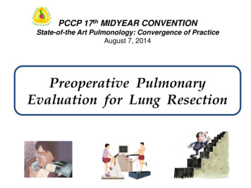
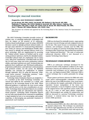
Endoscopic mucosal resectionFigure 2. Three examples of a target sign in the resected specimen. All 3 figures demonstrate resected polyps with resected muscularis propria appearingas a white circular disk surrounded by blue-stained submucosa, giving the appearance of a “target.” (Reprinted with permission from Swan et al.84 )Perforation after EMR of colonic lesions is also rare( 1%).15,16,76,83 A perforation can be identified by carefullyexamining the resection defect. Also, the transected surfaceof the resected specimen should be examined for a “target”sign. If muscularis propria has been inadvertently resected,the transected surface will have a white to gray central circulardisk surrounded by blue-stained submucosal tissue (if thesubmucosal injectate contained blue dye), giving the appearance of a “target” (Figs. 2 and 3).84 Small perforationsrecognized during the procedure can be successfully sealedby using endoscopic clips.84-87 Larger perforations mayrequire urgent salvage surgery to prevent peritonitis.Esophageal EMR. Bleeding after EMR in the esophagus is uncommon.21,88-92 In the largest single-center studythat included 681 patients who underwent 2513 EMRs, significant bleeding requiring intervention, transfusion, orhospitalization was noted in only 1.2% of patients.92Reported perforation rates during EMR for esophageallesions are relatively low at less than 0.5% for physicianswho are experienced in performing EMR.53,58,92-100 However, 1 study demonstrated a perforation rate of 5% in thefirst 120 esophageal EMRs performed by 6 physicians whowe
submucosal injection in Japan (MucoUp; Johnson & John-son, Tokyo, Japan) and has been demonstrated to sustain mucosal lifting longer than saline solution, reducing the vol-