Transcription
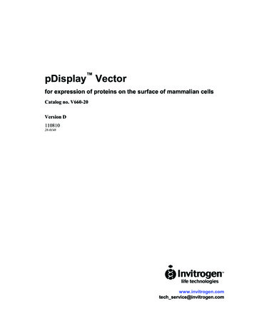
pDisplay Vectorfor expression of proteins on the surface of mammalian cellsCatalog no. V660-20Version D11081028-0148www.invitrogen.comtech service@invitrogen.com
ii
Table of ContentsTable of Contents. iiiImportant Information. vIntroduction. 1Overview. 1Methods. 2Cloning into pDisplay . 2General Guidelines for Transformation and Transfection. 4Troubleshooting . 6Appendix . 7pDisplay Vector. 7Technical Service. 9References. 11iii
iv
Important InformationContents20 µg of pDisplay , lyophilized in TE, pH 8.0.Shipping/StorageLyophilized plasmids are shipped at room temperature and should be stored at -20 C.ProductQualificationThe pDisplay vector is qualified by restriction digest. Restriction digests mustdemonstrate the correct banding pattern when electrophoresed on an agarose gel. Thetable below lists the restriction enzymes used to digest the vector and the expectedfragments.Restriction EnzymeExpected Fragments (bp)BamH I170, 5155Bgl II5325Pst I5325v
vi
IntroductionOverviewIntroductionpDisplay is a 5.3 kb mammalian expression vector that allows display of proteins on thecell surface. Proteins expressed from pDisplay are fused at the N-terminus to the murineIg κ-chain leader sequence, which directs the protein to the secretory pathway, and at theC-terminus to the platelet derived growth factor receptor (PDGFR) transmembrane domain,which anchors the protein to the plasma membrane, displaying it on the extracellular side.Recombinant proteins expressed from pDisplay contain the hemagglutinin A and mycepitopes for detection by western blot or immunofluorescence. To get started with cloninginto pDisplay , see page 4.TestedApplications ofpDisplay The pDisplay vector has been used to express c-jun and a single chain antibody againstphOx hapten (Chesnut et al., 1996). Both proteins were shown to be correctly expressed atthe cell surface by incubation with anti-myc antibody followed by incubation with amagnetic bead-conjugated secondary antibody. Transfected cells were selected by using amagnet.For your convenience, Anti-myc Antibody is available from Invitrogen, Catalog no.R950-25. In addition, Anti-myc Antibody is available as a horseradish peroxidase (HRP)conjugate or an alkaline phosphatase (AP) conjugate for one-step westerns (Catalog nos.R951-25 and R952-25, respectively).1
MethodsCloning into pDisplay IntroductionThe following section provides general guidelines for cloning your gene of interest intothe pDisplay vector. For help with DNA ligations, E. coli transformations, restrictionenzyme analysis, purification of single-stranded DNA, DNA sequencing, and DNAbiochemistry, please see Molecular Cloning: A Laboratory Manual (Sambrook et al.,1989) or Current Protocols in Molecular Biology (Ausubel et al., 1994).Maintenance ofpDisplay In order to propagate and maintain pDisplay , we recommend that you resuspend thelyophilized vector in 20 µl sterile water to make a 1 µg/µl stock solution. Store at -20 C.Use this stock solution to transform an E. coli strain that is recombination deficient (recA)and endonuclease A deficient (endA) such as TOP10F (Catalog no. C615-00) orequivalent.For your convenience, TOP10F is available from Invitrogen as chemically competent orelectrocompetent cells.ItemElectrocomp Ultracomp TOP10F TOP10F (chemically competent cells)One Shot TOP10F (chemically competent cells)Cloning into thepDisplay VectorQuantityCatalog no.5 x 80 µlC665-555 x 300 µlC665-0321 x 50 µlC3030-03pDisplay vector is a fusion vector requiring that you clone your gene of interest in framewith the initiation ATG of the N-terminal Ig κ-chain leader sequence and the C-terminalmyc epitope/PDGFR-TM. It may be necessary to use PCR to create a fragment with theappropriate restriction sites to clone in frame at both ends. Carefully inspect your geneand the multiple cloning site before cloning your gene of interest. A diagram of themultiple cloning site is included on the next page.continued on next page2
Cloning into pDisplay , ContinuedBelow is the multiple cloning site for pDisplay . Restriction sites are labeled to indicatethe cleavage site. The multiple cloning site has been confirmed by sequencing andfunctional testing.Multiple CloningSite of pDisplay 5 end of hCMV promoter/enhancer1GCGCGCGTTG ACATTGATTA TTGACTAGTT ATTAATAGTA ATCAATTACG GGGTCATTAG61TTCATAGCCC ATATATGGAG TTCCGCGTTA CATAACTTAC GGTAAATGGC CCGCCTGGCT121GACCGCCCAA CGACCCCCGC CCATTGACGT CAATAATGAC GTATGTTCCC ATAGTAACGC181CAATAGGGAC TTTCCATTGA CGTCAATGGG TGGACTATTT ACGGTAAACT GCCCACTTGG241CAGTACATCA AGTGTATCAT ATGCCAAGTA CGCCCCCTAT TGACGTCAAT GACGGTAAAT301GGCCCGCCTG GCATTATGCC CAGTACATGA CCTTATGGGA CTTTCCTACT TGGCAGTACA361TCTACGTATT AGTCATCGCT ATTACCATGG TGATGCGGTT TTGGCAGTAC ATCAATGGGC421GTGGATAGCG GTTTGACTCA CGGGGATTTC CAAGTCTCCA CCCCATTGAC GTCAATGGGA481GTTTGTTTTG GCACCAAAAT CAACGGGACT TTCCAAAATG TCGTAACAAC TCCGCCCCATenhancer region (5 end)AP1enhancer region (3 end)CAAT541TATA3 end of CMVTGACGCAAAT GGGCGGTAGG CGTGTACGGT GGGAGGTCTA TATAAGCAGA GCTCTCTGGCPutative transcriptional startT7 promoter/priming site601TAACTAGAGA ACCCACTGCT TACTGGCTTA TCGAAATTAA TACGACTCAC TATAGGGAGA661CCCAAGCTTG GTACCGAGCT CGGATCCACT AGTAACGGCC GCCAGTGTGC TGGAATTCGGIg k-chain leader sequence721CTTGGGGATA TCCACC ATG GAG ACA GAC ACA CTC CTG CTA TGG GTA CTG CTGMet Glu Thr Asp Thr Leu Leu Leu Trp Val Leu Leuhemagglutinin A epitope773CTC TGG GTT CCA GGT TCC ACT GGT GAC TAT CCA TAT GAT GTT CCA GATLeu Trp Val Pro Gly Ser Thr Gly Asp Tyr Pro Tyr Asp Val Pro AspSfi I821Xma I Sma ISac IISal IPst I Acc ITAT GCT GGG GCC CAGCCGGCCA GATCTCCCGG GATCCGCGG CTGCAGGTC GACTyr Alamyc epitope874Bgl IIPDGFR transmembrane domain (5 end)924GAA CAA AAA CTC ATC TCA GAA GAG GAT CTG AATGCTGTGG GCCAGGACACGlu Gln Lys Leu Ile Ser Glu Glu Asp Leu .GCAGGAGGTC ATCGTGGTGC CACACTCCTT GCCCTTTAAG GTGGTGGTGA TCTCAGCCAT984CCTGGCCCTG GTGGTGCTCA CCATCATCTC CCTTATCATC CTCATCATGC TTTGGCAGAA1044GAAGCCACGT TAGGCGGCCG CTCGAGATCA GCCTCGACTG TGCCTTCTAG TTGCCAGCCA1104TCTGTTGTTT GCCCCTCCCC CGTGCCTTCC TTGACCCTGG AAGGTGCCAC TCCCACTGTC1164CTTTCCTAAT AAAATGAGGAPDGFR (3 end)BGH poly (A) addition site3
General Guidelines for Transformation and TransfectionThe following guidelines and recommendations are provided for your convenience. If youneed more details about the techniques discussed, please refer to the general molecularbiology references in the References section.E. coliTransformationTransform your ligation mixtures into a competent recA, endA E. coli strain (e.g. TOP10,TOP10F , DH5α) and select on LB plates containing 50-100 µg/ml ampicillin. Select 1020 clones and analyze for the presence and orientation of your insert.MENDIONATRECOMIntroductionWe recommend that you sequence your construct with the T7 Promoter (Catalog nos.N560-02) and a gene specific reverse primer to confirm that your gene is correctly fusedto the Ig κ-chain leader sequence at the N-terminus and the myc/PDGFR-TM peptide atthe C-terminus.PlasmidPreparationOnce you have confirmed that your gene is correctly fused, prepare plasmid DNA fortransfection. Plasmid DNA for transfection into eukaryotic cells must be very clean andfree from phenol and sodium chloride. Contaminants will kill the cells and salt willinterfere with lipids, decreasing transfection efficiency. We recommend using theS.N.A.P. Miniprep Kit (Catalog no. K1900-01) for isolation of 10 - 15 µg of plasmidDNA or CsCl gradient centrifugation for isolation of 15 µg of plasmid DNA.Methods ofTransfectionFor established cell lines (e.g. HeLa, 293), please consult original references or thesupplier of your cell line for the optimal method of transfection. It is recommended thatyou follow exactly the protocol for your cell line. Pay particular attention to mediumrequirements, when to pass the cells, and at what dilution to split the cells. Furtherinformation is provided in Current Protocols in Molecular Biology (Ausubel et al.,1994).Methods for transfection include calcium phosphate (Chen and Okayama, 1987; Wigler etal., 1977), lipid-mediated (Felgner et al., 1989; Felgner and Ringold, 1989) andelectroporation (Chu et al., 1987; Shigekawa and Dower, 1988). Invitrogen offers theCalcium Phosphate Transfection Kit (K2780-01) and a large variety of reagents formammalian transfection. See our Web site at www.invitrogen.com for more details ontransfection products.continued on next page4
General Guidelines for Transformation and Transfection,ContinuedGeneticin (G418)ActivityGeneticin blocks protein synthesis in mammalian cells by interfering with ribosomalfunction. It is an aminoglycoside, similar in structure to neomycin, gentamycin, andkanamycin. Expression of the bacterial aminoglycoside phosphotransferase gene (APH),derived from Tn5, in mammalian cells results in detoxification of Geneticin (Southernand Berg, 1982).Geneticin SelectionGuidelinesGeneticin is available from Invitrogen in both 1 g (Catalog no. 11811-023) and 5 g(Catalog no. 11811-031) quantities. Use as follows: Prepare Geneticin in a buffered solution (e.g. 100 mM HEPES, pH 7.3). Calculate concentration based on the amount of active drug (check the lot label). Use 100 to 800 µg/ml of Geneticin in complete medium Test varying concentrations of Geneticin on your cell line to determine theconcentration that kills your cells (kill curve). Cells differ in their susceptibility toGeneticin Cells will divide once or twice in the presence of lethal doses of Geneticin , so the effectsof the drug take several days to become apparent. Complete selection can take up to3 weeks of growth in selective medium. For more information, see Ausubel et al. (1994)unit 9.5.Detection ofDisplayedProteinsYou may wish to confirm that your protein in being displayed on the cell membrane byusing in situ immunofluorescent labeling of cells with antibodies to c-myc or tohemagglutinin A. Alternatively, you may use an indirect magnetic selection procedurewith a magnetic bead-conjugated secondary antibody. For basic protocols, please seeAntibodies: A Laboratory Manual (Harlow and Lane, 1988, p. 359-421)or CurrentProtocols in Molecular Biology (Ausubel et al., 1994, p. 14.6.1-14.6.2)5
TroubleshootingIntroductionThe table below describes solutions to some possible problems.ProblemNo ExpressionProtein is not displayedReasonSolutionMethod of transfection isnot optimal.Optimize transfection orchange transfection method.Protein is not in frame withIg k-chain leader sequenceSequence your construct tocheck frame.Protein is not in frame withPDGFR-TMUse antibody tohemagglutinin or myc tocheck for expression.Sequence your construct tocheck frame.6
AppendixpDisplay VectorFeatures ofpDisplay pDisplay (5325 bp) contains the following elements. All features have been functionallytested.FeatureBenefitHuman cytomegalovirus (CMV)immediate-early promoter/enhancerPermits efficient, high-level expression ofyour recombinant protein (Andersson et al.,1989; Boshart et al., 1985; Nelson et al.,1987)T7 promoter/priming siteAllows for in vitro transcription in the senseorientation and sequencing through theinsertATG initiation codonPermits initiation of translation of thepDisplay fusion proteinMurine Ig κ-chain leader sequenceTargets protein to secretory pathway(Coloma et al., 1992)Hemagglutinin A epitope tagAllows detection of the fusion protein bymonoclonal antibody 12CA5 (Kolodziejand Young, 1991; Niman et al., 1983)(Tyr-Pro-Tyr-Asp-Val-Pro-Asp-Tyr-Ala)Multiple cloning region with eight unique Allows insertion of your gene andsitesfacilitates cloningmyc ows detection of pDisplay fusionprotein with the Anti-myc Antibodies(Evan et al., 1985)Platelet-derived growth factor receptortransmembrane domain (PDGFR-TM)Anchors the fusion protein to the plasmamembrane for display (Gronwald et al.,1988)Bovine growth hormone (BGH)polyadenylation signalEfficient transcription termination andpolyadenylation of mRNA (Goodwin andRottman, 1992)pUC originHigh-copy number replication and growthin E. coliSV40 early promoter and originPermits expression of the kanamycinresistance gene for Geneticin resistance inmammalian cellsAllows episomal replication in cellscontaining SV40 large T antigenKanamycin resistance geneConfers resistance to Geneticin inmammalian cellsTK polyadenylation signalEfficient transcription termination andpolyadenylation of kanamycin resistancegene mRNAAmpicillin resistance gene (β-lactamase)Selection in E. colif1 originAllows rescue of single-stranded DNAcontinued on next page7
pDisplay Vector, ContinuedSignalPeptideMVPCmycT7Sfi IBgl IIXma ISma ISac IIPst ISal IAcc IThe figure below shows the features of pDisplay . The complete sequence of pDisplay is available for downloading from our Web site (www.invitrogen.com) or by contactingTechnical Service (see page 9). Details of the multiple cloning site are shown on page 5.HAMap of pDisplay PDGFR Transmembrane DomainBGHpUP SVCMV promoter: bases 1-596T7 promoter: bases 638-657Murine Ig kappa-chain V-J2-C signal peptide: bases737-799Hemagglutinin A epitope: bases 800-826Multiple Cloning Site: bases 827-873myc epitope: bases 874-903PDGFR transmembrane domain: bases 907-1056Bovine growth hormone polyadenylation signal: bases1069-1288pUC origin: bases 1378-2051Thymidine kinase polyadenylation site: bases 2458-2187Neomycin/Kanamycin resistance gene: bases 3421-2366SV40 origin and promoter: bases 3797-34568n/NKaAmComments for pDisplayTM:5325 nucleotidespicillineo5.3 kbATKpf1 oriCpDisplayTMrio/40
Technical ServiceWorld Wide WebVisit the Invitrogen Web Resource using your World Wide Web browser. At the site, youcan: Get the scoop on our hot new products and special product offers View and download vector maps and sequences Download manuals in Adobe Acrobat (PDF) format Explore our catalog with full color graphics Obtain citations for Invitrogen products Request catalog and product literatureOnce connected to the Internet, launch your Web browser (Internet Explorer 5.0 or neweror Netscape 4.0 or newer), then enter the following location (or URL):http://www.invitrogen.com.and the program will connect directly. Click on underlined text or outlined graphics toexplore. Don't forget to put a bookmark at our site for easy reference!Contact UsFor more information or technical assistance, please call, write, fax, or email. Additionalinternational offices are listed on our Web page (www.invitrogen.com).Corporate Headquarters:Japanese Headquarters:European Headquarters:Invitrogen Corporation1600 Faraday AvenueCarlsbad, CA 92008USAInvitrogen Japan K.K.Nihonbashi Hama-Cho ParkBldg. 4F2-35-4, Hama-Cho, NihonbashiInvitrogen Ltd3 Fountain DriveInchinnan Business ParkPaisley PA4 9RF, UKTel: 1 760 603 7200Tel: 81 3 3663 7972Tel: 44 (0) 141 814 6100Tel (Toll Free): 1 800 955 6288Fax: 81 3 3663 8242Fax: 44 (0) 141 814 6287Fax: 1 760 602 6500E-mail: jpinfo@invitrogen.comE-mail: eurotech@invitrogen.comE-mail:tech service@invitrogen.comcontinued on next page9
Technical Service, ContinuedLimited WarrantyInvitrogen is committed to providing our customers with high-quality goods and services.Our goal is to ensure that every customer is 100% satisfied with our products and ourservice. If you should have any questions or concerns about an Invitrogen product orservice, please contact our Technical Service Representatives.Invitrogen warrants that all of its products will perform according to the specificationsstated on the certificate of analysis. The company will replace, free of charge, any productthat does not meet those specifications. This warranty limits Invitrogen Corporation’sliability only to the cost of the product. No warranty is granted for products beyond theirlisted expiration date. No warranty is applicable unless all product components are storedin accordance with instructions. Invitrogen reserves the right to select the method(s) usedto analyze a product unless Invitrogen agrees to a specified method in writing prior toacceptance of the order.Invitrogen makes every effort to ensure the accuracy of its publications, but realizes thatthe occasional typographical or other error is inevitable. Therefore Invitrogen makes nowarranty of any kind regarding the contents of any publications or documentation. If youdiscover an error in any of our publications, please report it to our Technical ServiceRepresentatives.Invitrogen assumes no responsibility or liability for any special, incidental, indirector consequential loss or damage whatsoever. The above limited warranty is sole andexclusive. No other warranty is made, whether expressed or implied, including anywarranty of merchantability or fitness for a particular purpose.10
ReferencesAndersson, S., Davis, D. L., Dahlbäck, H., Jörnvall, H., and Russell, D. W. (1989). Cloning, Structure, andExpression of the Mitochondrial Cytochrome P-450 Sterol 26-Hydroxylase, a Bile Acid BiosyntheticEnzyme. J. Biol. Chem. 264, 8222-8229.Ausubel, F. M., Brent, R., Kingston, R. E., Moore, D. D., Seidman, J. G., Smith, J. A., and Struhl, K. (1994).Current Protocols in Molecular Biology (New York: Greene Publishing Associates and Wiley-Interscience).Boshart, M., Weber, F., Jahn, G., Dorsch-Häsler, K., Fleckenstein, B., and Schaffner, W. (1985). A Very StrongEnhancer is Located Upstream of an Immediate Early Gene of Human Cytomegalovirus. Cell 41, 521-530.Chen, C., and Okayama, H. (1987). High-Efficiency Transformation of Mammalian Cells by Plasmid DNA. Mol.Cell. Biol. 7, 2745-2752.Chesnut, J. D., Baytan, A. R., Russell, M., M.-P.Chang, Bernard, A., Maxwell, I. H., and Hoeffler, J. P. (1996).Selective Isolation of Transiently Transfected Cells from a Mammalian Cell Population with VectorsExpressing a Membrane Anchored Single-Chain Antibody. J. Imm. Methods 193, 17-27.Chu, G., Hayakawa, H., and Berg, P. (1987). Electroporation for the Efficient Transfection of Mammalian Cellswith DNA. Nuc. Acids Res. 15, 1311-1326.Coloma, M. J., Hastings, A., Wims, L. A., and Morrison, S. L. (1992). Novel Vectors for the Expression ofAntibody Molecules Using Variable Regions Generated by Polymerase Chain Reaction. J. Imm. Methods152, 89-104.Evan, G. I., Lewis, G. K., Ramsay, G., and Bishop, V. M. (1985). Isolation of Monoclonal Antibodies Specific forc-myc Proto-oncogene Product. Mol. Cell. Biol. 5, 3610-3616.Felgner, P. L., Holm, M., and Chan, H. (1989). Cationic Liposome Mediated Transfection. Proc. West. Pharmacol.Soc. 32, 115-121.Felgner, P. L., and Ringold, G. M. (1989). Cationic Liposome-Mediated Transfection. Nature 337, 387-388.Goodwin, E. C., and Rottman, F. M. (1992). The 3 -Flanking Sequence of the Bovine Growth Hormone GeneContains Novel Elements Required for Efficient and Accurate Polyadenylation. J. Biol. Chem. 267, 1633016334.Gronwald, R. G., Grant, F. J., Haldeman, B. A., Hart, C. E., O'Hara, P. J., Hagen, F. S., Ross, R., Bowen-Pope, D.F., and Murray, M. J. (1988). Cloning and Expression of a cDNA Coding for the Human Platelet-DerivedGrowth Factor Receptor: Evidence for More Than One Receptor Class. Proc. Natl. Acad. Sci. USA 85, 34353439.Harlow, E., and Lane, D. (1988). Antibodies: A Laboratory Manual (Cold Spring Harbor, NY: Cold Spring HarborLaboratory).Kolodziej, P. A., and Young, R. A. (1991). Epitope Tagging and Protein Surveillance. Meth. Enzymol. 194, 508519.Nelson, J. A., Reynolds-Kohler, C., and Smith, B. A. (1987). Negative and Positive Regulation by a Short Segmentin the 5 -Flanking Region of the Human Cytomegalovirus Major Immediate-Early Gene. Mol. Cell. Biol. 7,4125-4129.Niman, H. L., Houghten, R. A., Walker, L. E., Reisfeld, R. A., Wilson, I. A., Hogle, J. M., and Lerner, R. A.(1983). Generation of Protein-reactive Antibodies by Short Peptides is an Event of High Frequency:Implications for the Structural Basis of Immune Recognition. Proc. Natl. Acad. Sci. USA 80, 4949-4953.continued on next page11
References, ContinuedSambrook, J., Fritsch, E. F., and Maniatis, T. (1989). Molecular Cloning: A Laboratory Manual, Second Edition(Plainview, New York: Cold Spring Harbor Laboratory Press).Shigekawa, K., and Dower, W. J. (1988). Electroporation of Eukaryotes and Prokaryotes: A General Approach tothe Introduction of Macromolecules into Cells. BioTechniques 6, 742-751.Southern, P. J., and Berg, P. (1982). Transformation of Mammalian Cells to Antibiotic Resistance with a BacterialGene Under Control of the SV40 Early Region Promoter. J. Molec. Appl. Gen. 1, 327-339.Wigler, M., Silverstein, S., Lee, L.-S., Pellicer, A., Cheng, Y.-C., and Axel, R. (1977). Transfer of Purified HerpesVirus Thymidine Kinase Gene to Cultured Mouse Cells. Cell 11, 223-232. 1997-2002, 2010 Invitrogen Corporation. All rights reserved.12
Introduction pDisplay is a 5.3 kb mammalian expression vector that allows display of proteins on the cell surface. Proteins expressed from pDisplay are fused at the N-terminus to the murine Ig κ-chain leader sequence, which directs the protein to the secretory pathway, and at the
