Transcription
RADIATION SAFETY IN THE LABORATORYTRAINING MANUALA Short Course Offered by:Radiation Safety OfficeUniversity of South CarolinaPhone: 777-5269Fax: 777-5275E-Mail: Radsafe@mailbox.sc.eduWebsite: http://ehs.sc.edu/Radiation.htmRadiation Safety in the LaboratoryPage 1
LESSON 1:ATOMIC COMPOSITION AND STRUCTUREThe atom consists of two regions: the nucleuswhich contains positively charged particles calledprotons and electrically neutral particles calledneutrons and the area around the nucleus calledthe orbital region where electrons are found. In anelectrically stable atom, there are an equalnumber of protons and electrons. Atoms can beeither stable or unstable. Unstable atoms have thecapacity to release radioactive emissions from itsnucleus as particles in the form of alpha particles,beta particles or neutrons. They can also releaseelectromagnetic energy from the nucleus in theform of gamma rays. Radioactive emissions can also originate from the electron orbitals in theform of x-rays.The nucleus is made up of positively charged protons and neutrons with no charge. Themajority of the mass of an atom is found in the nucleus. The atom could not exist if it containedonly protons, since the positive nature of the protons would cause them to repel each otherjust like the same poles of two magnets repel each other. Neutrons have an affinity for justabout anything and they impart the stability to the nucleus. For small atoms, there are an equalnumber of protons to neutrons. In larger atoms, there are more neutrons than protons. This isnecessary to have proper stability. However, there are times when too many neutrons arepresent and thus the atom becomes unstable or as we call it – radioactive. The second law ofthermodynamics states that matter in the universe moves from unstable states to stable states.In the case of an unstable atom, matter or energy is released to achieve a more stableconfiguration and we call this a radioactive emission. This phenomenon can be exploited byresearchers in many ways such as using them in tracer studies or for irradiating things.The electrons that orbit the nucleus have negative charges but are of much lesser mass thanthe protons or neutrons. When electrons are displaced from their normal predicted locations,x-radiation will be released. X-rays are a form of electromagnetic radiation and differ fromgamma rays only in their point of origin.Radiation Safety in the LaboratoryPage 2
TERMS TO KNOW Atomic Number (Z): The atomic number refers to the number of protons in the nucleus.It is often abbreviated with a capital “Z”. The atomic number defines the elementalnature of the particular atom. An atom with an atomic number of 1 is always hydrogen.An atom with an atomic number of 6 is always carbon.Mass Number (A): The atomic mass refers to total number of neutrons and protons inan atom. It is abbreviated with a capital “A”. When we speak of a specific type ofradioactive atom, we include the atomic mass. For instance, we can identify an atom ofphosphorus-32 with 32 being the atomic mass.Neutron Number (A-Z N): The number of neutrons in the nucleus can be calculated.X Whenever you see a radioactive material or nuclide written in this way, the “X”represents the element (hydrogen, phosphorus etc.). The subscript is the atomic numberand the superscript is the atomic mass.Examples: Here are some examples of radioactive atoms – hydrogen-3(also known astritium), carbon-14, cobalt-60 and cesium-137. Each nuclide is referred by its atomicmass. Isotope: Isotopes are a group of nuclides having the same atomic number (Z). I-123, I125, I-131 are examples of isotopes of Iodine, with a Z of 53. Iodine-127 is the stableform of iodine, which means it has no radioactive emissions. But iodine has isotopesranging from I-117 to I-139. Now in your research, you can use any of the radioactiveiodine isotopes. They all react chemically the same way. However, their physicalproperties can be quite different. For instance, Iodine 131 emits a number of gammarays with various energies. However, the principle gamma that is emitted 82% of thetime is one that is 364 kilo-electron volts. (That’s 364 thousand electron volts). It alsoemits beta particles less than 1% of the time. Now iodine-125 emits a gamma ray of 35kev with no beta particles emitted.Radioactive Decay: Another difference between isotopes is the rate at which they emitradiation. We refer to this as the rate at which the radioactive material “decays”. Thedecay rate is also called the half-life of an isotope. The half-life refers to the amount oftime it takes for the original amount of radiation to decay by one-half. Half-lives ofisotopes can vary from a less than a second to millions of years. The half-life of I-131 is 8days and the half-life of I-125 is 60 days. I mention these two isotopes because they arereadily available commercially. Most people working in laboratories will buy I-125because it has a long half-life and will last a lot longer. Since the gamma ray that it emitsis relatively weak, it can be shielded with a very thin piece of lead. Shielding is simplymaterial that is placed between the radiation source and yourself to absorb the radiationbefore it has a chance to be absorbed by you. Now I-131 is not used in research so muchanymore because it has a shorter half-life and the gamma rays it emits require shieldingRadiation Safety in the LaboratoryPage 3
with a piece of lead that is the thickness of a standard sized brick. However, I-131 is usedin the diagnosis and treatment of human diseases in the nuclear medicine departmentsof hospitals. The short half-life is ideal since it reduces the amount of overall dose to thepatient.IONIZING RADIATIONIonizing radiation consists of either electromagnetic or particulate radiation that when it comesin contact with a target atom is capable of removing an electron from the atom which results inthe formation of an ion pair, that is one negatively charged electron and a slightly positive atom.This is the only type of radiation that can do this. Now if that atom I am talking about iscomponent of an hereditary gene in your chromosome, it can result the that particular genebeing expressed differently than what nature had intended. This could result in the cell in whichthis gene resides to become cancerous. We know through studies that ionizing radiation is acarcinogen.Two Types of Ionizing Radiation: Particulate radiation- Energy emitted from an unstable atom or nucleus in theform of particles. These particles have mass and a charge. Particulate radiationconsists of alpha particles (a ). Alpha particles have a plus 2 charge and theyhave a significant amount of mass. Beta particles (B or B-) can either have aminus one or a plus one charge. They have the mass of an electron. The mostprevalent type is the negatively charged particle also referred to as a negatron.Another type of radioactive emission is the uncharged neutron. Neutrons can bespontaneously emitted from an atom like an alpha or beta emitter but that is arare occurrence. Most neutrons are emitted by combining an alpha emitter or aRadiation Safety in the LaboratoryPage 4
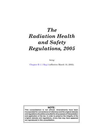
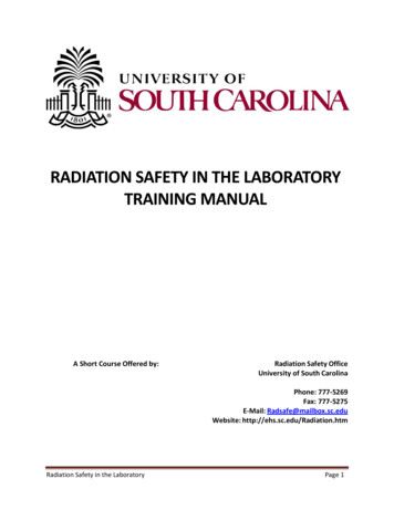
gamma emitter with a non-radioactive target material which then results in theemission of neutrons. You probably heard the expression of someone exposed toradiation as “glowing in the dark”. This can only happen if you are exposed to alarge neutron field. Remember what I said in an earlier slide that neutrons havean affinity for anything so they would attach to the atoms of your body thusmaking them unstable or radioactive. This is how many of the isotopes used inyour lab are made. They will take a target material and bombard them with astrong neutron field and produce the isotopes for your research. Electromagnetic radiation is radiation that is emitted from a nucleus or electronorbitals in the form of waves. These waves are made up of a basic unit calledphotons. There are 2 types of electromagnetic radiation discussed– gamma raysand x-rays. Remember that these types of radiation have no mass or charge andgenerally are more penetrating than particulate radiation of comparable energy.ELECTROMAGNETIC SPECTRUMAbove is a chart of the entire electromagnetic spectrum. The top of the chart is a scale forwavelength. Electromagnetic energy travels in waves. The number of wave crests that pass acertain point would be a measure of its frequency. At the bottom of the slide is a scale forphoton energies in electron volts. Notice that the photon energy will increase as the wavelengthdecreases and the frequency increases. At about 100 electron volts, you are in the upperultraviolet range or the lower x-ray energy range. This is where ionization begins to take place.Radiation Safety in the LaboratoryPage 5
As the photon energies increase on the chart to the right, the degree of ionization will alsoincrease. This is the area where gamma rays are located. This is the part of the spectrum wherethe principle biological effect to humans would be cancer.Other parts of the electromagnetic spectrum can have potential biological effects. The upperedge of the ultra-violet region can cause ionization. The two primary biological effectsassociated with the UV range are skin cancer and skin aging. Skin aging is probably the result ofbeing exposed to the lower ranges of the UV spectrum.Notice that visible light is located to the right of center on the chart. The photon energies ofvisible light are around the 1-2 electron volt range. This is about 100 – 100,000 times lessenergy that ionizing radiation. There are no known biological effects from exposure to visiblelight although if you stare at an intense light source for an extended period of time, you will doserious damage to the retina of your eye. Part of the damage could be attributed to the UVcomponent of light.Infra-red radiation, located to the left of visible light, has a photon energy range of 0.1 to 1electron volt. Exposure to infra-red radiation can cause the molecules in the exposed area tovibrate and produce heat. Infra-red lamps are often used in the treatment of injured muscles.They are also used in industry for the heating and drying of glues.Microwave ovens emit photons in the range of one one-thousandth of an electron volt. In thisrange, the water molecules of the affected material will begin to actually rotate and cause aheating effect.The photon energy of a standard cell phone is one one-millionth of an electron volt or one onethousandth the energy of a microwave oven. In this range, no rotation of water moleculesoccurs thus no heating can occur. There is not known biological effect in this range. Ionizingeffects cannot occur in this range. It would take anywhere from 1 to 10 million photons of cellphone radiation hitting the same molecule instantaneously to cause the same effect of onephoton of x-radiation. Quantum mechanics does not support the notion that energy from cellphones can cause cancer.RADIOACTIVE QUANTITYThere are many different terms for radioactive quantity. Probably the most familiar quantity toyou is the curie named after Madame Curie, an early researcher with radioactive materials. Acurie is equal to 3.7 x 10¹⁰ disintegrations per second. If you have a large amount of radioactivematerial, it is going to decay and give off some form of radiation. If the number of atomsreleased from this large amount of radioactive materials is 3.7 x 10¹⁰ per second, then a curie ofradiation is present. This number is actually based on the decay of one gram of Radium-226which decays at this rate. But anything that decays at this rate is considered a curie.A Becquerel is a new international unit that is replacing the curie. One Becquerel is equal to onedisintegration per second. This unit is slowly being adopted by the United States and it will bebeneficial to learn the conversation rates.Radiation Safety in the LaboratoryPage 6
Conversion Charts:1 Curie (Ci) 3.7 x 10¹⁰ disintegrations per second (dps)1 Becquerel (Bq) 1 dps 27 pCi1 Ci 37 GBq 37 x 109 BqIn laboratory research at USC, we do not often use curies of radiation. Another common unit ofradiation quantity is the millicurie which is one one-thousandth of a curie. Researchers oftenpurchase tritium and sulfur-35 in millicurie quantities. Also, the microcurie is a common unit. Amicrocurie is equal to one one-millionth of a curie or one one-thousandth of a millicurie.Phosphorus-32 is often ordered in microcurie quantities.Some useful conversions of Curies to Becquerels:1 TBq 1012 Bq1 pCi 37 mBq91 GBq 10 Bq1 nCi 37 Bq61 MBq 10 Bq1 uCi 37 kBq1 mCi 10-3 Ci1 mCi 37 MBq-61 uCi 10 Ci1 Ci 37 GBq-91 nCi 10 Ci1 kCi 37 TBq1 pCi 10-12 CiRADIOACTIVE DECAY AND DECAY RATEThe decay property of radioactive materials was discussed in a previous section. The decay rateor half-life formula can be of some benefit to you in your daily work with radioactive materials.It can be a helpful planning tool in determining if you have enough radiolabel to successfullycomplete your experiments. For example, if you plan to label bacteria cells that are growing in alarge flask and you know for a fact that the optimum growth period for the cells is going to be aweek or so from now, will you have enough of radiolabel in the freezer to label those cellssuccessfully? This formula can be a useful tool for you in such a situation.The formula is A Aoe-.693t/T1/2Ao the activity or number of atoms originally presentA the activity or number of atoms after a certain time (t) has elapsedt the time elapsed from Ao to AT1/2 half-life of the isotopeEXAMPLE:On April 1, you have 10mCi of P-32. What will be the activity of May 1?Radiation Safety in the LaboratoryPage 7
First of all, the time elapsed (t) is 30 days. The half-life of P-32 is 14.3 days. You can look up thehalf-lives of various isotopes or perhaps use the search engine on your computer.A (10mCi)e-.693(30)/14.3A (10mCi)(.233)A 2.33 mCiThe general decay formula can be used to get an exact number but an estimate can also be . Youknow for a fact that on April 1, you had 10 mCi of P-32. On April 14 (one half-life) you wouldhave 5mCi. On April 28(another half-life passes) you would have 2.5 mCi. So after 30 days, youwould have a little less than 2.5 millicuries.INTERACTION OF RADIATION WITH MATTERHow radiations interact with matter after they are released from an unstable atom Alpha ParticlesWhen an alpha particle is released, its range is rather short since it has a plus 2 chargeand a relatively large mass. Its effect is considered local. Alpha particles are notconsidered much of an external hazard. By external hazard, I am referring to a sourcelocated outside of the human body that emits radiation and causes one to receive adose from the shine coming off of the source. It would require an alpha particle of 7.5million electron volts just to penetrate the dead layers of skin. If you would get somealpha particles on the skin, this could result in some “hot spots” that could give theaffected area a large dose to an extremely small area. Normally, the residence time ofalpha particles would not be very long since repeated washing of the affected area andthe sloughing off of the dead skin cells would cause the particles to be removed.The greater concern with alpha particles is if the sources of alpha particles happen toend up inside of your body. For instance, if you were to inhale an alpha emitting particlethat was of a size that it would end up in the deeper recesses of the lung in the alveolarregions, these particles are considered below the level at which your body can easilyremove them and they stay there for years. The cells immediately surrounding the alphaparticles would receive a significant radiation dose over time and could possibly becomecancerous. So, you would want to avoid inhaling alpha particles. Individuals, who workwith this type of material, normally perform their work in a glove box and might alsohave further protection of the breathing zone by wearing a respirator.In summary, alpha particles:1. Have large Mass and a 2 charge2. Range is short and effect is local3. Not an external Hazard4. Can be a serious internal hazard5. Avoid inhalation of alpha particlesRadiation Safety in the LaboratoryPage 8
Beta ParticlesBeta particles are generally more penetrating than alpha particles of comparable energy.Betas can have either a minus one charge or a plus one charge. Now the low energy betaparticles such as H-3, C-14, S-35 and Ca-45 are not considered an external hazard. Noshielding is required to work with these isotopes. However, they can be an internalhazard if inhaled, ingested or absorbed. These isotopes will accumulate in the bodywherever these elements are normally found. Tritium and C-14 will end up in just aboutevery cell since these two elements are a component of most cells. S-35 will gravitate toproteins. If you recall from your basic biology instruction, the tertiary structure ofproteins are held together by disulfide bonds. Ca-45 is a bone seeker. It would alsobecome a component of your blood since calcium is important in the clotting process.High energy beta particles such as P-32 or Sr-90, are considered both an external and aninternal hazard. Internally, P-32 and Sr-90 would seek out the bones of your body. P-32could also end up as part of the backbone of the DNA molecules. P-32 has a very highspecific activity which means that just a small amount can be very radioactive.Whenever it is handled, P-32 can impart a significant local dose to the skin andsubcutaneous regions. Great care should be taken to avoid prolonged exposure to P-32.In summary, beta particles:1. More penetrating than alpha particles2. Low energy particles- not an external hazard (H-3,C-14,S-35,Ca-45)3. High energy particles- both internal and external hazard (P-32, Sr-90) Gamma and X-ray InteractionsGamma rays and x-rays are more highly penetrating since they are forms ofelectromagnetic energy and have no mass or charge like alpha or beta particles. Verylow energy gamma rays and x-rays would not penetrate the body very much and canimpart a significant local dose much like high energy beta particles. Higher energy raysare obviously more deeply penetrating and can deposit dose easily to the internalregions of the body.In summary, gamma and x-ray radiations:1. Highly penetrating (no mass or charge)2. Very low energy rays can impart a significant local dose3. Higher energy rays are much more penetrating4. Greater range in airRadiation Safety in the LaboratoryPage 9
PROPERTIES OF COMMONLY USED RADIOISOTOPESEach radionuclide is unique in terms of what types of radiation it emits. Some give off just betaparticles such as phosphorus-32. Some give off alpha particles and gamma rays such asAmericium-241. And some can give off alpha, beta and gamma radiations like uranium-235. Youcan categorize each radionuclide by the type of radiation it emits, the energy of the particles orphotons in electron volts, kilo-electron volts(thousand) or mega-electron volts(million). Andeach radionuclide has a rate of emission that we refer to as the half-life.Two videos are available on our web site titled “A Demonstration of the Various Types ofRadioactive Materials” and “Safety Precautions when Using Specific Types of Isotopes”. Thesevideos were created to actually show you the various types of radioactive materials that werejust discussed and present some of the safety precautions that are taken when working withthese radioisotopes.It is important for you to know that which isotopes are considered weak or high energy betaemitters. By knowing the category an isotope, you can then apply certain safety precautionswhen working with that material. For instance, if you are working with Phosphorus-32, youshould know that it is a high energy beta emitter and that Plexiglas shielding is needed as wellas a whole body and ring dosimeter. You would also need to have your Geiger counter handy tosurvey your work area during and after your experiment.Radiation Safety in the LaboratoryPage 10
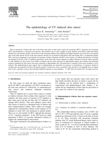
LESSON 2:Dose Units and Biological EffectsAs you recall, the units that are used for radiation quantity are the Curie and the Becquerel.There are a few other terms related to radiation field strength and radiation dose that you needto be familiar when working with radiation.The first term is the Roentgen (R) which was named after William Conrad Roentgen whodiscovered x-rays in 1895. A roentgen is a measure of radiation exposure in air. It specificallyrefers to x-rays or gamma rays but is often used to quantify other types of radiation, as well. Aroentgen measures the strength of a radiation field. In other words, if you had a source thatwas emitting radiation, you could measure that field using a Geiger counter. The units on atypical Geiger counter are milli-roentgens (mR) per hour. A milli-roentgen is a thousandth of aroentgen. The roentgen unit does not correlate directly with the amount of dose that a humancan receive from a radiation source.Another unit was developed called the radiation absorbed dose or the RAD. The energy of theradiation and the density of the material that absorbs the radiation are two important factors indetermining dose. Radiation energy is directly correlated with the degree of penetration in ahuman body. More energetic radiations are more penetrating. The denser tissues of the bodywill absorb more radiation.The RAD is a unit of absorbed dose, considers the energy of the radiation and the density of theabsorbing material. 1RAD absorbed dose when 100 ergs of energy per gram is transferred tothe absorbing material.When x-rays come in contact with photographic film, theyhave the ability to darken the film. Notice in this slide thatthe ribs come out lighter on the film than the areas inbetween the ribs. The ribs are much denser than the softtissue between the ribs and so therefore, they absorb moreradiation, get more dose and appear lighter on the film.Radiation Absorbed Dose (RAD) – Proportionality Constantskv100250400Filtermm of Al5.53nilRadiation Safety in the Laboratoryair0.870.870.87constant 1.421.11Page 11
If you could measure the radiation quantity at the surface of the body in roentgens, you couldthen determine the amount of absorbed dose in RADS using the proportionality constants inthis table. The very far left column lists the energy of the radiation in kv (kilovolts) from 100 to400 kv. Now as you move from left to right in the table you seen columns for increasing materialdensities from air (least dense) to bone (most dense). In summary, you can take the number forskin entrance exposure and multiply it by the appropriate proportionality constant to obtain avalue for absorbed dose in RADS.Another term associated with dose is the roentgen equivalent man or the REM. A REM is ameasure of radiation dose equivalent. As discussed, radiations can differ: some have pluscharges, some have negative charges, some have no charges or mass. As the result of thesedifferences, they affect living systems differently. For example, if you had a 1 MEV alpha particleand a 1 MEV gamma ray penetrating your body, each would behave differently as it passedthrough you. The 1 MEV gamma ray, since it has no mass or charge, would pass through yourbody rather easily. If you could measure the gamma ray as it exited your body, it would probablybe less than 1MEV in energy because of collisions with atoms. If you look at the path of the 1MEV alpha particle, it would only penetrate possibly a few layers of skin. Now if you couldmeasure the amount of radiation that is deposited in each cell from both types of radiation, youwould discover that there is far more energy deposition from the alpha particle since itdeposited most of its energy over a much shorter range. As a result of this phenomenon, wecan say that the alpha particle has a higher biological effectiveness than does the gamma ray.The table provides the QF for the different type radiations.Radiation TypeX-rays or gamma raysBeta ParticlesNeutronsAlpha ParticlesQuality Factor111020NEW INTERNATIONAL UNITSIn 1975, the International Commission on Radiological Units (ICRU) adopted new unitsapplicable to radiation protection that are consistent with the International System of Units (SISystem). The Gray (Gy) has replaced the RAD. One gray is equal to 100 RAD’s. And the Seivert(Sv) has replaced the REM. One seivert is equal to 100 REM. Most countries of the world haveadopted the new units. However, they are still not widely used in the United States and Russia,but there is much movement to adopt these units. So, it is important to know both sets ofterms. If you look at the scientific literature, you will find the Gray and Seivert used. If you visitmost countries, you will find the new units used in discussions about radiation dose.Gray (Gy) replaces RAD1 Gy 1 joule/kg 100 RADRadiation Safety in the LaboratorySeivert (Sv) replaces REM1 Sy 100 REMPage 12
DOSE LIMITS- TOTAL EFFECTIVE DOSE EQUIVALENT (TEDE)Everyone is exposed to a certain amount of radiation from the environment (cosmic, radon,terrestrial ) which is called the background radiation dose. In SC, the average backgroundradiation level is between 100- 500 mrem per year. Once you begin to use radiation in ourlaboratories, you will be considered a “radiation worker” by the regulatory authorities. And as aradiation worker, it is expected that you will receive some dose from your activities. Theregulatory authorities allow radiation workers to receive a certain amount of dose related totheir work activities. The dose limit for radiation workers is referred to as the Total EffectiveDose Equivalent or TEDE. The TEDE includes radiation shine from external sources such as an xray machine or a bottle of radiolabel. It is referred to as the Deep Dose Equivalent or DDE. Thedeep dose equivalent is obtained from radiation dosimeters worn by workers. The secondcomponent of dose is what can be received from internal deposition of radiation in the body.This is referred to as the Committed Effective Dose Equivalent of CEDE. The CEDE really tries toaddress the deposition of long-lived isotopes that may have a long residence time in your body.There are 3 main routes of entry for radioactive materials in the body: ingestion, inhalation,and absorption. If you can get a measure of the amount of radiation that is depositedinternally, then you can project how much dose that source of radiation will cause over a 50year work period. It would be rare to have this type of radiation deposition by working with theisotopes that we normally use at USC. The only isotope that is capable of causing an internaldose under normal conditions is I-125 in a free form of sodium iodide. If you were to open a vialof this form of Iodine-125, it has a relatively high vapor pressure and will be volatile. Theradioactive atoms could easily permeate your work space just by opening the vial. Employeeswho work with I-125 for the purpose of tagging another organic molecule will be required towork in the laboratory fume hood. This protects the worker from inhaling the radioactiveatoms. Now if you happen to be boiling a radioactive solution, this could possibly result inairborne atoms and this activity should also be performed in a fume hood. We measure internaldeposition of iodine by measuring radiation emissions from the thyroid since iodine isaccumulated almost exclusively in the thyroid.If you are working with just an x-ray machine, there is no possibility of having an internal dose –only an external dose. So, your TEDE would just consist of DDE readings from the personneldosimeter.Summary:Total effective dose equivalent – sum of the deep dose equivalent (DDE) from externalexposures and the committed effective dose equivalent (CEDE) from internal doses to any organor tissue.TEDE DDE CEDEThe annual limits are:5 rem - total effective dose equivalent50 rem - total organ dose equivalent to any single organ or tissue15 rem – total dose to lens of the eyeRadiation Safety in the LaboratoryPage 13
It should be pointed out that these limits do not imply a safe level of radiation dose. Thebiological effects from low doses of radiation are not well known. As a result, the regulatorybodies have taken the position that radiation doses must be kept as low as reasonablyachievable (ALARA) which will be discussed below.LIMITS DURING PREGNANCYThe dose to the embryo/fetus over the entire gestation period (9 months) must not exceed 0.5rem. The dose should be delivered at a fairly uniform rate over the entire period of pregnancyand not delivered in a few large doses. The rule permits an additional 0.05 rem if the pregnantwoman has greater than 0.45 rem at the time of notification. Abdominal dosimeters will beissued to the employee, if applicable, who has declared her pregnancy in writing to theRadiation Safety Office. A more detailed discussion of this topic can be found later in thismanual.ALARAIt is important to note that the dose limits listed are believed to be a safe level based on studiesof the biological effects of ionizing radiation. However, we cannot say beyond the shadow of adoubt that they are safe for everybody. Humans have varying degrees of sensitivity to radiation.For example, the very young are more sensitive to radiation. Young folks are growing rapidly andthey experience rapid cell reproduction. Cells that are actively diving are more sensitive toradiation therefore there are more stringent limits during pregnancy. Also, older humans aremore radiosensitive because their immunity systems are not as strong and radiation exposureor exposure to any kind of physical stressor could upset the homeostatic balance of the body.As a result of this, the regulatory authorities state that even though there is an establishedradiation limit, we should strive to keep all do
The atomic mass refers to total number of neutrons and protons in an atom. It is abbreviated with a capital "A". When we speak of a specific type of radioactive atom, we include the atomic mass. For instance, we can identify an atom of phosphorus-32 with 32 being the atomic mass. Neutron Number (A-Z N)