Transcription
RADIATION SAFETY EXAMSTUDY GUIDEFOR USERS OFRADIOACTIVE MATERIALWashington University in St. Louis, andWashington University Medical CenterRADIATION SAFETY DIVISIONEnvironmental Health and SafetyWashington University in St. LouisSt. Louis, MissouriVersion 2022
TABLE OF CONTENTSPreface . 31.Nature of Radioactivity and Radiation . 42.Interaction of Radiation with Matter . 73.Radiation Exposure and Dose . 114.Biological Effects of Low Level Radiation . 135.Sources of Radiation Exposure . 176.Radiation Dose Recommendations and Regulations . 197.Radiation Safety Instruments and Devices . 218.Control of Radiation Exposure . 249.Specific Rules that Apply to the Use of Radioactive Materials . 2810.Obligations and Rights of Personnel Handling Radioactive Materials . 4011.Existence and Role of Regulatory Agencies . 41Radiation Safety Exam Study GuideFor Users of Radioactive Material2February, 2022
PREFACEThis manual is intended to provide the research and laboratory medicine staff ofWashington University in St. Louis and Washington University Medical Center aconvenient summary of the basic essentials of radioactivity and radiation safetypractices necessary to pass the institutional radiation safety examination, a requirementof personnel prior to their working with radioactive materials without the directsupervision of their Authorized User or designated Lab Contact.The Nuclear Regulatory Commission, the federal regulatory agency with authority overthe use of radioactive materials at Washington University and the Medical Center,specifies that personnel must be instructed concerning the following: Radioactive decay and radiation Potential hazards or risks Radiation signs, symbols and labels Radioactive materials used Locations where radioactive materials are use or stored ALARA program Protective measures to keep personnel exposure low Emergency actions Specific radiation safety program requirements Existence and location of NRC license Existence and role of regulatory agencies Each worker's obligation to report unsafe conditions Worker's rights Who to contact if there are questionsEach of these topics is addressed in this manual.Radiation Safety Exam Study GuideFor Users of Radioactive Material3February, 2022
1.NATURE OF RADIOACTIVITY AND RADIATIONRadioactivity is a spontaneous process characteristic of atoms with unstable nuclei inwhich the nucleus releases energy either as a particle with kinetic energy or aselectromagnetic energy. Upon release of this energy the nucleus may be stable or itmay still be unstable and subsequently transform again. The original isotope prior toradioactive decay is termed the "parent" with the isotope after the transformation calledthe "daughter". A given transformation, that is, one parent/daughter transition, is calleda "decay" or a "disintegration".1.1.Radioactive DecayThe rate of radioactive decay, also called the disintegration rate or decay rate, is usedto indicate the "radioactivity" of a sample and is termed the “activity”. The traditionalunit of activity is the curie (Ci) defined as the amount of radioactive material having adisintegration rate of 3.70 X 10¹⁰ (37 billion) disintegrations per second (dps). It iscommon to express activities in curies (Ci), millicuries (1 thousand mCi 1 Ci),microcuries (1 million Ci 1 Ci), nanocuries (1 billion nCi 1 Ci), picocuries (1 trillionpCi 1 Ci), etc. It is important to note that activity denotes the rate of disintegration; itprovides no information regarding the kind of radiation emitted during the radioactivedecay.In most of the world, the curie has been replaced by new units based on the metricsystem. The unit of activity in the System International (SI) is the becquerel (Bq). Thebecquerel is defined as a disintegration rate of one per second, that is, 1 Bq 1 dps.For example, a millicurie of activity (3.7 x 10⁷ dps 37. x 106 dps) is equivalent to 37megabecquerel (37 MBq) of activity. It is common to see activity levels in the literatureexpressed in both units, usually with one in parenthesis. However, the NuclearRegulatory Commission (NRC) uses the traditional unit, the curie, to denote activitylevels, so for radiation safety purposes we do as well.1.2.Half-LifeThe “half-life” is an important characteristic of any radionuclide. (Any specific nuclearcombination of neutrons and protons comprise a nuclide; an unstable nuclide is termeda radionuclide.) The half-life corresponds to the time necessary for one-half of theradioactive atoms of a sample to decay. The fraction of atoms (or activity) remaining asa function of the number of elapsed half-lives is as follows:Radiation Safety Exam Study GuideFor Users of Radioactive Material4February, 2022
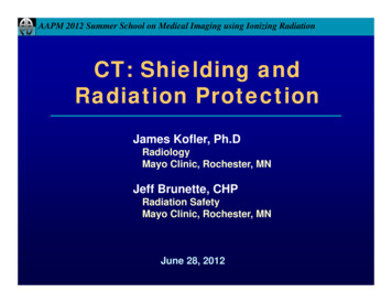
Number of ElapsedHalf-LivesFraction ofActivity )40.0625(6.25%)100.000977(0.1%)In general, the fraction remaining is expressed by the relationship, where n is thenumber of half-lives that have passed:12𝑓𝑟𝑎𝑐𝑡𝑖𝑜𝑛 𝑟𝑒𝑚𝑎𝑖𝑛𝑖𝑛𝑔Thus, 1/2 (50%) of the activity remains after 1 half-life, 1/4 (25%) after 2 half-lives, 1/8(12.5%) after 3 half-lives, and so on.With a starting activity of 40 mCi, the activity left after 4 half-lives would be 2.5 mCi (40mCi x (1/2)4 40/16 2.5 mCi). The following illustration shows a very easy way todetermine the activity remaining for the same example.½40 mCin (T1/2’s) ½½20 mCi10 mCi12½5 mCi32.5 mCi4The half-lives of several radionuclides commonly used at our institution are:RadionuclideHydrogen-3 (tritium)Half-Life(3H)12.3 yearsCarbon-14 (14C)5700 yearsPhosphorus-32 (32P)14.3 daysSulfur-35(35S)Iodine-125 (125I)1.3.88 days60 daysTypes of Radioactive DecayOnly a limited number of types of transformation have been observed in the decay ofradionuclides. The four most common types of radioactive decay are (1) alpha decay,(2) beta decay, (3) electron capture and (4) isomeric transition. Prior to discussingRadiation Safety Exam Study GuideFor Users of Radioactive Material5February, 2022
these types of radioactive decay it is helpful to introduce a unit of energy, the "electronvolt". This unit is used to denote particle and photon energies in atomic physics and itrepresents the energy change experienced by an electron while undergoing a potentialenergy change of one volt. It is common to express certain energies in kiloelectronvolts (keV one thousand eV), megaelectron volts (MeV one million eV), etc. Forcomparative purposes, the kinetic energy of an air molecule at typical room temperatureis about 0.025 eV.1.3.1. Alpha DecayAlpha decay is generally limited to isotopes of the heavy elements (isotopes aredifferent nuclear varieties of an element – same atomic number, Z, but different massnumbers, A). In alpha decay, a charged particle (alpha particle) consisting of twoprotons and two neutrons, is ejected from the nucleus with high kinetic energy –typically, about 4 MeV. The alpha particle, although its initial energy is great, is stoppedby a few centimeters of air or by a few microns of tissue. After the emission of the alphaparticle, the daughter atom may be in an excited level of that particular isotope. Theresulting transition to the ground state is generally accompanied by the emission of agamma-ray, a photon of electromagnetic energy that is emitted from the nucleus of theexcited atom.1.3.2. Beta DecayBeta decay is the name of a transition that results in the emission of an electron fromthe nucleus. The emitted electron may be negatively charged, that is, an ordinaryelectron referred to as a beta particle ( -particle) or it may be positively charged, that is,a positron, depending on the particular radioactive isotope.There are some radionuclides that exhibit "simple -decay", and the daughter nucleus isat the "ground state" subsequent to the decay. Important examples of suchradionuclides are tritium (hydrogen-3, 3H), carbon-14 (14C), phosphorus-32 (32P) andsulfur-35 (35S). In the case of simple beta decay, the only detectable radiation emittedduring the decay process is the beta particle.However, the more common situation is for the daughter to be an "excited isomericstate" of the nuclide with subsequent emission from the nucleus (usually promptly) of aphoton of electromagnetic radiation, called a "gamma ray". Either the gamma rays orthe -particles can be detected to identify the presence of the radionuclide.Radiation Safety Exam Study GuideFor Users of Radioactive Material6February, 2022
1.3.3. Electron CaptureAn unstable nucleus of certain isotopes can convert a proton to a neutron by a processtermed electron capture. An orbital electron is "captured" while passing through thenucleus of these unstable nuclei. The captured electron combines with a nuclear protonto yield a neutron. A rearrangement of the orbital electrons results to fill the vacancy leftby the captured electron. This rearrangement of orbital electrons is generallyaccompanied by the emission of characteristic x-rays. In addition, if the decay leavesthe daughter at an excited level, then -rays are likely to be emitted in the subsequenttransition(s) to the ground state of the daughter nucleus. The presence of radionuclidesthat decay by electron capture, chromium-51 (51Cr) and iodine-125 (125I) are examples,can often be identified by either the x-rays or gamma-rays or both.1.3.4. Isomeric TransitionThe excited levels and the ground state level of a radionuclide differ only in nuclearenergy content. The various energy levels are called nuclear "isomers" and thetransitions between them are called isomeric transitions. These transitions normallyoccur promptly after formation of the excited level (promptly usually means de-excitationtimes of the order of nanoseconds) and generally result in -rays of energy equal to thedifference of the energies of the two isomeric levels involved in the transition.Occasionally, there are instances in which the excited state of a daughter nucleus isrelatively long-lived with the transition to the ground level, being delayed not bynanoseconds but by minutes or hours. The protracted excited level is termed ametastable level and is denoted by adding the letter m to the mass number. Anexample is technetium-99m (99mTc), which has a half-life of the metastable state of 6hours.2.INTERACTION OF RADIATION WITH MATTER2.1.1. - and -Radiation (x- and -rays) -rays and -rays are electromagnetic radiation. The terms x-ray and -ray distinguishthe origin of the radiation; x-rays are emitted by electrons, whereas -rays are emittedby the nucleus of an atom involved in radioactive decay. X-rays and -rays of the sameenergy are identical in every respect except origin. Electromagnetic radiation, such asx- and -rays can interact with matter by more than ten different processes. In someinteractions the electromagnetic radiation behaves like energy waves, and in others itbehaves like particles, called photons. As electromagnetic radiation, x- and -rays travelwith the speed of light and are characterized by their energy. For radiation safetyRadiation Safety Exam Study GuideFor Users of Radioactive Material7February, 2022
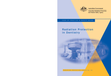
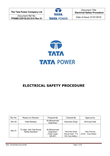
purposes, there are only three important interactions: photoelectric absorption,Compton scattering, and pair production. The following sections summarize these threeimportant photon interactions with matter.2.1.1.1. Photoelectric AbsorptionIn this process the incident photon is absorbed by a bound electron present in aconstituent atom of the material with which the photon interacts, and the photon ceasesto exist. The electron is ejected from its previously bound state and carries away mostof the absorbed energy as kinetic energy.The probability of photoelectric absorption is very much dependent upon the energy ofthe radiation and upon the atomic number of the absorbing material. In general,photoelectric absorption is the dominant absorption process at low radiation energiesand it occurs, for a given energy, much more in materials of high atomic number than inmaterials with low atomic number. Thus, lead with the high atomic number of 82, is anexcellent material for absorbing radiation by the photoelectric process.2.1.1.2. Compton ScatteringIn this interaction the incident photon undergoes a billiard-ball type collision with anelectron of the interacting material. The photon is deflected, or “scattered”, by thecollision and continues on with reduced energy. Compton interactions occur to aboutthe same extent in unit masses of different materials, that is, a gram of wood is about aseffective as a gram of lead if the attenuation process is Compton scattering. Theprobability of Compton scattering in any material steadily decreases as the energy ofthe radiation increases.2.1.1.3. Pair ProductionIn this attenuation process, the incident photon materializes as a pair of particles (anelectron and a positron) in the vicinity of a nucleus. The minimum or threshold energyfor this interaction is 1.02 MeV (This corresponds to the rest mass energy equivalenceof the 2 created particles). The probability of pair production increases above thethreshold and it occurs more in matter with high atomic number constituents than matterof low atomic number. Pair production is an important way to stop or attenuateelectromagnetic radiation only for high energy radiation, that is, for photon energiesgreater than several MeV.In general, photoelectric absorption is the dominant interaction at low energies whilepair production is the dominant interaction at very high energies. Compton scatter is theimportant mechanism for intermediate energies. The relative importance of eachRadiation Safety Exam Study GuideFor Users of Radioactive Material8February, 2022
process is shown below as a function of photon energy and absorbers of differentatomic number.Predominating (most probable) interaction versus photon energyfor absorbers of different atomic number.2.1.1.4. Half-Value-LayerIt is frequently convenient to express the attenuation or "absorption" properties of amaterial in terms of its half-value-layer, HVL. The HVL is defined as the thickness of thematerial required to reduce the transmitted radiation to one-half of the incident value.The transmitted fraction (f ) through n half-value-layers is given by the expression12𝑓Thus, 1/2 (50%) of the incident radiation is transmitted through one HVL, 1/4 (25%)through 2 HVL, 1/8 (12.5%) through 3 HVL, and so on.Radiation Safety Exam Study GuideFor Users of Radioactive Material9February, 2022
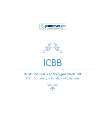
The following illustration shows a very easy way to determine the amount of activityremaining after transmission through a given number of HVL’s. Assume a startingactivity of 10 mCi. The activity left after transmission through 3 HVL’s would be 1.25mCi.½10 mCin (HVLs) ½½5 mCi2.5 mCi1.25 mCi1232.1.2. Particle InteractionsAs a charged particle passes among the atoms of an absorbing material it may (1)dislodge electrons from atoms to form positive and negative ions (ionization), (2) exciteelectrons to higher energy levels in atoms, (3) set up vibrations of molecules in the path,(4) break molecular bonds, or (5) produce electromagnetic radiation subsequent to asudden change in its course (bremsstrahlung). It is the ionization process that isresponsible for the radiobiological effects of radiation and it also makes possible thedetection of radiation with various types of equipment.The energy that a particle loses to its surroundings for each unit of path length is calledthe linear energy transfer (LET). Think about running a race: for every mile you run,you lose a little more energy. The average energy per mile that you lost would be your“linear energy transfer”.The distance that a charged particle travels from the point of its origin to the placewhere it no longer acts as a destructive particle is called its range. Think again of ourrace: at the finish line, you are completely spent you can’t go one step farther andhave reached your “range”.You need to understand the process of bremsstrahlung. When electrons are diverted orstopped by their interactions with matter, some of their energy can go into creating xrays. For electrons of a given energy, (MeV), the fraction of their energy, f, that is lostby the emission of x-rays (called bremsstrahlung), is given by the expression:f /1400where the absorber atomic number. This means that the higher the absorber’satomic number (Z), the greater the energy lost by the electron which then appears asbremsstrahlung (x-rays). Thus, it is preferable to shield a pure beta emitter of highenergy, for example, 32P, with low atomic number (low “Z”) plastic rather than highatomic number lead in order to minimize the production of the penetrating x-raysreferred to as bremsstrahlung.Radiation Safety Exam Study GuideFor Users of Radioactive Material10February, 2022
3.RADIATION EXPOSURE AND DOSEIt is important to know the "radiation level" to which workers are exposed. Everyradiation worker must not only consider his or her own radiation level, but be cautious tominimize the radiation level affecting others in the area.There are three different technical concepts used to discuss radiation levels the ambient radiation field in an area, or “radiation exposure”; the energy deposited by radiation in a material, the “absorbed dose”; and the biological relevance of the energy deposited by radiation – specifically inliving tissue – called the “dose equivalent”.The terms “dose” and “exposure” are commonly, or casually, used as if they areinterchangeable, and it is hard to avoid doing so. However, when we specifically usethe three terms “radiation exposure”, “absorbed dose”, and “dose equivalent”, they havevery specific and conceptually different meanings:Radiation exposure – This term applies to x- and -radiation fields only. This concept isbased on the ability of photon (electromagnetic) radiation to ionize air. Specifically, theamount of electrical charge liberated in a unit volume of air is used to define the level ofradiation exposure. The traditional unit of radiation exposure is the roentgen, R, whichis approximately 2.08x10⁹ ion pairs per cm3 of dry air at standard temperature andpressure (or equivalently, 2.58 x10⁻⁴ coulomb per kilogram of air).Absorbed dose – This term applies to all forms of ionizing radiation, that is, both photon(electromagnetic) and particulate (α, β, neutron, etc.) radiation. The absorbed dosedenotes the amount of energy imparted to any kind of matter by ionizing radiation, perunit mass of the irradiated material at the point of interest. The traditional unit ofabsorbed dose is the rad. The rad is defined as the energy absorption of 100 ergs pergram of material (or equivalently, 0.01 joule per kilogram of material).Dose equivalent – This term applies to all forms of ionizing radiation. The “doseequivalent” denotes the potential biological effect of the ionizing radiation. It is definedas the product of the absorbed dose and certain “modifying factors”. The modifyingfactors are intended to “adjust” the absorbed dose for the relative effectiveness withwhich different types of radiation produce more or less damage in biological tissue forthe same amount energy deposited. The traditional unit of this "product", that is, theunit of dose equivalent, is the rem. Although several modifying factors have beenproposed, only the "quality factor" (QF) has been extensively used. The quality factor isa subjective measure of the "relative hazard or biological effect" of a given type ofradiation and is strongly related to the linear energy transfer (LET) of the radiation.Radiation Safety Exam Study GuideFor Users of Radioactive Material11February, 2022
The quality factors for common types of radiation are: x-ray, -ray, or β-particle, QF 1; thermal neutron, QF 5; and α-particle or fast neutron, QF 20.An estimate of biological effect, that is, the dose equivalent in rem, is computed bymultiplying the absorbed dose in rad by the appropriate QF. Example: An individualreceives a fast neutron absorbed dose of 2 rad. The computed dose equivalent is 40rem.Dose equivalent is used to indicate the radiation dose due to internal radioactivecontamination as well as external exposure. The dose pattern from organ to organ thatresults from internal radioactivity is normally very uneven. A computational method ofhandling non-uniform organ and tissue doses is the effective dose equivalent (EDE). Acomputed EDE, obtained by adjusting (weighting) designated organ doses according totheir relative sensitivity to harm by radiation, is a single quantity that indicates thepotential harm or risk of the non-uniform dose pattern. Specifically, the EDE is thecomputed value of a uniform whole body dose that theoretically imparts the samenumerical risk as the actual non-uniform dose situation.The Nuclear Regulatory Commission employs several dose equivalent variations,including the following: Deep dose equivalent – the dose due to external radiation computed for a tissuedepth of 1 cm. Eye dose equivalent – the dose to the lens of the eye due to external radiationcomputed for a tissue depth of 0.3 cm. Shallow dose equivalent – the dose due to external radiation computed for atissue depth of 0.007 cm (representative skin thickness). Committed organ dose equivalent – the dose due to internal radioactivity to aspecific organ during the 50-year period following the intake. Committed effective dose equivalent – the computed effective dose due tointernal radioactivity during the 50-year period following the intake. Total effective dose equivalent – the sum of the deep dose due to externalradiation and the committed effective dose equivalent.Many scientific units have been redefined in order to achieve a world-wide consistencyof usage. The new units are based on the metric system and are referred to as SI units(Systeme International d’Unites). Two radiation units that are affected are those forRadiation Safety Exam Study GuideFor Users of Radioactive Material12February, 2022
absorbed dose and dose equivalent. The SI units and their relationship to the traditionalunits of absorbed dose and dose equivalent are as follows:QuantitySI UnitNameAbsorbed DosegrayGy100 rad per grayDose EquivalentsievertSv100 rem per sievertSymbolRelationship ToTraditional UnitThus, a milligray (mGy) 100 millirad and a microsievert ( Sv) 0.1 millirem, asexamples. The Nuclear Regulatory Commission, the federal agency that has authorityover most radioactive materials in the United States, has not yet (2015) adopted SIunits, and so our radiation safety program also continues to use the traditional units.4.BIOLOGICAL EFFECTS OF LOW LEVEL RADIATIONRadioactive material emits ionizing radiation whose interaction with human tissues mayresult in biological damage. The biological damage to tissues is primarily due tosecondary charged particles that result when the tissues are exposed to the ionizingradiation. The energetic secondary particles yield highly reactive free radicals thatinteract with molecules in the tissues, breaking chemical bonds and causing otherchemical changes. Some of the resulting damage is repairable, some is not. Biologicaleffects of low-level radiation (Note: low-level radiation means acute whole body dosesof 10 rem or less or substantially larger doses if received over an extended length oftime) can be classified into the three categories below:Somatic – Effects occurring in the exposed person. The manifestation of low doseeffects is delayed. The period of time between exposure and demonstration of thedelayed effect is referred to as the latent period.Genetic – Abnormalities occurring in the future children of exposed persons and insubsequent generations.Developmental or teratogenic – Effects observed in children who were exposed duringthe fetal or embryonic stages of development.At the low levels observed from occupational exposure it is impossible to demonstrate arelationship between dose and effect. The changes induced by radiation often requiremany years or decades before being evident and, thus, a very long follow-up period isnecessary to define risks. Studies of human populations exposed to low-level radiationare the appropriate basis for defining risk, but it is rare that they have the neededstatistical power.Radiation Safety Exam Study GuideFor Users of Radioactive Material13February, 2022
We can, instead, look for effects from higher-level radiation doses and try to extrapolatedown into the low-level dose range. However, the number of situations from which therelationship between high radiation dose and response can be estimated is limited, thebest being those of the A-bomb survivors in Nagasaki and Hiroshima. There isconsiderable uncertainty and controversy regarding the best estimates of the radiationrisk of low level doses.4.1.Summary of Current Radiation Risk EstimatesAs used in this section, "risk" is the probability or chance of severe harm or death fromradiation exposure.4.1.1. Somatic EffectsThe somatic effects of interest are cataract and cancer induction.Cataract Induction – The lens of the eye differs from other organs in that dead andinjured cells are not removed. Single doses of a few hundred rem can result inopacities that interfere with vision within a year. When the dose is fractionated over aperiod of a few years, larger doses are required and the cataract appears several yearslater. The 2006 BEIR VII Report (a report prepared by a special committee of theNational Research Council) concludes that cataract induction is not a concern at thedoses currently permitted for radiation workers, which are limited to 15 rem per year bythe NRC (see section 6.1, below)Cancer Induction – The 2006 BEIR VII Report concludes that cancer arising in a varietyof organs and tissues is the only somatic effect possible at low levels of radiationexposure.Various organs and tissues differ greatly in their susceptibility to cancer induction byradiation at high doses. For example, red bone marrow, the lungs and thegastrointestinal (GI) tract exhibit relatively high susceptibility to induced harm byradiation while skin, muscle, and bone surfaces are relatively insensitive to radiationharm. The following is known regarding radiation-induced cancer: The manifestations are delayed after exposure to radiation with latent periods ofa few years for leukemia development and, perhaps, as many as 20 years ormore for solid tumor formation. The resulting forms of cancer are indistinguishable from similar cancersspontaneously occurring in people.Radiation Safety Exam Study GuideFor Users of Radioactive Material14February, 2022
NRC regulatory guidance states that a cautious, conservative estimate of the risk offatal cancer due to ionizing radiation is approximately 4 chances in 10,000 per rem ofwhole body dose (or effective dose equivalent) when averaged over a representativepopulation of radiation workers (or 1 chance per 2,500 rem). This information ispresented in NRC Regulatory Guide 8.29 (1996) and is based on the 1990 BEIR VReport.The more recent 2006 BEIR VII Report states that a more conservative estimate isapproximately 1 chance in 1,000 per rem of whole body dose (or effective doseequivalent) when averaged over a representative population of radiation workers.Although this may be considered a more recent scientific evaluation, NRC guidance hasnot been updated, Thus for radiation safety purposes we use 4 chances in 10,000 remas the basis for risk estimates in our program.For perspective, the following are thought to result in comparable fatal risk (one in amillion chance of death): 1 millirem of whole body dose or effective dose equivalent smoking 1.4 cigarettes eating 40 tablespoons of peanut butter driving a car 40 miles spending two days in New York CityAnother comparison is to look at estimates of the average number of days of lifeexpectancy lost from occupational exposure to radiation and to compare this numberwith days lost, on the average, for various types of industry. The following resultssuggest that the health risks from occupational radiation exposure are smaller than thecollective risk associated with various industries.Industry TypeAverage Days of LifeExpectancy LossMining & ring43Occupational Radiation Exposure(340 mrem per year for 30 years)49Finally, some scientists today believe that low doses of ionizing radiation actually have anet health benefit. This concept is called "hormesis". The net beneficial or hormeticeffect due to low doses of radiation is attributed to various adaptive processes thatRadiation Safety Exam Study GuideFor Users of Radioactive Material15February, 2022
appear to be initiated by exposure to ionizing radiation. This view is controversial at thepresent time.The view that regulators and most advisory groups take is to assume a linear, nothreshold model to describe the relationship between harm and dose at low levels. Thiscautious approach assumes that any level of radiation exposure carries a proportionalrisk, that is, there is no threshold for potential harm.4.1.2. Genetic EffectsA mutation is an inheritable change in the genetic material within
Radiation Safety Exam Study Guide 3 February, 2022 For Users of Radioactive Material PREFACE This manual is intended to provide the research and laboratory medicine staff of Washington University in St. Louis and Washington University Medical Center a convenient summary of the basic essentials of radioactivity and radiation safety