Transcription
RSC AdvancesView Article OnlineOpen Access Article. Published on 23 December 2020. Downloaded on 4/16/2021 3:28:31 PM.This article is licensed under a Creative Commons Attribution 3.0 Unported Licence.PAPERView Journal View IssuePericardial fluid proteomic label-free quantificationof differentially expressed proteins in ischemicheart disease patients with systolic dysfunction bynano-LC-ESI-MS/MS analysis†Cite this: RSC Adv., 2021, 11, 320Junaid Ullah,a Satwat Hashmi,b Arslan Ali, *c Faisal Khan,c Shahid Ahmed Sami,dNageeb Basir,e Syeda Saira Bokhari,e Hasanat Sharif,d Hesham R. El-Seedifgand Syed Ghulam Musharraf *acLeft ventricular systolic dysfunction (LVSD) is common in patients with pre-existing ischemic heart disease(IHD) and myocardial infarction. An untargeted proteomic approach is used to improve the understandingof the molecular mechanisms associated with LVSD and to find out potential proteomic signatures inpericardial fluid. The pericardial fluid of IHD (n ¼ 45) patients was grouped into two categories accordingto the left ventricular ejection fraction, LVEF 45 (n ¼ 33) and LVEF 45 (n ¼ 12), and analyzed by usingnano-liquid chromatography–mass spectrometry (nano-LC-MS/MS) technique. The nano-LC-MS/MSanalysis resulted in the identification of 709 pericardial fluid (PF) proteins in both normal and impairedsystolic functional groups (LVEF 45 vs. LVEF 45). Sixteen proteins were found to be differentiallyexpressed (p 0.05, fold change 2) including 12 down-regulated and 4 up-regulated in the impairedsystolic functional group (LVEF 45) compared to the normal group (LVEF 45). Among the differentiallyReceived 1st October 2020Accepted 30th November 2020expressed proteins the inflammatory marker albumin, atherosclerosis marker apolipoprotein A-IV andDOI: 10.1039/d0ra08389ehedgehog-interacting protein marker of angiogenesis were predominantly associated with the impairedLVEF 45 group. KEGG pathway analysis revealed that the hedgehog (Hh) signalling pathway is up-rsc.li/rsc-advancesregulated in LVSD reflecting the underlying molecular and pathophysiological processes.IntroductionCardiovascular disease (CVD) is one of the main causes ofmortality and disability worldwide. According to the WorldHealth Organization (WHO), in 2008 approximately 17.3 millionpeople died from cardiovascular disease and this will, expectedly, rise to 23.3 million annually by 2030.1 Ischemic heartaH.E.J. Research Institute of Chemistry, International Center for Chemical andBiological Sciences, University of Karachi, Karachi-75270, Pakistan. E-mail:musharraf1977@yahoo.com; Fax: 92 213 4819018; 92 213 4819019; Tel: 92 2134824924; 92 213 4824925; 92 213 4819010bDepartment of Biological and Biomedical Sciences, Agha Khan University, Karachi74800, PakistancDr Panjwani Center for Molecular Medicine and Drug Research, International Centerfor Chemical and Biological Sciences, University of Karachi, Karachi-75270, Pakistan.E-mail: arslanali1986@gmail.comdDepartment of Surgery, The Aga Khan University Hospital, Karachi-74800, PakistaneDepartment of Medicine, The Aga Khan University Hospital, Karachi-74800, PakistanfPharmacognosy Group, Department of Pharmaceutical Biosciences, BMC, UppsalaUniversity, SE-751 23 Uppsala, SwedengInternational Research Center for Food Nutrition and Safety, Jiangsu University,Zhenjiang 212013, China† Electronic supplementary10.1039/d0ra08389einformation320 RSC Adv., 2021, 11, 320–327(ESI)available.SeeDOI:disease (IHD) is the most prevalent condition among the CVDs.Le ventricular systolic dysfunction (LVSD) is common inpatients with IHD.2 LVSD refers to an impairment of the le ventricular contractility that results in reduced le ventricularejection fraction (LVEF) and cardiac output. Patients with LVSDwere at increased risk of death compared to subjects witha normal LVEF.3 A 10% reduction in LVEF predicts a 39%increase in all-cause mortality, hospitalization and an increasedheart failure risk.4 Prevalence of LVSD increases substantiallywith age and is more prevalent in men (7.6%) as compared towomen (2.6%)5The measurement of proteins in biological samples playsa key role in the diagnosis and prognosis of patients that are athigh risk of diseases. Pericardial uid has been used tounderstand the molecular mechanism of disease in a number ofstudies.6 Pericardial uid (PF) is an ultra ltrate of the plasmaand a biochemical window into the heart by which informationrelated to the pathophysiological status of the heart can beobtained.7 Pericardial uid is present between the two layers;parietal and visceral of the pericardium and is produced by thevisceral pericardium of the heart. Under normal conditions, itmeasures about 15–50 mL in volume. Many diseases includingischemic heart disease, infections, and idiopathic causes can 2021 The Author(s). Published by the Royal Society of Chemistry
View Article OnlineOpen Access Article. Published on 23 December 2020. Downloaded on 4/16/2021 3:28:31 PM.This article is licensed under a Creative Commons Attribution 3.0 Unported Licence.Paperincrease the volume and change its composition.8 The proteomic analysis of pericardial uid may be an important tool to nd out the pathophysiological mechanisms underlying cardiacdiseases and to nd out potential therapeutic targets.9 Severalproteomic signatures have been discovered and used in themanagement of LVSD such as Brain Natriuretic Peptide (BNP),which is released in the pericardial uid and serum by theventricles in response to increased wall tension or stretch ofcardiac chambers.10 The proteomic analysis of pericardial uidin patients with lung bullae disease resulted in the identi cation of 1007 proteins which provided valuable insight intodisease mechanism.11 To the best of our knowledge, this is the rst study constituting the proteomic pro ling of the pericardial uid in patients with LVSD with established IHD.The present study aimed to uncover the differentiallyexpressed proteins in the pericardial uid by a label-freequantitative proteomic approach in IHD patients withimpaired (LVEF 45) and normal (LVEF 45) systolic function.The study will help to improve the understanding of themolecular mechanisms associated with LVSD and to nd outpotential proteomic signatures.Material and methodsExperimental design and statistical analysisFor the present study fourty ve patients (43 men and 2 women)were recruited with con rmed ischemic heart disease (IHD) atSouth city and Aga Khan University Hospital Karachi, Pakistan.A written informed consent was taken from each patient beforecoronary artery bypass gra (CABG). In this study, patients withrenal insufficiency, metabolic bone diseases, valvular diseasemoderate to severe, constrictive pericarditis and in ltrative,hypertrophic or restrictive cardiomyopathies, establishedpulmonary disease and malignancy were excluded. Twodimensional and Doppler echocardiography was used for theassessment of le ventricular systolic function following ASEguideline 2009. Patients were further categorized into twogroups according to the LV ejection fraction: group A was 33patients with normal le ventricular systolic function (LVEF 45), and group B was 12 patients with impaired le ventricularsystolic function (LVEF 45) as depicted in Table 1. The studywas approved by the Ethical Review Board (ERB) of the AgaKhan University Hospital and Institutional Review Board (IRB)of International Centre for Chemical and Biological Sciences(ICCBS), University of Karachi, Pakistan. All procedures followed in this study were in accordance with the ethical principles of the 1964 declaration of Helsinki.Sample collection and processingPericardial uid samples were collected from each subject inpre-labeled clean and sterile falcon tubes. Samples werecentrifuged at 3500 rpm, for 15 min at 4 C to remove cellularcomponents. For further analysis aliquoted samples werestored at 80 C in microcentrifuge tubes. Bicinchoninic acid(BCA) procedure was followed for measurement of total proteinconcentration according to the Kit protocol.12 2021 The Author(s). Published by the Royal Society of ChemistryRSC AdvancesTable 1Baseline characteristicsa(LVEF 45%)(LVEF 45%)NAge [years; mean (SD)]Body weight [kg; mean (SD)]3359.7 7.172.7 11.31258.0 9.676.6 12.7Medical tension18/1523/109/37/5Renal functionCreatinine [mg dL 1; mean (SD)]BUN [mg dL 1; mean (SD)]1.0 0.216.0 3.41.0 0.319.2 11.3Echocardiographic parametersLVEF [%; mean (SD)]58.2 4.936.6 8aSD standard deviation, BUN blood urea nitrogen, LVEF le ventricularejection fraction.FASP digestionEach pericardial uid sample equivalent to 200 mg protein, weredigested using lter-aided sample preparation (FASP) method.13In short, protein concentrates in 0.5 mL 10 kDa cutoff centrifuge lters were mixed with 500 mL of 6 M urea in 0.1 M tris/HCland the samples were centrifuged at 14 000g for 25 min at 20 C.A er centrifugation, 10 mL of 0.1 M Dithiotreitol (DTT) wasadded to the lters, and incubated at 56 C for 30 min. Then, 10mL of 0.3 M iodoacetamide (IAA) was added to the lters, a erwhich the samples were incubated for 30 min in darkness.Finally, 2 mg of trypsin (MS Grade, Thermo Scienti c) in 50 mL of100 mM NH4HCO3 was added to each lter. The protein toenzyme ratio was 50 : 1. Samples were incubated overnight at37 C and peptides were released and collected by centrifugation at 14 000g at 20 C for 25 min.LC-MS/MS analysisThe protein digest were dissolved in 0.1% TFA and 1 mL(equivalent to 2 mg) was injected on a C18-reverse-phase trapping column (C18, Acclaim PepMap 100, 75 mm 2 cm,particle size 3 mm, nanoViper, Thermo Fisher Scienti c) andwas separated by C18-reverse-phase nano-LC column (75 mm 15 cm Acclaim PepMap RSLC C18 column with 2 mmparticle size and 100 A pore size, NanoViper, Thermo FisherScienti c), using Nano-UHPLC, (Dionex, UltiMate 3000RSLCnano System, Thermo Scienti c, USA) coupled to MaXis IIESI-QTOF Mass spectrometer (Bruker Daltonics, Bremen, Germany). Buffers A and B contained 0.1% formic acid in ultra-purewater and 0.1% formic acid in acetonitrile (ACN), respectivelywere used for peptide elusion. ACN gradient at 300 nL min 1was used to separate peptides a er 6 min sample load at a owrate of 5 mL min 1 with 2% B. Elution of the peptides wereachieved by applying a linear gradient of 1–35% B over 29 min,with subsequent gradient increase to 95% B over 2 min, hold Bfor 7 min at 95%, and then decreased to 2% B for 4 min followedby 12 min equilibration at 2% B. Data dependent acquisitionRSC Adv., 2021, 11, 320–327 321
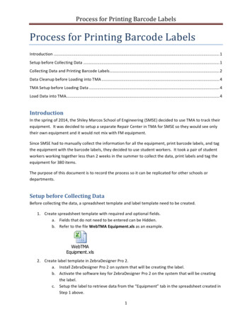
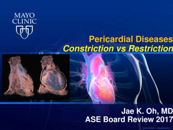
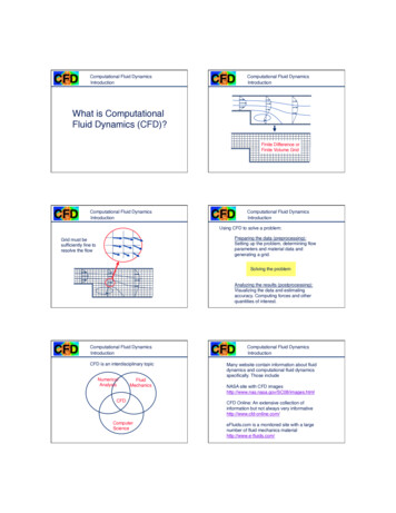
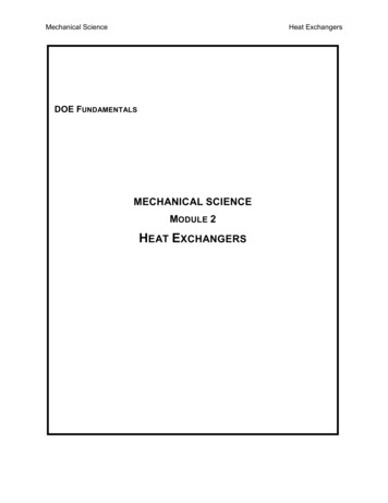
View Article OnlineRSC Advances(DDA) was carried out in positive ion mode. The parameterswere set as follows: end plate offset voltage was 500 V, capillaryvoltage was 4500 V, dry gas ow rate was 4.0 L min 1 and drytemperature was 180 C. Scan mode for MS and MS/MS werebetween 150 to 2200 m/z. The threshold for MS/MS was 2500and cycle time of 3 s.Open Access Article. Published on 23 December 2020. Downloaded on 4/16/2021 3:28:31 PM.This article is licensed under a Creative Commons Attribution 3.0 Unported Licence.Label-free quanti cationBruker Daltonics Compass Data Analysis So ware (version 4.4)was used to generate the molecular feature list for each LC-MS/MS run. The resulting molecular features were transferred toPro leAnalysis So ware to calculate buckets. A bucket is characterized by its MH mass value, retention time, and intensityvalues. The parameters for bucket calculation were set as,retention time range 6–45 min, mass range 150–2200 m/z,normalization with a quantile, value count of group attributewithin bucket were set as 2 and value count of bucket 30. A erbucket generation, peptide regulation ratios were calculatedusing T-test and transferred to ProteinScape so ware.To link peptide regulation output with the identi cationresults, the LC-MS/MS raw data les of each run were convertedto (.xml) using Bruker Daltonics Compass Data Analysis So ware (version 4.4). The resulting xml les were imported toProteinScape so ware for the Mascot Database search. A er theprotein database search, peptide identi cation was linked totheir peptide regulation ratios by selecting the retention timecorrection option and p-value 0.05 as assignment criteria.Finally, protein regulation ratios were calculated from peptideregulation ratios and a list of proteins includes quanti cationinformation such as the number of peptides ratios, coefficientof variation (CV) values, and the regulation ratios weregenerated.14MS/MS data analysis using ProteinScapeNano-LC-MS/MS raw data les of each run were converted to(.xml) and were searched using the Mascot search engine(Version 2.6, Matrix Science, London, UK) triggered by ProteinScape against Swiss-Prot database with the followingparameters: taxonomy lter was set to homo sapiens, forproteolytic cleavages, trypsin was used with maximum missedcleavages up to 1, oxidation of methionine as variable modi cations and carbamidomethyl of cysteine as xed modi cationswere allowed. Peptide mass tolerance was set at 0.08 Da andfragment mass tolerance was set at 0.2 Da. Peptide charges wereset at 2, 3, and 4. The accepted FDR was set to 1%.PaperLength cutoff 33 and expect value 1 10 3 were set as parameter for blastp-fast. Parameters for the gene ontology (GO)annotations were set as; Hit lter 500, GO weight 5, HSP-Hitcoverage cutoff 0, annotation cutoff 55 and E-value-hit- lter 1 10 6. The level 2 pie/bar chart con guration was used forshowing annotated sequences in GO graphs. The associationbetween differentially expressed quanti ed proteins with eachother and their interaction with other proteins were assessed bythe STRING EMBL so ware (version 11.0, https://string-db.org/). The medium con dence score of 0.400 was used.16ResultsFor the present study, work ow is shown in Fig. 1. The dataacquisition stage consisted of pericardial uid sample analysisthrough nano-LC-ESI-MS/MS by using the data dependentacquisition (DDA) strategy. The statistical analysis stage wasused to nd out signi cantly differentially expressed proteins inboth groups (LVEF 45 vs. LVEF 45) followed by the bioinformatics analysis (GO and KEGG) to understand the biological role of these proteins.Label-free proteomics and identi cation of differentiallyexpressed proteinsA total of 709 pericardial uid proteins were identi ed using theDDA strategy in both groups (LVEF 45 vs. LVEF 45) (TableS1†). The annotated spectra of all the identi ed proteins alongwith the differentially expressed proteins are given in the ESI le.† For a better look, the double fold change for the log 2based values derived from the quotient (LVEF 45/LVEF 45) ispresented in Fig. 2 as a volcano plot, statistically determined bya P-value 0.05, at 2.0-fold change. The identi ed proteins in thetwo groups of the experiment (LVEF 45/LVEF 45) wereanalyzed and compared, 16 proteins were found differentiallyexpressed, including 12 down-regulated and 4 up-regulatedproteins in impaired (LVEF 45) systolic function group asBioinformatic analysisLabel-free quanti ed proteins were further subjected to bioinformatic analysis, including gene ontology (GO) and KEGGpathway analysis. The 16 differentially expressed proteinssequence information was extracted from the online UniProtknowledgebase database (https://www.uniprot.org/) andretrieved in FASTA format. Functional classi cation of proteinswas done by gene ontology on the basis of biological process,molecular process and cellular component using OmicsBox(version 1.2, https://www.biobam.com/omicsbox/).15 HSP322 RSC Adv., 2021, 11, 320–327Fig. 1 Workflow of the present study. First stage is the data acquisitionstage; it consisted of pericardial fluid (PF) sample preparation and itsnano-LC MS/MS analysis using DDA strategy. The second stage is thedata analysis stage; it consisted of statistical analysis and bioinformaticanalysis (GO and KEGG pathway analysis). 2021 The Author(s). Published by the Royal Society of Chemistry
View Article OnlinePaperRSC Advancesand hedgehog-interacting protein were the most up-regulatedproteins. The results with protein name, accession number,isoelectric point, sequence coverage, molecular weight, foldchange and t-test p-values are presented in Table 2.Open Access Article. Published on 23 December 2020. Downloaded on 4/16/2021 3:28:31 PM.This article is licensed under a Creative Commons Attribution 3.0 Unported Licence.Functional classi cation of differentially expressed proteinsVolcano plot of peptide areas, derived from the quotient (LVEF 45/LVEF 45) with log 2 fold-change (x-axis) versus log 10 p-value(y-axis). The orange dots, located in the upper-left and upper-rightsections, corresponding to high fold changes and low p-values, indicate significantly regulated signals while the gray dots indicate thenon-significant differentially expressed signals. For the differentiallyexpressed signals, a p-value of 0.05 and fold change of 2 are set asthe significant thresholds.Fig. 2compared to normal (LVEF 45) systolic function group. Serumalbumin, mesothelin, E3 ubiquitin-protein ligase SH3RF3, RhoGTPase-activating protein 44 and serotransferrin were the mostdown-regulated while AT-rich interactive domain-containingprotein 4A, zinc nger protein GLI2, zinc nger protein 831,A GO classi cation was performed for the 16 signi cant differentially expressed proteins in patients with normal (LVEF 45)and impaired (LVEF 45) systolic function group as shown inthe pie chart at graph level 2 (Fig. 3 and Table S2†). The GOanalysis of differentially expressed protein revealed involvementin multiple biological processes in both groups. The highestproportion of (18%, 7 proteins: MTUS1, TRFE, PHF1, MSLN,SH3R3, OSBL6, RHG44) sequences were found to be involved incellular processes followed by biological regulation (15%, 6proteins: MTUS1, TRFE, PHF1, SH3R3, OSBL6, RHG44), andmetabolic processes (13%, 5 proteins: PHF1, MSLN, SH3R3,OSBL6, APOA4), while the remaining functions such asresponse to a stimulus; localization, signaling, developmentalprocess, cellular component organization, locomotion, immunesystem process biological adhesion and multicellular organismal process, were observed in less proportions in both groups(LVEF 45/LVEF 45). Differentially expressed, proteins werealso classi ed based on their molecular functions, such asprotein involved with binding (81%, 13 proteins: MTUS1, PHF1,MSLN, EFC4B, GLI2, RHG44, TRFE, SRBP2, ARI4A, SH3R3,Table 2 Differentially expressed proteins identified in ischemic heart disease patients with normal LV systolic function (LVEF 45) and impairedLV systolic function (LVEF 45)AccessionProteinALBU HUMANTRFE HUMANAPOA4 HUMANMSLN HUMANRHG44 HUMANSerum albuminSerotransferrinApolipoprotein A-IVMesothelinRho GTPase-activating protein44Oxysterol-binding proteinrelated protein 6Zinc nger protein GLI2Sterol regulatory elementbinding protein 2PHD nger protein 1Zinc nger protein 831EF-hand calcium-bindingdomain-containing protein 4BNuclear pore complex proteinNup205Microtubule-associated tumorsuppressor 1Hedgehog-interacting proteinE3 ubiquitin-protein ligaseSH3RF3AT-rich interactive domaincontaining protein 4AOSBL6 HUMANGLI2 HUMANSRBP2 HUMANPHF1 HUMANZN831 HUMANEFC4B HUMANNU205 HUMANMTUS1 HUMANHHIP HUMANSH3R3 HUMANARI4A HUMANMW[kDa]MascotpI scoreSequencecoverage#Peptides [%]RMS90[ppm]Fold change log 2(LVEF 45):p(LVEF 45)Value Regulation69.377.045.468.989.25.9 3565.86.8 2660.85.3 461.26.0 76.26.1 90.0001 Down167.7123.66.98.737.734.2211.51.0664.681.51 4.471.500.005 Up0.0147 6.495.9116.971.42 4.031.500.0001 Down0.0009 Up0.0147 Down227.85.820.010.4922.751.080.0116 Down141.37.319.610.88.451.500.0147 Down78.892.78.29.117.617.0112.01.06.7110.22 2.343.170.0184 Up0.0001 Down142.75.015.611.03.54 4.490.0042 Up 2021 The Author(s). Published by the Royal Society of ChemistryRSC Adv., 2021, 11, 320–327 323
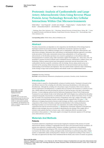
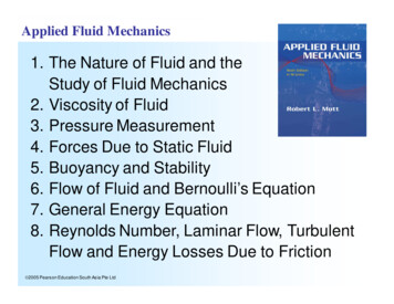
View Article OnlineOpen Access Article. Published on 23 December 2020. Downloaded on 4/16/2021 3:28:31 PM.This article is licensed under a Creative Commons Attribution 3.0 Unported Licence.RSC AdvancesPaperFig. 3 Go annotation of “biological process” “molecular function” and “cellular component” analyze by OmicxBox (version 1.2, https://www.biobam.com/omicsbox/).OSBL6, APOA4, ZN831), transporter activity (12%, 2 proteins:TRFE, OSBL6) and catalytic activity (6%, 1 protein: HHIP).OmicsBox analysis, also classi ed the differentiallyexpressed proteins according to their cellular component. Mostof the proteins were assigned to the organelle (12%, 4 proteinsMTUS1, MSLN, NU205, OSBL6), followed by extracellular region(12%, 4 proteins: TRFE, MSLN, APOA4, ALBU) and membrane(9%, 3 proteins: MTUS1, MSLN, OSBL6), while the remainingcomponent such as extracellular region part, membrane part,membrane-enclosed lumen, supramolecular complex andprotein-containing complex were observed in fewerproportions.KEGG pathway analysisSTRING was used to gain an insight into the interactive linksknown to be associated with the 16 proteins expressed differentially in the dataset. Interactive links between the differentially regulated proteins could be traced as shown in Fig. 4. Atotal of 8 RCTM/KEGG pathways were observed in the presentstudy. The suggested pathways of these differentially regulatedproteins are listed in Table 3, along with term ID, term de nition, observed gene count, background gene count, FDR andmatching proteins within a network.324 RSC Adv., 2021, 11, 320–327Interactive network analysis and association of 16 differentiallyexpressed pericardial fluid proteins obtained from STRING (version11.0, https://string-db.org/).Fig. 4 2021 The Author(s). Published by the Royal Society of Chemistry
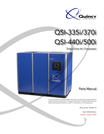
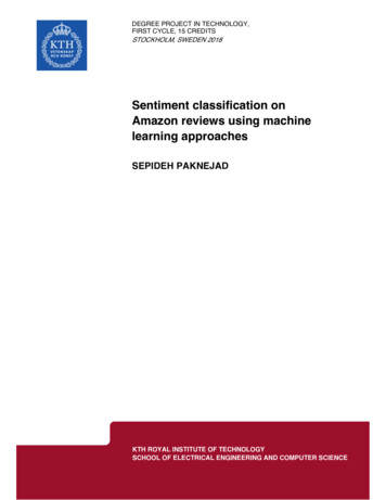
View Article OnlinePaperRSC AdvancesTable 3 Pathways of differentially expressed protein identified in ischemic heart disease patients with normal LV systolic function (LVEF 45) andimpaired LV systolic function (LVEF 45)Open Access Article. Published on 23 December 2020. Downloaded on 4/16/2021 3:28:31 PM.This article is licensed under a Creative Commons Attribution 3.0 Unported Licence.KEGG/RCTM pathways#Term IDTerm descriptionObserved gene countBackground genecountFalse discoveryrateMatching proteins inyour network (labels)HSA04340HSA05217HSA-381426Hedgehog signaling pathwayBasal cell carcinomaRegulation of insulin-like growth factor(IGF) transport and uptake by insulin-likegrowth factor binding proteins (IGFBPs)Post-translational proteinphosphorylationMetabolism of steroidsPlasma lipoprotein remodelingBile acid and bile salt metabolismHedgehog ‘on’ state22346631230.0090.0090.012GLI2, HHIPGLI2, HHIPALB, MSLN, TF31060.012ALB, MSLN, TF32221463042820.0120.0120.01570.0405ALB, OSBPL6, SREBF2ALB, APOA4ALB, OSBPL6GLI2, -5632684DiscussionProteomics is used to perform quantitative as well as qualitativeanalyses of a large array of proteins in biological samples.17Recently, label-free quantitative proteomics (LFQP) has gainedmore popularity as mass spectrometer's efficiency has signi cantly improved. The LFQP becomes a widely applied approachfor identifying as well as quantifying differentially expressedproteins. It increases the dynamic range of proteins 3 to 4 timeas compared to 2DE and is more sensitive and rapid than manyother proteomic methods.18In the present study, we have found 16 signi cantly differentially expressed proteins in LVEF 45 vs. LVEF 45. Amongthe differentially expressed proteins 12 proteins down-regulatedin impaired (LVEF 45) systolic function group, includingserum albumin, serotransferrin, apolipoprotein A-IV, mesothelin, Rho GTPase-activating protein 44, sterol regulatoryelement-binding protein 2, oxysterol-binding protein-relatedprotein 6, PHD nger protein 1, nuclear pore complex proteinNup205, EF-hand calcium-binding domain-containing protein4B, Microtubule-associated tumor suppressor 1, and E3ubiquitin-protein ligase SH3RF3. While, the remaining 4proteins were found up-regulated in impaired (LVEF 45)systolic function group including, zinc nger protein 831, zinc nger protein GLI2, AT-rich interactive domain-containingprotein 4A and hedgehog-interacting protein. One of thedown-regulated protein in patients with impaired (LVEF 45)group is albumin. Albumin is a 66.5 kDa protein which is themost abundant protein in plasma. Albumin plays a signi cantrole in regulating plasma oncotic pressure and uid distribution throughout the body compartment.19 Serum albumin'sphysiological properties include anti-in ammatory, antioxidant, anticoagulant activity and regulating the transport ofcholesterol.20 Normal pericardial uid contains between onequarter and one third of the protein of plasma and demonstrates a far higher proportion of albumin to other proteins inthe pericardial uid.21,22 Hypoalbuminemia, with serumalbumin levels less than 3.5 g dL 1 (ref. 23) has been linked to 2021 The Author(s). Published by the Royal Society of Chemistryseveral cardiovascular diseases such as coronary artery disease,congenital heart disease, stroke and heart failure.24 Hypoalbuminemia has been reported in one-third of heart failurepatients with reduced ejection fraction (LVEF 45).25 Inconcordance with the ndings in the serum, the present ndings suggest that the down-regulation of albumin in the pericardial uid is associated with impaired systolic function of thele ventricle. Hence, it could be used as a potential risk markerin identifying the patients with le ventricular systolicdysfunction.Another down-regulated protein in impaired (LVEF 45)group is serotransferrin. It is an iron binding protein, responsible for transporting iron from sites of heme degradation andabsorption to those of utilization and storage.26 The serum levelof serotransferrin decreases in in ammatory conditions such asdiabetes mellitus which in turn increases the risk of CVD.27 Ithas been demonstrated that decrease level of plasma serotransferrin is associated with rheumatic valvular heart disease(RVD),28 and coronary artery dilation (CAD) caused by Kawasakidisease.29 The down-regulation of serotransferrin in impaired(LVEF 45) group may be due to the in ammatory response ofthe body as its level also decreases in patients with CKD andESRD.30Apo A-IV is another protein that is down-regulated inimpaired (LVEF 45) group. Apo A-IV, is an oligosaccharidecontaining protein with a molecular weight of 46 kDa.31 Itsconcentration in plasma is 15–37 mg dL 1.32 Apo A-IV issynthesized in the intestine and is incorporated into thechylomicrons and secreted into the intestinal lymph during fatabsorption.33 Apo A-IV plays a signi cant role in the lipid andglucose metabolism.34 It also acts as anti-in ammatory agentand potentially exert its anti-atherogenic effect35 by reversecholesterol pathway, which removes cholesterol from peripheral cells and transports it to the liver.36 Clinical studies havedemonstrated an inverse relationship between apo A-IV andcoronary artery diseases.37 Apo A-IV de ciency is associated withatherosclerosis, cardiovascular diseases, chronic pancreatitis,malabsorption disorders38 and diabetes mellitus.39 Low Apo A-RSC Adv., 2021, 11, 320–327 325
View Article OnlineOpen Access Article. Published on 23 December 2020. Downloaded on 4/16/2021 3:28:31 PM.This article is licensed under a Creative Commons Attribution 3.0 Unported Licence.RSC AdvancesIV levels in these patients with systolic dysfunction as comparedto patients with preserved EF may point towards a diseasemechanism underlying the progression from preserved EF toreduced EF.Among the up-regulated differentially expressed proteins inpatients with impaired (LVEF 45) group is AT-rich interactivedomain containing protein 4A. It is also known as ARID4A orretinoblastoma – binding protein 1 is encoded by the geneARID4A.40 Studies shows that ARID4A play a signi cant role invarious types of cancer such as breast cancer and leukemia.ARID4A binds with other proteins and play a key role in cellproliferation and differentiation.41 Zinc nger protein 831belongs to a large class of zinc nger proteins (ZNFs) is upregulated in impaired (LVEF 45) group. Zinc nger proteins(ZNFs) have a key role in tissue development and differentiation. Alterations in ZNFs are involved in the development ofseveral diseases such as congenital heart disease, defects of thecardiac out ow tract and diabetes.42KEGG network analysis with pathway enrichment identi edthat the hedgehog signaling pathway is up-regulated inimpaired (LVEF 45) group as compared to the normal (LVEF 45) group. Network analysis revealed that two up-regulatedproteins zinc nger protein GLI2 and hedgehog-interactingprotein are involved in hedgehog signaling (Hh) pathway. Hhsignaling pathway was rst identi ed as a key mediator oforganogenesis in invertebrates by Nusslein–Volhard and Wieschaus in 1980.43 Three Hh homologues have been discoveredin mammals in the early 1990s, including Indian hedgehog(Ihh), Sonic (Shh) and Desert (Dhh). Sonic is one of the beststudied and most widely expressed during embryonic development as compared to Dhh and Ihh.44 Hh signaling pathway isknown to regulate cell differentiation and migration duringembryonic development but in post-natal life, the Hh signalingcan be recapitulated under several pathological conditions,including cardiac ischemia.45 The absence of tissue speci c Hhsignalling components reduces proangiogenic gene expressionand thus loss of coronary vasculature that leads to cardiomyocyte cell death and ventricular failure. The presence ofthe Hh signalling pathway may protect and limit the extent ofmyocardial ischemic damage.46 Many theories are postulated toelucidate the regulation mechanism of Hh signaling pathw
and a biochemical window into the heart by which information related to the pathophysiological status of the heart can be obtained.7 Pericardial uid is present between the two layers; parietal and visceral of the pericardium and is produced by the visceral pericardium of the heart. Under normal conditions, it measures about 15-50 mL in volume.