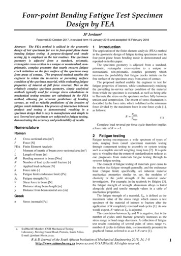
Transcription
Horisberger et al. Arthritis Research & 019) 21:80RESEARCH ARTICLEOpen AccessThe Fatigue Assessment Scale as a simpleand reliable tool in systemic lupuserythematosus: a cross-sectional studyAlice Horisberger1* , Delphine Courvoisier2 and Camillo Ribi1AbstractBackground: The vast majority of patients with systemic lupus erythematosus (SLE) complain about fatigue. Theyalso report fatigue as one of their most debilitating symptoms. Yet, in clinical practice, fatigue is only rarely assessedand remains poorly understood. The purpose of this study is to validate the Fatigue Assessment Scale (FAS) andassess the impact of disease activity on fatigue in SLE.Methods: A cross-sectional single-center study of patients was included in the Swiss SLE Cohort Study. The FASand the Short Form 36 (SF-36) were administered to SLE patients and controls with primary Sjogren’s syndrome(pSS) and healthy volunteers (HV) attending our clinic. Disease activity in SLE was captured at the same time aspatient-reported outcomes using the SLE Disease Activity Index score with the Safety of Estrogens in SLE NationalAssessment modification (SELENA-SLEDAI) and the physician’s global assessment. We explored the internal consistency,reproducibility, construct validity, and convergence of the FAS, in comparison to the vitality subscale (VT) of the SF-36. Weexamined the association of FAS with demographics, disease type, SLE disease activity, and clinical features.Results: Of the 73 SLE subjects, 89% were women and 77% were Caucasians. The median age was 43 years, and 23 (32%)patients had active SLE. Demographics in pSS and HV were similar. Within the SLE group, FAS displayed good internalconsistency (Cronbach’s alpha 0.93), unidimensionality, and test-retest reliability (ICC 0.90). FAS and VT correlated well.The total FAS was highest in active SLE and pSS and higher in non-active SLE compared to HV.Conclusion: The FAS is a promising tool to measure fatigue in SLE. Patients with SLE display a significantly higher level offatigue than HV, which is even more pronounced in active disease and comparable to fatigue levels measured in pSS.Keywords: Systemic lupus erythematosus, Fatigue Assessment Scale, Disease activityKey messages This study is the first to validate the FatigueAssessment Scale (FAS) as a reliable and simple toolto assess fatigue in systemic lupus erythematosus. Fatigue assessed by FAS correlates with globaldisease activity in systemic lupus erythematosus. Fatigue levels in active systemic lupus erythematosusare comparable to those in matched controls withSjogren’s syndrome.* Correspondence: alice.horisberger@gmail.com1Division of Immunology and Allergy, Department of Medicine, CentreHospitalier Universitaire Vaudois, CHUV BH09-627, CH-1011 Lausanne,SwitzerlandFull list of author information is available at the end of the articleBackgroundSystemic lupus erythematosus (SLE) is a chronic autoimmune disease with a wide spectrum of clinical andbiological manifestations. The multiple dysfunctions inthe innate and adaptive immune system that ultimatelylead to autoimmunity and inflammation are thought tobe triggered by various environmental factors in genetically susceptible individuals [1]. The vast majority of SLEpatients complain about fatigue [2, 3] and report it asone of the most debilitating disease features [4, 5].Whether fatigue reflects SLE disease activity is still amatter of debate [6]. Lupus fatigue has important repercussions on daily activities and is associated with poorquality of life (QoL) [7]. By using a qualitative interviewtool, Sterling et al. highlighted its negative impact on The Author(s). 2019 Open Access This article is distributed under the terms of the Creative Commons Attribution 4.0International License (http://creativecommons.org/licenses/by/4.0/), which permits unrestricted use, distribution, andreproduction in any medium, provided you give appropriate credit to the original author(s) and the source, provide a link tothe Creative Commons license, and indicate if changes were made. The Creative Commons Public Domain Dedication o/1.0/) applies to the data made available in this article, unless otherwise stated.
Horisberger et al. Arthritis Research & Therapy(2019) 21:80emotional, cognitive, professional, and social status [8].Given its major role on patient’s morbidity, fatigue is anessential aspect to consider in SLE management. Yet, fatigue remains poorly understood by both health careproviders and the patient’s entourage [9].Multiple instruments for assessing fatigue and variousdefinitions are available, without one standing out for itssimplicity and reliability. Thus, fatigue is difficult toevaluate in daily practice and to standardize across studies for research purposes. The Fatigue Assessment Scale(FAS) is a simple 10-item self-reported questionnaire designed by Michielson et al. to assess fatigue in the general population and validated subsequently in thesarcoidosis setting [10, 11]. The FAS is derived from apool of 40 items selected in four previous valid questionnaires: the Fatigue Scale, the Checklist IndividualStrength, the Emotional Exhaustion subscale of theDutch version of the Maslach Burnout Inventory, andthe Energy and Fatigue subscale of the World HealthOrganization Quality of Life assessment instrument. Theface validity was studied through a semantical analysis inorder to guide the selection of items. The FAS is reportedly a unidimensional scale measuring fatigue independently from depression [12]. It has proven to be a reliableand valid tool as well as sensitive to change in sarcoidosis patients [13]. Owing to its good psychometric properties in this specific disease, this instrument was thenused in a placebo-controlled randomized clinical trialevaluating the effect of N-acetylcysteine on fatigue inSLE [14, 15]. However, the reliability and validity of FASin SLE patients have yet to be demonstrated. In thisstudy, the primary objective was to study the construct,the convergent validity, and test-retest correlation of theFAS in SLE patients. Secondary objectives were to compare the fatigue score between SLE patients, non-SLEpatients, and healthy volunteers; to measure perceivedfatigue in SLE patients with active and inactive disease;and to determine whether other factors contribute tofatigue.MethodsStudy populationParticipants were aged 18 years old and attended theCentre Hospitalier Universitaire Vaudois (CHUV) inLausanne between June 2015 and July 2016. All were included in the Swiss Systemic Lupus Erythematosus Cohort Study (SSCS) [16, 17]. All SLE patients fulfilled therevised American College of Rheumatology (ACR) criteria and/or the Systemic Lupus International Collaborating Clinics (SLICC) criteria [18, 19]. Control groupsconsisted of patients with primary Sjogren syndrome(pSS) meeting the 2002 American-European Classification Criteria [20] and age- and gender-matched healthyvolunteers (HV). Patients with completed FAS andPage 2 of 8SF-36 forms were retained for the cross-sectional study.Patients with pSS were asked to participate in the cohortstudy during their regular clinical follow-up. The HVwere recruited by public notice in the CHUV and wereevaluated medically to confirm the good health and absence of autoimmune disease or immunomodulatorytreatment. The protocol was approved by the CantonVaud ethical committee. All participants gave their written informed consent, and the study was carried out incompliance with the Helsinki Declaration.Data collection and instrumentsData on patient’s age, sex, ethnicity, educational status,tobacco use, body mass index (BMI), disease durationsince diagnosis, activity and damage, and treatment modalities were collected during the medical visit. Diseaseactivity in SLE was assessed by the SLE Disease ActivityIndex with the Safety of Estrogens in Lupus Erythematosus National Assessment modification (SELENA-SLEDAI) [21]. This score is based on 24 clinical andbiological items, which reflect disease activity within thepast month. Disease activity was also evaluated using thePhysician’s Global Assessment score (PGA) with a4-point-Likert-scale ranging from 0 (inactive) to 3 (veryactive disease) [21]. Patients with a SELENA-SLEDAI 4and a PGA 1 were considered to have active disease[22]. Damage was assessed in SLE with the SLICC/ACRDamage Index (SDI) [23]. Disease-modifying treatment(DMARD) at study visit and in the 4 weeks before wasclassified into three groups: systemic glucocorticoids, antimalarials, and immunosuppressants. Data onhealth-related quality of life (HRQoL) and fatigue wereassessed with the Short Form 36 (SF36) and the FAS inall participants. The SF36 is a widely used health surveyform measuring 8 dimensions of QoL; each of themranges from 0 to 100 with lower scores reflecting poorerhealth. The SF-36 vitality subscale (VT-SF36) is used forconvergent validity of fatigue questionnaires [24]. Themental health subscale of the SF-36 (MH-SF36) wasused for the discriminant validity. The FAS comprises 10questions with answers varying from never to always ona 5-point-scale. The total FAS score ranges from 10 to50, increasing proportionally to fatigue. Both questionnaires were completed by participants during the studyvisit. A sample of 30 SLE patients was asked to completethe FAS 2 weeks after the first assessment. These patients were reminded by text messages to complete andsend back the questionnaire after 2 weeks with apre-stamped envelope. Eligibility for this test-retest studywas a completed form, and the absence of importantintercurring events reported by the patient that wouldinfluence the state of fatigue during the test-retestperiod. FAS, SF-36, demographical data, and clinical data
Horisberger et al. Arthritis Research & Therapy(2019) 21:80were collected in the two control groups of pSS patientsand HV.Statistical procedureDescriptive statistics were presented as absolute count andpercent for qualitative data and as median and interquartilerange (IQR) for quantitative. Difference between groupsand correlations were evaluated using non-parametric tests.The psychometric properties of the FAS were evaluatedusing the following methods. Cronbach’s alpha was calculated to measure the reliability of the tool by looking atinter-item consistency. This measure is generally considered satisfactory if the alpha value is above 0.7 forgroup-level analysis but a value above 0.9 is desirable for individual patients in clinical application [25]. A factor analysis was performed using the principal component analysis(PCA) based on Kaiser criteria (eigenvalue 1) and visualinspection of the scree plot. A PCA was used with the FASand the MH-SF36 in order to examine the divergent validity (construct validity) of the FAS. Factor analysis extractionwas presented with an oblimin rotation. A coefficient factorabove 0.3 was considered significant. As there were slightlyless than 5 (4.87) patients per item for this measure, weconfirmed the results on a larger sample using SLE, pSS,and HV. The convergent validity was examined with aSpearman’s rho correlation between FAS and VT-SF36.The test-retest reliability was measured using intraclass correlations (ICC). A linear regression model was used formultivariable analysis, with total FAS score as dependentvariable and disease activity and corticoid use as independent variables. P values 0.05 were considered significant.All statistics were performed on IBM SPSS statistics 24(IBM Corp Armonk, NY) and GraphPad Prism 7 (GraphPad Software, La Jolla, CA, USA).Page 3 of 8ResultsGroup characteristicsSeventy-three patients with SLE, 23 patients with pSS,and 23 healthy volunteers were included (Table 1). Therewas no statistical difference in demographic features between groups. SLE subjects were predominantly women(89%), of Caucasian origin (77%) and had a median ageof 43 years at assessment. Median disease duration fromdiagnosis was 7 (3–15) years in patients with SLE compared to 1 (0–2) in those with pSS (p 0.001). Patientswith SLE were more frequently treated with systemiccorticosteroids and immunosuppressant drugs comparedto those with pSS. Among SLE patients treated with immunosuppressants, four had received cyclophosphamidein the month preceding the study. Within the SLEgroup, 23 (32%) had active disease (SELENA-SLEDAI 4 and PGA 1). Table 2 compares the characteristics ofSLE patients according to their disease activity.Psychometric analysis of FASThe internal consistency of the FAS was measured at0.93 (Cronbach’s alpha coefficient), and none of theitems would have improved the internal consistency ifremoved. Principal component analysis favored aone-component solution, confirmed by visual inspectionof the scree plot (Fig. 1). The factor analysis of FASitems extracted a unique factor explaining 64% of thevariance with the items loading between 0.68 (item 3)and 0.88 (item 5).Of the 73 SLE subjects, 30 (41%) had a test-retest assessment. Seven patients were excluded because of aself-reported significant event between the first and thesecond FAS assessment. There was no difference in age,sex, ethnicity, and disease activity between the wholeTable 1 Patient’s and volunteer’s characteristicsAge, median (IQR) yearsAll SLE (n 73)pSS (n 23)HV (n 23)p value§43 (34–57)44 (33–58)34 (22–49)0.44Female sex (%)65 (89)21 (91)20 (87)0.89Caucasian, no. (%)56 (77)19 (83)20 (87)0.53Higher education*, no./total (%)30/59 (51)10/22 (45)10/23 (43)0.80Active smoker, no. (%)17 (23)4 (17)8 (35)0.37Body mass index, median (IQR)23 (20–28)25 (22–28)22 (20–24)0.48Use of psychiatric medication , no. (%)14 (19)8 (34)3 (13)0.16Time from diagnosis, median (IQR) years7 (3–15)1 (0–2)– 0.01Immunomodulators past month, no. (%)64 (88)16 (70)–0.04Antimalarials, no. (%)52 (71)14 (61)–0.35Systemic corticosteroids, no. (%)39 (53)5 (22)–0.01Immunosuppressants, no (%)35 (48)5 (22)–0.03SLE systemic lupus erythematosus, pSS primary Sjogren syndrome, HV healthy volunteers, IQR interquartile range*Defined as higher professional or technical school or university degree Defined as any antidepressants, antipsychotics, and anxiolytics/hypnotics§p value 0.05 is considered significant
Horisberger et al. Arthritis Research & Therapy(2019) 21:80Page 4 of 8Table 2 Clinical characteristics of systemic lupus erythematosus patients according to global disease activity (n 73)Inactive SLE (n 50)Active SLE (n 23)p value§Age, median (IQR) years46 (35–59)39 (30–52)0.20Female sex (%)44 (88)21 (91)0.68Caucasian, no. (%)38 (76)18 (78)0.83Higher education*, no./total (%)20/39 (51)10/20 (50)0.93Active smoker, no. (%)9 (18)8 (35)0.12Body mass index, median (IQR)24 (20–29)22 (19–24)0.35Use of psychiatric medication , no. (%)9 (18)5 (22)0.75Disease duration, median (IQR) years7 (3–15)6 (3–15)0.42History of lupus nephritis, no (%)19 (38)6 (26)0.31History of neurolupus, no (%)6 (12)2 (7)0.67SELENA-SLEDAI, median (IQR) score2 (0–2)6 (4–10) 0.001PGA score, median (IQR) score0 (0–0)1 (1–2) 0.001Immunomodulators past month, no. (%)Antimalarials, no. (%)44 (88)20 (87)0.9037 (74)15 (65)0.44Systemic corticosteroids, no. (%)26 (52)13 (57)0.72Immunosuppressants, no. (%)25 (50)10 (43)0.60IQR interquartile range, SLE systemic lupus erythematosus*Active disease is defined as a Systemic Lupus Erythematosus Disease Activity Index score with the Safety of Estrogens in Lupus Erythematosus NationalAssessment modification (SELENA-SLEDAI) 4 and a Physician’s Global Assessment score (PGA) 1 Defined as any antidepressants, antipsychotics, and anxiolytics/hypnotics§p value 0.05 is considered significantSLE sample and the test-retest subjects (data notshown). The test-retest correlation was good with anICC of 0.90 (95%CI 0.80–0.95, p 0.001) for a 2-weekinterval. The FAS correlated strongly with the VT-SF36subscale (rs 0.85, p 0.001). The FAS correlation withthe MH-SF36 was rs 0.65 (p 0.001). The PCA on thecombined pool of the FAS and the MH-SF36 items favored a two-component solution (eigenvalue factor I,8.4; factor II, 1.9; percentage explained variance, 69%).One statement of the ten FAS items (FAS-7) loadedhigher on the mental health scale. All other items loadedhigher on the fatigue factor. All items of the MH-SF36loaded higher on the mental health factor (Table 3).These results were similar using a larger sample including SLE, pSS, and HV (data not shown) except for theitem FAS-7 showing a higher loading on the fatigue factor (0.48) than on the mental factor (0.42).Comparison of fatigue levels between groupsFig. 1 Scree plot of the Fatigue Assessment Scale (FAS) andits itemsThe FAS score was significantly increased in both SLEand pSS compared to healthy subjects. Median (IQR)FAS was 23 (17–32) in SLE, 27 (20–34) in pSS, and 16(14–18) in HV (p 0.001). These fatigue findings wereconfirmed using the VT-SF36 subscale (median VT score[IQR] 45 [23–58] in SLE, 35 [15–50] in pSS, and 70[50–75] in HV, p 0.001). There was a good correlationbetween the FAS and the VT-SF36 score among allgroups of participants (Table 4). There was a lesser butsignificant correlation between FAS and the other subscales of the SF36 in both SLE and pSS. In contrast, HVshowed modest to the non-significant correlation between FAS and SF-36 subscales other than VT. In thethree groups of participants, no correlation was found
Horisberger et al. Arthritis Research & Therapy(2019) 21:80Page 5 of 8Table 3 Factor loading of the FAS and the MH-SF36 obtained in 73 patients with SLE in a two-factor solutionItemsFactor 1 - fatigueFactor 2 - mental healthI am bothered by fatigue (FAS-1)0.94†0.10I get tired very quickly (FAS-2)0.94†0.08†I do not do much during the day (FAS-3)0.750.08I have enough energy for everyday life (FAS-4) 0.74† 0.13†Physically, I feel exhausted (FAS-5)0.890.01I have problems starting things (FAS-6)0.62† 0.32†I have problems thinking clearly (FAS-7)0.37† 0.51†I feel no desire to do anything (FAS-8)† 0.25†0.65Mentally, I feel exhausted (FAS-9)0.62 0.25When I am doing something, I can concentrate quite well (FAS-10) 0.77† 0.20Have you been a very nervous person? (SF09B)0.180.97†Have you felt so down in the dumps that nothing could cheer you up? (SF09C) 0.050.87†Have you felt calm and peaceful? (SF09D)0.20 0.67†Have you felt downhearted and low? (SF09F)0.020.91†Have you been a happy person? (SF09H)0.17 0.68†FAS Fatigue Assessment Scale, MH-SF36 mental health component of the Short Form 36 health survey questionnaire†A coefficient factor above 0.3 was considered significantbetween the total FAS score and demographic featuressuch as age, sex, ethnicity, educational status, and BMI(data not shown). Tobacco use was weakly associatedwith a higher degree of fatigue in the pSS group (p 0.024). The use of psychiatric medication was associatedwith higher fatigue levels in both SLE (p 0.002) andpSS (p 0.047) patients, but not in HV.Association of fatigue and disease activity in SLE patientsThe median (IQR) FAS score was 31 (20–36) in patientswith active SLE, compared to 22 (15–27) in those withinactive disease (p 0.005). In healthy volunteers, theFAS score was slightly lower than in patients with inactive SLE (p 0.05). In contrast, patients with activedisease presented with a significant higher level ofTable 4 Correlation coefficient between the FAS and the SF-36subscoresFASSF-36 subscoresSLE (n 73)pSS (n 23)HV (n 23)Physical functioning 0.58* 0.73* 0.25Role physical 0.59* 0.58* 0.32Role emotional 0.60* 0.64*0.00Bodily pain 0.68* 0.67*0.23General health 0.71* 0.70* 0.50*Vitality 0.85* 0.78*0.85*Social functioning 0.67* 0.47*0.52*Mental health 0.65* 0.60* 0.50*FAS Fatigue Assessment Scale, SF-36 Short Form 36, SLE systemic lupuserythematosus, pSS primary Sjogren’s syndrome, HV healthy volunteers*Correlations are significant at p value 0.05fatigue than HV (p 0.001) (Fig. 2). No association wasfound between fatigue and other clinical parameterssuch as the time elapsed from diagnosis to assessment,number of ACR criteria fulfilled at inclusion, presence ofrenal involvement at any time during disease course,presence of auto-antibodies (anti-dsDNA and anti-SSA/Ro) at study visit, and damage accrual (SDI score). Therewas no difference in levels of fatigue between SLE patients with or without a history of renal disease (median[IQR] FAS was 24 [17–32] and 23 [16–31], respectively).Fatigue was higher in the patients treated with corticosteroids (r 0.27, p 0.022). There was no differencewhen comparing fatigue with the use of other DMARDs.In a linear regression model, after adjusting for corticosteroids and psychiatric medication use at visit, activedisease remained significantly associated with fatigue sothat FAS increased on average by 6.2 points (95%CI 2.2–10.3, p 0.003) for each additional point in diseaseactivity.DiscussionTo the best of our knowledge, ours is the first study thatvalidates the FAS as a simple and reliable tool to assessfatigue in SLE. The FAS measures fatigue by means of10 items including two reverse questions. This fatiguescale was previously shown to be useful and valid in thegeneral population, in the working population, and insarcoidosis patients. Fatigue is a prominent feature ofSLE, a disease known for its wide range of symptoms.Brain fog is a common complaint in SLE patients, whichrefers to periods of impaired cognition [26] without anysigns of neurolupus. Indeed, only a very few of the
Horisberger et al. Arthritis Research & Therapy(2019) 21:80Page 6 of 8Fig. 2 Fatigue Assessment Scale (FAS) scores in 73 patients with systemic lupus erythematosus (SLE) according to disease activity and controlswith primary Sjogren’s syndrome or in good health. SLE systemic lupus erythematosus, FAS Fatigue Assessment Scales (ranges from 10 – nofatigue to 50 – extreme fatigue), pSS primary Sjogren’s syndrome, Healthy healthy volunteers. Plots represent the individual values (diamonds),the median score, and the IQR for each group. *p 0.05; ** 0.001patients in this study had overt neurological disease. Fatigue is also a major complaint in other autoimmunediseases such as pSS. The thin line between cognitivedysfunction and depression in SLE and pSS makes fatigue assessment a particular challenge in this population. Our study shows that the FAS displays solidpsychometric abilities, with an excellent internalconsistency and test-retest reliability. Its convergent validity is supported by the good correlation with theVT-SF36. Concerning discriminant validity, it is revealedthat fatigue and mental disorders such as depression arerelated but distinct constructs.This cross-sectional study also shows that fatigue measured by FAS is significantly increased in both SLE andpSS patients compared to healthy controls. Several authors reported similar results for chronic inflammatorydiseases, including SLE and pSS [27, 28]. In previousstudies, the prevalence of fatigue in SLE subjects variedfrom 76 to 90% [3, 29, 30]. By comparing FAS to theSF-36 subscales, we confirm the relationship between fatigue and various aspects of HRQoL, such as perceivedmental health, emotional state, bodily pain, and socialfunctioning in both SLE and pSS. We found that fatiguelevels were higher in patients using psychiatric medication. Whether fatigue is the cause or the consequence ofmental health issues and prescribed psychiatric medication in these patients remains to be established. SLE patients in our study differed from those with pSS in termsof disease duration from diagnosis, which was significantly shorter for pSS. On the other hand, pSS has amore insidious disease course than SLE. Diagnosis ofpSS is often delayed by years and the duration of symptoms difficult to establish. This may explain why thetime elapsed since pSS and SLE diagnosis in our studyhad no influence on the measured fatigue levels and reflects our clinical impression of long-standing fatigue inmost patients suffering from these conditions. Moreover,we show that patients with active SLE have significantlymore fatigue than healthy controls, whereas this difference is much less pronounced in those with inactiveSLE. The association between fatigue and disease activityin SLE is controversial. Some authors reported a lack ofassociation between disease activity and the Fatigue Severity Score [30–32]. Others, however, were able to showthat fatigue increases with SLE activity, although to various degrees [3, 14, 33]. In the present study using twoglobal scores (SELENA-SLEDAI and PGA) to ascertainSLE disease activity, we demonstrate a clear positive correlation with fatigue. Interestingly, patients with activeSLE displayed a similar degree of fatigue than those withpSS. This observation amplifies the need for further investigation of immune factors that could contribute tofatigue in both active pSS and SLE. Recently, Petri et al.demonstrated a significant decrease in fatigue and disease activity in SLE patients treated with blisibimod, aselective inhibitor of B cell activating factor, in a phase2b study. The authors found a weak correlation betweendisease activity and fatigue and concluded that thissymptom appears to be closely related to immune dysfunction [33, 34]. Others have found that the use of IL-6
Horisberger et al. Arthritis Research & Therapy(2019) 21:80Page 7 of 8blocking agents has a favorable impact on fatigue in SLE[35]. These findings could be explained by the impact ofpro-inflammatory cytokines on the central nervous systemic with induction of illness behavior, expressed insymptoms such as fatigue and anhedonia [36]. The reliefof fatigue after the use of various biological agents supports a relationship between fatigue and inflammationdue to ongoing disease activity. We did not find any association of fatigue with traditional markers, such asauto-antibodies. This underlines the need for furtherstudies to assess fatigue levels in relation to circulatingcytokines and novel biomarkers in autoimmune disease.Our study has some limitations. We did not assessadditional factors potentially contributing to fatigue,such as sleep disorders, depression, and physical activity.Our study is cross-sectional and does not allow the determination of the sensitivity to change. Longitudinalstudies with FAS are needed to assess variations in fatigue, and its causes and its impact on health-relatedquality of life and working capacity both in SLE and pSS.Authors’ contributionsAH, DC, and CR analyzed and interpreted the patients’ data. All authors readand approved the final manuscript.ConclusionIn conclusion, this study validates the FAS as a simpleand reliable tool to assess fatigue in patients with SLEand shows a close and positive correlation of fatiguewith disease activity, independently from corticosteroiduse and psychiatric medication.References1. Zhernakova A, van Diemen CC, Wijmenga C. Detecting shared pathogenesisfrom the shared genetics of immune-related diseases. Nat Rev Genet. 2009;10(1):43–55.2. Moses N, Wiggers J, Nicholas C, Cockburn J. Prevalence and correlates ofperceived unmet needs of people with systemic lupus erythematosus.Patient Educ Couns. 2005;57(1):30–8.3. Wysenbeek AJ, Leibovici L, Weinberger A, Guedj D. Fatigue in systemiclupus erythematosus. Prevalence and relation to disease expression. Br JRheumatol. 1993;32(7):633–5.4. Krupp LB, LaRocca NG, Muir J, Steinberg AD. A study of fatigue in systemiclupus erythematosus. J Rheumatol. 1990;17(11):1450–2.5. Ahn GE, Ramsey-Goldman R. Fatigue in systemic lupus erythematosus. Int JClin Rheumtol. 2012;7(2):217–27.6. Cleanthous S, Tyagi M, Isenberg DA, Newman SP. What do we know aboutself-reported fatigue in systemic lupus erythematosus? Lupus. 2012;21(5):465–76.7. Pettersson S, Lovgren M, Eriksson LE, Moberg C, Svenungsson E,Gunnarsson I, et al. An exploration of patient-reported symptoms insystemic lupus erythematosus and the relationship to health-related qualityof life. Scand J Rheumatol. 2012;41(5):383–90.8. Sterling K, Gallop K, Swinburn P, Flood E, French A, Al Sawah S, et al.Patient-reported fatigue and its impact on patients with systemic lupuserythematosus. Lupus. 2014;23(2):124–32.9. Golder V, Ooi JJY, Antony AS, Ko T, Morton S, Kandane-Rathnayake R, et al.Discordance of patient and physician health status concerns in systemiclupus erythematosus. Lupus. 2018;27(3):501–6.10. De Vries J, Michielsen HJ, Van Heck GL. Assessment of fatigue amongworking people: a comparison of six questionnaires. Occup Environ Med.2003;60(Suppl 1):i10–5.11. De Vries J, Michielsen H, Van Heck GL, Drent M. Measuring fatigue insarcoidosis: the Fatigue Assessment Scale (FAS). Br J Health Psychol. 2004;9(Pt 3):279–91.12. Michielsen HJ, De Vries J, Van Heck GL. Psychometric qualities of a brief selfrated fatigue measure. The Fatigue Assessment Scale J Psychosom Res.2003;54(4):345–52.13. de Kleijn WP, De Vries J, Wijnen PA, Drent M. Minimal (clinically) importantdifferences for the Fatigue Assessment Scale in sarcoidosis. Respir Med.2011;105(9):1388–95.14. Garcia RJ, Francis L, Dawood M, Lai ZW, Faraone SV, Perl A. Attention deficitand hyperactivity disorder scores are elevated and respond to Nacetylcysteine treatment in patients with systemic lupus erythematosus.Arthritis Rheum. 2013;65(5):1313–8.AbbreviationsBMI: Body mass index; CHUV: Centre Hospitalier Universitaire Vaudois;DMARD: Disease-modifying treatment; FAS: Fatigue Assessment Scale;HRQoL: Health-related quality of life; HV: Healthy volunteers; ICC: Intraclasscorrelation coefficient; IQR: Interquartile range; MH-SF36: Mental healthsubscale of the SF-36; PCA: Principal component analysis; PGA: Physician’sGlobal Assessment score; pSS: Primary Sjogren’s syndrome; QoL: Quality oflife; SDI: SLICC/ACR Damage Index; SELENA-SLEDAI: SLE Disease ActivityIndex score with the Safety of Estrogens in SLE National Asse
order to guide the selection of items. The FAS is report-edly a unidimensional scale measuring fatigue independ-ently from depression [12]. It has proven to be a reliable and valid tool as well as sensitive to change in sarcoid-osis patients [13]. Owing to its good psychometric prop-erties in this specific disease, this instrument was then