Transcription
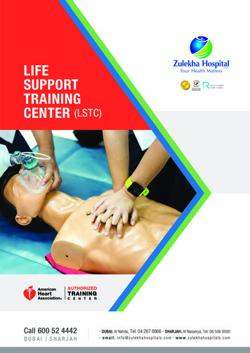
Dear ACLS Student:Please Read this letter carefullyThis letter is to confirm your registration in the Advanced Cardiac Life Support (ACLS) course.Thank you for choosing Fast Response. We appreciate the opportunity to provide you withyour clinical and professional educational needs.Please plan to be on time because it will be difficult for late students to catch up once we start.All classes start at 9:00 am sharp. If you are more than 15 minutes late, you may be turnedaway, as required by the American Heart Association (AHA). Students are expected to attendand participate in the entire course.Be prepared to pass the adult 1-rescuer CPR with AED skills test. Please note that we do notrenew your BLS card based on this CPR test, which is a requirement of the ACLS courseitself. All renewal (1-day) participants must bring their current American Heart Associationissued ACLS card to class. There are no exceptions for expired cards.ACLS cards and Continuing Education Units (CEU’s) will be issued at the end of class.How to Get ReadyThe ACLS Course is designed to teach you the lifesaving skills required to be both a teammember and a team leader in either an in-hospital or an out-of-hospital setting. Because theACLS Course covers extensive material in a short time, you will need to prepare for the coursebeforehand.The ACLS Course does not teach CPR, ECG rhythm identification, pharmacology, or ACLSalgorithms. The course format requires all students to be fully prepared prior to comingto class. If you do not review CPR, learn and understand ECG’s or the pharmacologyinformation in the Pre-course Self-Assessment, it is unlikely that you can successfullycomplete the ACLS Course.**Fast Response offers the AHA’s “ECG & Pharmacology” course as a preparatory class forACLS; please call us for more information.1
Pre-course RequirementsYou should prepare for the course by doing the following:1.Complete the precourse preparation checklist that came with your ACLS ProviderManual. Bring the checklist with you to the course.2.Review the course agenda.3.Review and understand the information in your ACLS Provider Manual. Payparticular attention to the 10 cases in Part 5.4.The resuscitation scenarios require that your BLS skills and knowledge are current.Review and understand all BLS 2010 guidelines. You will be tested on 1-rescuer adultCPR and AED skills at the beginning of the ACLS Provider Course. You must knowthis in advance, since you will not be taught how to do CPR or how to use an AEDduring the course.5.Review, understand, and complete the ECG and Pharmacology Precourse Selfwill not beAssessment on the Student Website (www.heart.org/eccstudent). Youtaught how to read or interpret ECGs in the course, nor will you be taught detailsabout ACLS pharmacology.6.Print your scores for the Precourse Self-Assessment and bring them with you to class.What to Bring and What to WearBring your ACLS Provider Manual to each class. You will need it during each lesson in thecourse. You may wish to purchase the AHA’s 2010 Handbook of Emergency CardiovascularCare for Healthcare Providers (optional), which you may bring to the course to use as areference guide during some of the stations in the course.Please wear loose, comfortable clothing to class. You will be practicing skills that require youto work on your hands and knees, and the course requires bending, standing, and lifting. If youhave any physical condition that might prevent you from engaging in these activities, pleasetell an instructor. The instructor may be able to adjust the equipment if you have back, knee, orhip problems.2
Please be aware:Reschedule Policy No refunds will be issued. All registrations are final. You may reschedule your course by calling us at least 2 business days prior to yourscheduled course date. You will be charged a rescheduling fee of 5.00. If you reschedule your course fewer than 2 business days prior to the course startdate, you will be charged a rescheduling fee of 50% of the course fee. If you “no show” to your scheduled class, you will be charged 50% of the coursecost to reschedule. Course must be rescheduled and attended within 30 days from the original start date.No additional rescheduling requests will be honored. Only one reschedule request will be honored per course. Our Administrative Offices are closed on weekends and holidays. We do not acceptrescheduling requests on weekends or holidays. We do not accept requests left on the answering machine.Cancellation Policy We do not issue refunds for course fees. All registrations are final. If you cancel or do not attend the class you have registered for, you will forfeit yourentire course fee.Late Arrival Our classes start on time. Please plan your trip accordingly and remember to allowtime for parking. If you are late for your scheduled class, you will be not be admitted into class and youmust reschedule. You will be charged a 5.00 rescheduling fee.Lisa Dubnoff, MICP, R.N.ALS Program Director3
Dear Student,In order for us at Fast Response to be able to provide you with a quality program, there areAmerican Heart Association (AHA) guidelines we must follow. Outlined below are the FastResponse policies that enact the AHA’s requirements for possession of student / providermanuals.Each student must have the 2010 American Heart Association ACLS Provider Manualavailable to them before, during, and after the course in order to comply with AHAguidelines. These books are available at Emergencystuff.com for a discounted rate. Ifyou show up to your class without the required manual, there are two options:A: You must purchase the book to attend the class. Books can be purchased atthe reception desk. The current cost (as of July 2011) is 52.75 IncludingAlameda County sales tax for the Advanced Cardiac Life Support provider manual.B: You may reschedule of a 5 fee. To attend your rescheduled class, you mustpurchase the book. If you arrive to your rescheduled class without the book, youwill be asked to purchase the book or be turned away from class.If you are attending this class from a contracted hospital provider, you are required to obtainthe ACLS Provider Manual from your education department, if possible. If you failed to dothis, you will be required to purchase the book prior to being granted entrance into class.Fast Response would like to apologize for any inconvenience this may cause, but theguidelines set forth by the AHA are very specific in how the class literature must be handled.Thank you,John Greene, NREMT-PDirector of Continuing EducationFast Response School of Health Care Education4
American Heart LinksThere are several resources available to you on the American Heart Association website atwww.americanheart.org . Here are some helpful kinks: You can find information on cardiovascular diseases and risk factors ur-Risk-of-Heart-Attack UCM 002040 Article.jsp You can access information on the warning signs of heart attack and stroke tack/Heart%C2%AD%E2%80%90Attack UCM 001092 SubHomePage.jsp You can find out how to lead a healthy lifestyle tingHealthy UCM 001078 SubHomePage.jspAmerican Heart Association Heart Disease and stroke Update UCM 423970 Article.jsp5
ACLS Course Agenda ProviderDay 10900-0920Welcome / IntroductionsCourse overview / OrganizationPrecourse Self-Assessment Review0920-0940BLS and ACLS Surveys: Video0940-1005Importance of CPR: Lecture1005-1030EKG Review: Lecture1030-1040Break1040-1120Management of Respiratory Arrest (Group 1),CPR and AED (Group 2): Learning Stations1120-1200Management of Respiratory Arrest (Group 2),CPR and AED (Group 1): Learning Stations1200-1300Lunch1300-1335Stroke: Video and Discussion1335-1410The Megacode and Resuscitation Team Concept: Video andDiscussion1410-1420Break1420-1700Cardiac Arrest (VF / Pulseless V-Tach): Learning Station6
ACLS Course Agenda ProviderDay 20900-0935Acute Coronary Syndromes: Video and Discussion0935-1035PEA / Asystole / Bradycardia,Tachycardia - Stable and Unstable: Learning Station1035-1045Break1045-1145Megacode Practice: Learning Station1145-1245Lunch1245-1445Megacode; Testing1445-1500ACLS Review: Jeopardy Game1500-1700Written Exam: Testing7
ACLS Course Agenda Renewal0900-0915Welcome / IntroductionsCourse overview / OrganizationPrecourse Self-Assessment Review0915-0940ACLS Science Update: Video0940-1010Importance of CPR: Lecture1010-1020Break1020-1100Management of Respiratory Arrest (Group 1),CPR and AED (Group 2): Learning Stations1100-1140Management of Respiratory Arrest (Group 2),CPR and AED (Group 1): Learning Stations1140-1200Stroke: Video1200-1300Lunch1300-1330The Megacode and Resuscitation Team Concept: Video andDiscussion1330-1430Megacode Practice: Learning Station1430-1440Break1440-1540Megacode: Testing1540-1600ACLS Review: Jeopardy game1600-1700Written Exam: Testing8
Patient AssessmentIn ACLS, the specific treatment of a given dysrhythmia or condition depends on the patient’shemodynamic status. In general, patients can be divided into four categories to determinetreatment priorities: AsymptomaticSymptomatic – StableSymptomatic – UnstablePulselessAsymptomatic patients do not receive treatment, but should be monitored for changes incondition. Any patient with symptoms (even apparently mild symptoms such as palpitations)should be assessed to determine if they are Stable or Unstable. Determination of a patient’slevel of hemodynamic compromise can include several factors: General Appearance: The first indication of hemodynamic status comes from apatient’s general appearance, including skin signs, level of activity, and work ofbreathing. If a patient shows signs of compensation (such as pale, cool, or diaphoreticskin) or acute distress, they are unstable. Level of Consciousness: Interaction with the patient allows the provider to evaluatethe patient’s level of consciousness based on the patient’s activity, awareness of theirsurroundings, and ability to provide information. If a patient shows any level of mentaldeficit, family or friends should be consulted to determine if this state differs from thepatient’s baseline. If the mental deficit is acute, the patient should be consideredunstable. Vital signs: Vital signs provide a diagnostic evaluation of the patient. Blood pressureis the primary indicator. A systolic blood pressure above 90 mm usually indicates thatthe patient is stable (although the provider should be alert for changes in bloodpressure that might indicate an unstable patient even if blood pressure is normal).Other vital signs may be useful; however, the provider should remember that variousconditions (CO2 poisoning) can mask changes in blood oxygen levels, and that a highO2 saturation may be present in unstable patients (those in shock). Additionally, heartrate is of no use in determining if a patient is stable or unstable – a patient with a heartrate of 80 can be severely unstable, while a patient with a heart rate of 210 can bestable if they are still perfusing well.9
If a patient’s General Appearance, Level of Consciousness, and Vital Signs are all normal,the patient is stable. If possible, treatment should be rendered starting with the least invasivethat is appropriate for that patient’s hemodynamic status. In ACLS, the preferentialtreatment for symptomatic, but stable patients is generally medications. The preferentialtreatment for unstable patients is generally Electrical Therapy.Once treatment is rendered, the provider must reassess the patient. If the patient remainssymptomatic, the appropriate treatment (medications or electricity) should be given againdepending on the patient’s heart rhythm and current hemodynamic status. Thus, if a patientwas stable before, but becomes unstable after administration of a drug, the patient shouldreceive electrical therapy to continue treating the dysrhythmia rather than additional doses of amedication.If a patient’s General Appearance indicates they may be unconscious, you should check forresponsiveness. If the patient is Unresponsive, get help (send someone to call 911 and bringback an AED, call a code, etc.). Then assess Circulation by checking for a pulse. If thepatient has a pulse, assess breathing next. If the patient is not breathing, or breathinginadequately, rescue breathing should be initiated. If the patient is pulseless, rescuers shouldbegin CPR.Once you determine that a patient is Pulseless, an AED or EKG monitor should be attachedas soon as possible. CPR should be continued with minimal interruptions. After each rhythmcheck, the patient should be defibrillated if appropriate (rhythm Ventricular Fibrillation orPulseless Ventricular Tachycardia). Regardless of the heart rhythm, medications should begiven as soon as possible after CPR is resumed. The specific medication should bedetermined by the patient’s exact status and heart rhythm.Remember: Treat the patient not the monitor!!10
EKG and Electrical Therapy ReviewThe EKG tracing represents electrical activity through the heart. The P wave representsdepolarization of the atria; the QRS complex represents depolarization of the ventricles; andthe T wave represents the repolarization of the ventricles. The interval from the first deflectionof the P wave to the beginning of the QRS complex is the P-R Interval (PRI), and should bebetween 0.12 and 0.20 seconds. A patient's QRS complex has duration of 0.12 seconds orless; a longer duration (wide QRS) indicates delayed conduction through the ventricles, oftenas the result of a ventricular pacemaker focus. The horizontal axis of the EKG stripsmeasures time. Each large box represents 0.20 seconds; each small box represents 0.04seconds.To obtain a 3-lead EKG tracing, place the white (RA) electrode on the right chest just belowthe clavicle; the black electrode (LA) on the left chest just below the clavicle; and the redelectrode (LL) laterally on the lower left abdomen. Pacer pads generally are applied to theanterior/posterior positions; however, defibrillation pads can either be applied in the traditionallocation of sternum and apex, or they can also be placed in the anterior/posterior positions.Rhythm Disturbances: Treat the patient, not the dysrhythmia. Always assess your patientfor pulses, perfusion, and level of consciousness – is the patient Stable, Unstable, orPulseless? Next, assess the rhythm: Is it fast or slow? Is it life threatening? As you treat thepatient, try to discover the cause of the dysrhythmia – for many patients, their only chance ofsurvival is if you can identify and treat a reversible cause. There are many possible causesof rhythm disturbances, especially bradycardia or PEA. Although lab draws can be useful, ahistory of the patient and the current event obtained from a parent or caregiver is often moreuseful.11
Defibrillation (Unsynchronized Shock)Fibrillation is a disorganized rhythm that, if present in the ventricles, is life threatening.Immediate CPR combined with early defibrillation is critical to survival from sudden cardiacarrest. Defibrillation terminates all electrical activity in the pulseless heart in the hopes it willresume beating in a coordinated fashion. A shock should be delivered about once every 2minutes if the patient remains in Ventricular Fibrillation. With a monophasic defibrillator, therecommendation is to deliver the first shock at 360 joules. If a biphasic defibrillator is used,the recommended dosage is machine dependent and should appear on the front of themachine. If optimal shock dosage is not known, the default setting is 200 joules.Synchronized CardioversionIn stable patients with narrow-complex tachycardia (i.e., SVT), attempt vagal maneuvers firstand then administer adenosine. For stable patients experiencing wide-complex tachycardia,consider adenosine if the rhythm is regular and monomorphic. For any unstable tachycardia(characterized by hypotension, acute alteration of mental status, shock, etc.), synchronizedcardioversion is the treatment of choice. This is especially true in the absence of an IV / IO. Ifan IV / IO is available, and the tachycardia consists of regular narrow complexes, adenosinecan be considered. If the patient is conscious, consider sedation prior to cardioversion;however, synchronized cardioversion should not be delayed while waiting for sedationin severely symptomatic patients.With a biphasic monitor, the initial dose is delivered at 120 to 200 joules for atrial fibrillation.Cardioversion of atrial flutter and other SVT’s generally require less energy; an initial energy of50 joules to 100 joules is often sufficient. Deliver additional shocks in stepwise fashion.Monomorphic V-Tach with a pulse responds well to initial energies of 100 joules. PolymorphicV-Tach should be treated like V-Fib. That is, deliver a high energy unsynchronized shock.Transcutaneous Pacing (TCP)External cardiac pacing may be useful for the treatment of symptomatic bradycardia,especially for unstable patients who do not respond to Atropine. If the patient is conscious,consider sedation. However, pacing should not be delayed while waiting for sedation. Acommon pacing protocol is to begin pacing at zero milliamps, slowly increasing until capture isachieved. Then set the rate at 20 beats per minute (bpm) above the monitored heart rate, witha minimum rate of 50 bpm.12
Sinus BradycardiaNormal Sinus RhythmRhythmRegularRhythmRegularRate60 – 100Rate40 - 60P wavesNormal configuration & direction; one P wave precedes eachQRSPwavesNormal configuration & direction; one P wave precedes eachQRSPRINormal (0.12 – 0.20 seconds)PRINormal (0.12 – 0.20 seconds)QRSNormal (0.12 seconds or less)QRSNormal (0.12 seconds or less)Sinus TachycardiaRhythmRegularRate150 – 250 P wavesUnable to discern (usually hidden in preceding T wave).PRINot measurableQRSNormal (0.12 seconds or less)RhythmRegularRate100 - 160P wavesNormal configuration & direction; one P wave precedes eachQRSPRINormal (0.12 – 0.20 seconds)QRSNormal (0.12 seconds or less)Atrial Fibrillation (A(A-Fib)Atrial FlutterRhythmRegular or irregular (depends on AV conduction ratio)Rhythmirregular (often grossly irregular)RateAtrial Rate: 250-400Ventricular Rate: Varies, however slower than atrial rate.RateAtrial Rate: 350Ventricular Rate: Varies, however slower than atrial rate.P wavesV-shaped flutter waves (F waves) with a “sawtooth”appearanceP wavesIrregular fibrillatory waves; sinus P waves usually not presentPRINot measurablePRINot measurableQRSNormal (0.12 seconds or less)QRSNormal (0.12 seconds or less)FirstFirst-Degree AV BlockJunctional Escape RhythmRhythmRegularRate40 – 60P wavesUsually inverted in Lead II; may occur before or after the QRScomplex or be hidden within the QRS complexPRIUsually short(0.10 seconds or less); not measurable if P wavewithin or after QRSQRSNormal (0.12 seconds or less)RhythmRegularRateHeart rate is that of the underlying rhythm (usually sinus); bothAtrial and ventricular rates will be the sameP wavesNormal in configuration & direction; one P wave precedes eachQRSPRIProlonged ( 0.20 seconds); remains constantQRSNormal (0.12 seconds or less)13
SecondSecond-Degree AV Block Type ISecondSecond-Degree AV Block Type IIRhythmIrregular (may be Regularly Irregular, depending on the locationand severity of the block )Depends on the underlying rhythm; Ventricular rate is less thanatrial rateRateP wavesNormal in configuration & direction; one P wave precedes eachQRS until a P wave occurs with no following QRS complexAtrial: Rate of underlying rhythmVentricular: Rate depends on conduction through AV node; lessthan the atrial rateP wavesProgressively lengthens until a QRS is dropped, then the cyclebegins againNormal in configuration & direction; some P waves not followedby QRS complexesPRIPRIMay be normal or prolonged; remains constantQRSNormal (0.12 seconds or less)QRSCan be Normal or Wide (depending on location of block)RhythmIrregular (may be Regularly Irregular)RateThirdThird-Degree AV BlockRhythmIrregular (atrial and ventricular rhythms are each regular, butare disassociated)RateAtrial: varies (often 60-100)Ventricular: varies (often 20-40)RhythmUnderlying is usually regular. PVC’s may be unifocal (sameshape) or multifocal (different shape).P wavesUsually normal in configuration & direction; P waves and QRScomplexes have no relationshipRateDependent on the underlying rhythm. Maybe fast or slowP wavesNormal for the underlying rhythm. PVC may not have onePRIN/A (because QRS complexes and P waves are completelydisassociated)PRINormal for the underlying rhythm. PVC N/AQRSCan be normal but are often wide ( 0.12 seconds)QRSUnderlying rhythm normal. Wide in PVC ( 0.12 seconds)RhythmUsually regularRhythmUsually regularRate 100n(usually 140 to 250)Rate 100n(usually 140 to 250)P wavesSA node often still beats; however, the P wave is usuallyhidden in the QRSP wavesSA node often still beats; however, the P wave is usuallyhidden in the QRSPRIN/APRIN/AQRSWide (0.12 seconds or greater)QRSWide (0.12 seconds or greater)AsystoleRhythmIrregular; the baseline is totally chaotic.RhythmRegularRateCannot be determined (no discernible waves or complexes).RateNoneP wavesThere are no discernible waves.P wavesUsually absent, but may be presentPRIN/APRIN/AQRSThere are no discernible complexesQRSAbsent14
issued ACLS card to class. There are no exceptions for expired cards. ACLS cards and Continuing Education Units (CEU's) will be issued at the end of class. How to Get Ready The ACLS Course is designed to teach you the lifesaving skills required to be both a team member and a team leader in either an in-hospital or an out-of-hospital setting.