Transcription
ACLS STUDY GUIDE 2015WE HIGHLY RECOMMEND REVIEWING THE NEW ALGORYHMS FOUND IN THE 2010 ACLSTextbook availablethrough AHA and other retail outlets.The AHA also requests the following in preparation for your ACLS course:1.CPR/AED competency (this will be practiced and evaluated in the ACLS course).2.Understand the 10 cardiac cases found in the ACLS Provider Manual3.Understand the ACLS algorithms for the cases in the ACLS Provider Manual4.Complete the online ACLS Pre-course Self-Assessment on ACLS ECGs andpharmacology (ACLS Student Website www.heart.org/eccstudent )To successfully pass the ACLS course, AHA requires you successfully manage a megacode. A mega-code is hands-on, dynamic, in real time practice of treating a life-threateningcardiac emergency. The cardiac emergency will include that may include ventricular fibrillation,ventricular tachycardia with or without pulses, asystole, pulseless electrical activity, bradycardiaand more. In addition, you’re required to pass a written exam with a score of 84%.In managing a code or cardiac arrest, you will be required to recognize and correctlyidentify basic life-threatening rhythms or arrhythmias. You’ll be required to assess the patient’sgeneral condition and effectively treat the patient according to ACLS algorithms andrecommendations using the defibrillator and basic cardiac drugs.During this course, you will have to perform roles that may be outside your standardscope of practice during the mega-code practice and testing portion of the class. You’ll berequired to be a “team leader” in managing the code, requesting defibrillation, synchronizedcardioversion, CPR, and the correct cardiac drug and dosage when indicated according to ACLSguidelines. In managing the mega-code, the team leader will assign their team members tospecific roles and responsibilities including: CPR, respiratory management, use of thedefibrillator/monitor, selecting drugs out of the code cart, recorder, etc.Though you’ll have sufficient time to practice running mega-codes and you’ll watchaudio-visual programs presented by the AHA, course preparation is highly recommended tomake your experience valuable toward your education and your successful completion of the1
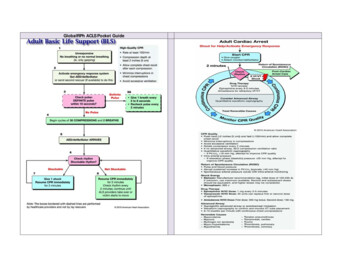
class. You’re encouraged to purchase or borrow an ACLS Provider Manual that offers directionto a web site and a pre-course assessment. The online pre-course assessment has shown to beof high value by those who have attended our classes. In addition, the provider manual willprovide you with ACLS algorhythm pocket guides that can be used during your mega-codetesting as a reference. The textbook itself is also a great resource in preparation for your courseand the written exam.This is not to be considered a replacement for the ACLS Student Manual, the onlinepre-course assessment and other resources offered by the AHA. Again—we highly encourageyou to get a provider manual and access the website found in the manual for study.BLS CPRBLS CPR changed in 2010. The primary change is from the “ABC” format to “CAB.”After establishing unresponsiveness and calling for a code, check for a pulse in 5 to 10 secondsthen begin compressions immediately, within 10 seconds of arriving at the patient’s side. Afterthirty compressions, give your first two breaths. The 30:2 ratio then will continue. “Look, listenand feel” for breathing has been removed from the new guidelines. One other significantchange in 2010, the use of an AED is now indicated for infants.Here are the basic steps in BLS:1. Check for responsiveness2. Call for help and an AED3. Check for pulse and simultaneously scan the chest for breathing.4. Begin the thirty compressions (within 10 seconds of arriving at thepatient)5. Give two breaths—continue 30:2 ratio.6. Apply the AED as soon as it arrivesNOTES:2
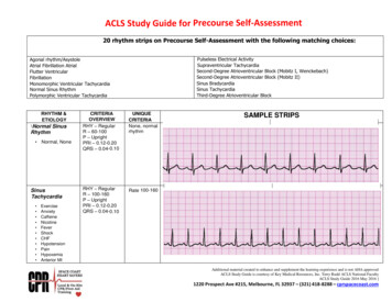
Sinus Bradycardia (sinus rhythm with a rate less than 50)Is your patient stable or unstable?Stable- monitor closelyUnstable/ Symptomatic – this patient is showing signs of poor perfusion(their heart rate is not fast enough to deliver an adequate volume of bloodto the body and requires treatment/ intervention) for example:hypotensive feels faint, decreased or altered mental status, cool orclammy/diaphoretic.Administer 0.5mg Atropine. If Atropine is ineffective you now havethree options:administer Dopamine 2-10 mcg/kg/minute ORAdminister Epinephrine infusion: 2 – 10 mcg per minutePrepare and provide external transcutaneous pacing.Note: if the patient is severely hypotensive,transcutaneous pacing may or may not capture and notbe able to produce an adequate profusing pressure. Theepinephrine drip or dopamine drip with transvenouspacing with expert consultation should now beconsidered.NOTES:3
Heart BlocksIs your patient stable or unstable?Stable- monitor closely, seek expert consultationUnstable/ Symptomatic – this patient is showing signs of poor perfusion(their heart rate is not fast enough to deliver an adequate volume of blood tothe body and requires treatment/ intervention) for example: low B/P, feelsfaint, decreased or altered mental status, cool or clammy/diaphoreticAdminister 0.5mg Atropine. But don’t rely on atropine in Mobitztype II, second-degree block or third-degree block with a new wideQRS complex.For an unstable Mobitz type II second-degree or third-degree heartblock patient be prepared for transcutaneous pacingNOTES:4
Supraventricular Tachycardia- SVT (SUSTAINED rapidnarrow complex tachycardia with a rate greater than 150)Is your patient stable or unstable?Stable- Attempt vagal maneuvers like bearing down or a good hard cough.If you’re a physician, try carotid massage.If vagal maneuvers aren’t successful in slowing their heart rate, administer6 mg. of Adenosine. What is unique about administering Adenosine is thatit is a fast-push and fast-acting drug. It may cause a second or two ofasystole. Patient also must be monitored.If the first dose of 6 mg isn’t successful, ACLS allow you to repeat theAdenosine providing 12 mg the second and third time if needed.Unstable/ Symptomatic – this patient is showing signs of poor perfusion(low B/P, feels faint, decreased or altered mental status, cool orclammy/diaphoretic) it may be due to their heart rate is too fast to deliveran adequate volume of blood to the body and requires rapid treatment/intervention. Provide synchronized cardioversion of 50 – 100 joules.NOTES:5
Monomorphic Ventricular Tachycardia- VT (SUSTAINED) 30 seconds rapid wide complex tachycardia)First and important question: Does your patient have a pulse with thisrhythm?If yes, he/she does have a pulse, then is you patient stable?o With a PULSE and UNSTABLE (low b/p, weak, clammy, cold, ashen,unresponsive or lethargic)Deliver immediate synchronized cardioversion at 100J.Evaluate the rhythm post cardioversion and consider a secondattempt at a higher energy level if needed.With a PULSE and STABLE, treat with adenosine only if regular andmonomorphic.Consider antiarrythmic infusion i.e. Procainamide or AmiodaroneIV drip.NOTES:6
Polymorphic Ventricular Tachycardia- VTTorsadesde PointesArrhythmias with a polymorphic QRS appearance, torsades de pointes forexample, will usually not permit synchronization. The patient will have tobe treated as V-fib: defibrillate at 200 joules.If there is doubt about whether an unstable patient has monomorphic orpolymorphic VT, do not delay treatment for further rhythm interpretation.Provide defibrillation (not synchronized cardioversion) at 200 joules.7
VFib is a chaotic and disorganized rhythm that generates absolutely noperfusion! The heart is quivering as it is dying and requires IMMEDIATEdefibrillation do not delay! The sooner the heart in VF can bedefibrillated, the higher the chances of successfully converting to anorganized rhythm.Quickly .Rapidly assemble your teamBegin chest compressionsApply defibrillator (hands-free) pads to patient, clear your coworkers from touching the patient or the bed and deliver 200Jshock as quickly as you can.Immediately after the shock is delivered, resume compressions andbag mask ventilations. (CPR should not stop for more than 10seconds.)You will continue CPR for 2 minutes (make sure your timer/recorderis tracking this for you) and prepare your first drug – your firstmedication will be Epinephrine 1mg, but do not administer it yet.This is also a good time to get IV or IO access if not alreadyestablished.At 2 minutes clear to reevaluate your rhythm- if VF persists chargeand defibrillate a second time at 200J, clear the patient and deliverthe shock. Immediately resume compressions (make sure to rotatecompressor and person bagging every 2 minutes for optimalcompressions- you will get tired quickly)During this 2 minute cycle administer the Epinephrine an preparethe second medication- Amiodarone 300mg.8
Again at the 2 minute mark clear to reevaluate your rhythm- if VFpersists charge and defibrillate at 200J, again resume compressionsimmediately after the shock is delivered.During this 2 minute cycle administer the Amiodarone 300mg andprepare your next dose of Epinephrine, 1 mg.These 2 minute cycles of rhythm check- shock if indicated- CPRadminister med will continue as long as VF or pulseless VT persists.NOTES:Asystole- Ventricular Standstill – Absolutely no electrical ormechanical activity!Asystole requires immediate interventionBegin compressions and airway management, good CPR.Administer Epinephrine 1mg IVP as soon as it’s available.1 mg of Epinephrine (1:10,000 used in cardiac arrest) is givingevery 3-5 minutes and there is no maximum dose.A critical step to restoring a perfusing rhythm is to quickly identifyone of the underlying/reversible causes that most frequently leadto asystole. The most common are known as the H’s & T’s! As ateam leader you should run through the list for consideration.H’s & T’s are as follows: Hypoxia, Hypothermia,Hypo/Hyperkalemia, Hydrogen Ion (acidosis), Hypovolemia,Toxins, Tension Pneumothorax, Tamponade, Thrombus(coronary or pulmonary)9
Pulseless Electrical Activity (PEA) – Electrical Activity withoutmechanical contractility, you have an organized rhythm, but the heart isn’tpumping .NO PULSEWhat do you do if you are in a code and you find an organized rhythm onthe monitor?CHECK FOR A PULSE! If you have a rhythm and no pulse you are inPEABegin compressions and airway management / good CPR at a ratioof 30:2.Administer Epinephrine 1:10,000 1mg IVPA critical step to restoring a perfusing rhythm is to quickly identifyone of the underlying/reversible causes that most frequently leadto PEA. The most common are known as the H’s & T’s! As a teamleader you should run through the list for consideration.H’s & T’s are as follows: Hypoxia, Hypothermia,Hypo/Hyperkalemia, Hydrogen Ion (acidosis), Hypovolemia, Toxins,Tension Pneumothorax, Tamponade, Thrombus (coronary orpulmonary)Remember- PEA is not always a Sinus Rhythm and it is not always aslow PEA. It can look like any organized rhythm.NOTES:10
During class you will have opportunity to get hands-on practice with each of theserhythms and their appropriate algorithms. If you study the content the applicationin the simulation labs will pull it all together for you and your instructor will helptie it all together and answer any additional questions you may have.This content is not intended to replace your ACLS provider manual- you are stillhighly encouraged to have the manual or borrow one provided by yourdepartment in preparation for and while attending class.11
ACLS PharmacologyDrugs for Pulseless Arrest VF/VTEpinephrine 1:10,000: 1mg IV/IO followed by 20 ml flush; repeat throughout code every3-5 minutes. There is no maximum dose.When is Epi administered? Asystole, PEA, VF, Pulseless VT .in other words .when there isn’t apulse, this is the first drug given. .Vasopressin: 40 units can replace the first or second dose of Epinephrine.Amiodarone: 300mg IV/IO push. Second dose (if needed) 150 mg IV/IO push.-Antiarrhythmic medication that has been shown to increase chance of return of pulse afterdefibrillation of a shockable rhythm, VF or pulseless VT.NOTES:Drugs for Pulseless Arrest PEA or AsystoleEpinephrine: 1mg IV/IO followed by 20ml flush; repeat throughout code every 3-5 minutes.There is no maximum dose.NOTES:12
Drugs for BradycardiaAtropine: 0.5mg IV/IO followed by 20ml flush; repeat every 3-5 minutes to a maximum totaldose of 3mg.-Increases heart rateDopamine drip: second line drug for Bradycardia, 2-10mcg/kg/min as an IV Infusion on aninfusion pump only. Titrate to patient response.- NEVER GIVE IV PUSH-Increases heart rate at lower doses, and increases B/P at higher dosesEpinephrine drip: Dilute 1mg Epinephrine in 500ml NS and run as an IV Infusion on aninfusion pump at a rate of 2-10mcg/min.-Titrate to patient response.-Increases heart rate and B/PNOTES:Drugs for Supraventricular Tachycardia and Stable Wide Complex VTAdenosine: 6mg rapid IV push followed by an immediate 2 ml flush; may repeat with a12mg dose twice if needed.-fast push and fast acting drug that may cause a short period of asystole.-Allows heart to return to a normal rhythmNOTES:13
Drugs for ACS (Acute Coronary Syndromes)MONAM MorphineO Oxygen (for oxygen saturation less than 94%)N NitratesA AspirinIt is preferable to do a 12 Lead ECG, if available, prior to the administrationof nitroglycerin to verify your patient’s cardiac rhythm status.Morphine: Initial dose is 2 to 4 mg IV over 1 to 5 minutes, administer slowly and only titrateto effect-May administer to patients with suspected ischemic pain unresponsive to oxygen and nitrates-Before administering morphine, be sure that the patient’s systolic blood pressure is 90 mm Hgand they are not hypovolemic-Remember to reassess and repeat vital signs between doses- Fentanyl may be an alternative pain medication to morphine for the unstable patient (ie,systolic blood pressure 90 mm Hg, hypovolemia, bradycardia, etc.)Nitroglycerin: 1 spray sublingually every 3-5 minutes up to a total of
4. Complete the online ACLS Pre-course Self-Assessment on ACLS ECGs and pharmacology (ACLS Student Website www.heart.org/eccstudent ) To successfully pass the ACLS course, AHA requires you successfully manage a mega- code. A mega-code is hands-on, dynamic, in real time practice of treating a life-threatening cardiac emergency. The cardiac emergency will include that may include ventricular