Transcription
World HeadquartersJones & Bartlett Learning5 Wall StreetBurlington, MA .comJones & Bartlett Learning books and products are available through mostbookstores and online booksellers. To contact Jones & Bartlett Learning directly,call 978-443-5000, fax 978-443-8000, or visit our website, www.jblearning.com.Substantial discounts on bulk quantities of Jones & Bartlett Learning publicationsare available to corporations, professional associations, and other qualifiedorganizations. For details and specific discount information, contact the specialsales department at Jones & Bartlett Learning via the above contact information orsend an email to specialsales@jblearning.com.Copyright 2014 by Jones & Bartlett Learning, LLC, an Ascend Learning CompanyAll rights reserved. No part of the material protected by this copyright may bereproduced or utilized in any form, electronic or mechanical, including photocopying,recording, or by any information storage and retrieval system, without writtenpermission from the copyright owner.Emergency & Critical Care Pocket Guide ACLS Version, Eighth Edition is anindependent publication and has not been authorized, sponsored, or otherwiseapproved by the owners of the trademarks or service marks referenced in thisproduct.The procedures and protocols in this book are based on the most currentrecommendations of responsible medical sources. The authors and the publisher,however, make no guarantee as to, and assume no responsibility for, thecorrectness, sufficiency, or completeness of such information or recommendations.Other or additional safety measures may be required under particular circumstances.This book is intended solely as a guide to the appropriate procedures to beemployed when rendering emergency care to the sick and injured. It is not intendedas a statement of the standards of care required in any particular situation,because circumstances and the patient’s physical condition can vary widelyfrom one emergency to another. Nor is it intended that this book shall in any wayadvise emergency personnel concerning legal authority to perform the activities orprocedures discussed. Such local determination should be made only with the aidof legal counsel.Production CreditsExecutive Publisher: Kimberly BrophyExecutive Acquisitions Editor—EMS:Christine EmertonAssociate Editor: Carly LavoieAssociate Production Editor: Nora MenziDirector of Marketing: Alisha WeismanVP, Manufacturing and InventoryControl: Therese ConnellComposition: diacriTechCover Design: Kristin E. ParkerDirector of Photo Research andPermissions: Amy WrynnPrinting and Binding: John P. PowCompanyCover Printing: John P. Pow CompanyISBN: 978-1-284-02370-16048Printed in the United States of America17 16 15 14 13 10 9 8 7 6 5 4 3 2 1
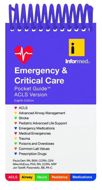
ACLSACLSACLS1ACLS CPR: Adult, Child, or Infant1. Unresponsive? (Not breathing, or only gasping?)2. Call for assistance—have someone get defibrillator/AED.3. Check pulse within 10 seconds (If present, give 1 breathevery 5–6 seconds; check pulse every 2 minutes).IF NO PULSE:4. Position patient supine on hard, flat surface.5. Begin chest compressions, 30:2, push hard and fast 100/minutes, allow full chest recoil—minimize interruptions.6. Open airway: head-tilt/chin-lift, ventilate 2* (avoid excessiveventilations).7. Attach AED to adult (and child 1 year old).SHOCKABLE RHYTHM?Yes8. Shock 1.9. Resume CPR immediately for 2 minutes.10. Check rhythm.IF SHOCKABLE:11. Shock 1; resume CPR.Lower half of sternumACLSNo8. Resume CPR immediately for 2 minutes.9. Initiate ALSinterventions.10. Check rhythm every 2minutes.Head-tilt/chin-lift
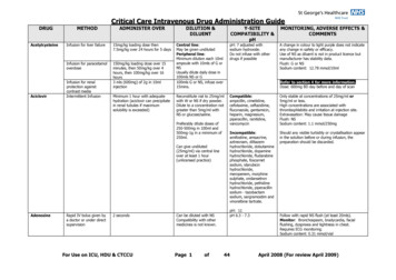
ACLSACLSACLSACLS2CPRAdult: 1 Person*Adult: 2 Person*Child: 1 PersonChild: 2 PersonInfant: 1 Depth 2 in. 2 in.2 in.2 in.1 3 cxInfant: 2 Person15:21001 3 cxNewborn: 2 Person3:11001 3 cxCheck rachial,femoralBrachial,femoral*Adult—once an advanced airway is placed, ventilate at 8–10 breaths/minute. Cardiac Arrest RhythmsCoarse Ventricular FibrillationNote the chaotic, irregular electrical activity. Treatment: Shock.Fine Ventricular FibrillationNote the low-amplitude, irregular electrical activity. Treatment: Shock.Ventricular TachycardiaNote the rapid, wide complexes. Treatment: Shock if no pulse.
AsystoleNote the absence of electrical activity. Treatment: Perform CPR.Pulseless Electrical Activity (PEA)Any organized ECG rhythm with no pulse. Treatment: Perform CPR.Other Common ECG RhythmsNormal Sinus RhythmNote the regular PQRST cycles.fibrillatorywavesAtrial FibrillationNote the irregular rate and atrial fibrillatory waves.3ACLSACLSACLSACLS
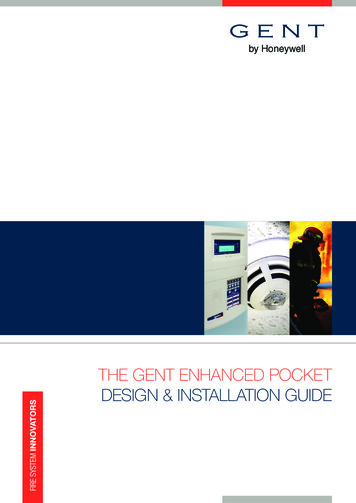
ACLSACLSACLSACLS4PACPVCPJCNormal QRSNormal QRS complex;Wide, bizarrecomplex; differentinverted or nocomplex;P waveP waveno P wavePremature Atrial, Junctional, and Ventricular ComplexesOther Common ECG RhythmsSupraventricular Tachycardia (SVT)Note the rapid, narrow QRS complexes.Inverted PInverted PJunctional RhythmNormal QRS complexes; inverted, or no P waves1 AV BlockProlonged PRInterval 0.20 secondsBundle Branch BlockWide QRS 0.12 seconds
PPPPPPdropped QRS2 Heart Block, Wenckebach, Mobitz Type IThe PR interval lengthens, resulting in a dropped QRS.Other Common ECG Rhythmsdropped QRS2 Heart Block, Mobitz Type IIThe PR interval does not lengthen, but a QRS is dropped.QRSPPQRSQRSPPPQRSQRSPPThird (Complete) Heart BlockThe P waves are dissociated from the QRS complexes.spikesElectronic Ventricular PacemakerNote the pacer spikes before each QRS.5ACLSACLSACLSACLS
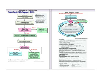
ACLSACLSACLSACLS6 Basic ECG Interpretation1 second0.040.20second secondRTP1 millivolt 1 cm. (std.calibration)Q S1 mm1 cmVentricularcontractionRAtrial contractionQVentricularrelaxationand passive fillingT WaveSP WaveECG Waves:P Wave: AtrialdepolarizationQRS Complex:Ventricular depolarizationT Wave: VentricularrepolarizationQRS
3-Lead and MCL1 Electrode PlacementLead IBlack-150 -30 avRMCL 1LavI120 90 0 IIavFIIadLeIII Lead IIIWhite60 Red (on thighi) 12-Lead Electrode PlacementV1V1:V2:V3:V4:V5:V6:MCL1:MCL6:MC4R:V5 V6V2 V3 V4 V5 V6Fourth interspace, just to the right of the sternumFourth interspace, just to the left of the sternumHalfway between V2 and V4Fifth intercostal space, midclavicular lineAnterior-axillary line, horizontal with V4Mid-axillary line, horizontal with V4Red lead on V1, black lead on left arm—monitor lead IIIRed lead on V6, white lead on right arm—monitor lead IIRed lead on fifth intercostal space right midclavicular line, black lead onleft arm—monitor lead III7ACLSACLSACLSACLS
ACLSACLSACLSACLS8 ACLS AlgorithmsNOTE: Not all patients require the treatment indicatedby these algorithms. These algorithms assume that youhave assessed the patient, started CPR where indicated,and performed reassessment after each treatment.These algorithms also do not exclude other appropriateinterventions that may be warranted by the patient’scondition.Treat the patient, not the ECG.
Cardiac Arrest Shout for help, begin CPR (30:2, push hard and fastat 100/min, minimize interruptions), give O2, attach ECG.YESShockable Rhythm?NOVF or VTAsystole/PEAa Defibrillate 120 J–200 JContinue CPR immediately 2 minutes. Start IV/IO.Epinephrine, 1 mg IV/IO, repeatevery 3–5 minutes, OR:Vasopressin, 40 Units IV/IO (singledose only), consider advancedairway (ET tube, supraglottic airway)Ventilate 8–10 breaths/minute withcontinuous compressionsUse waveform capnography:If PETCO2 15, improve CPRBiphasic (or 360 Jmonophasic, or AED) Continue CPR immediately 2minutes. Start IV/IO. VF/VT? a DefibrillateContinue CPR 2 minutesEpinephrine, 1 mg IV/IO,repeat every 3–5 minutes, OR:Vasopressin 40 Units IV/IO (singledose only)Consider advanced airway(ET tube, supraglottic airway)Ventilate 8–10 breaths/minute withcontinuous compressionsUse waveform capnography:If PETCO2 15, improve CPR Asystole/PEA?Continue CPR 2 minutesConsider reversible causes.* If ROSC (pulse, BP, PETCO2 40 mm Hg),see ROSC algorithm, next page.*Reversible Causes Hypoxia Hypovolemia Acidosis Hyper-hypokalemia Hypothermia Coronary thrombosis Pulmonary thrombosis Cardiac tamponade Tension pneumothorax Toxins VF/VT?a DefibrillateContinue CPR 2 minutes.Amiodarone, 300 mg IV/IO(may repeat once 150 mgin 5 minutes)Consider reversible causes.* If ROSC (pulse, BP, PETCO2 40 mm Hg),see ROSC algorithm, next page.9ACLSACLSACLSACLS
ACLSACLSACLSACLS10 Return of Spontaneous Circulation:Post-Cardiac Arrest CareOptimize ventilation/oxygenation(Start at 10–12 breaths per minute, but do not hyperventilate)Goal: PETCO2 35–40 mm HgUse minimum amount of FiO2 to keep SaO2 94%Consider waveform capnography Keep blood pressure 90 mm Hg (or MAP 65 mm Hg)IV fluid bolus: 1–2 Liter(s) NS or RL(May use cold [4 C] IV fluid if induced hypothermia)Consider vasopressor infusionEpinephrine: 0.1–0.5 mcg/kg/minuteDopamine: 5–10 mcg/kg/minuteNorepinephrine: 0.1–0.5 mcg/kg/minuteConsider reversible causes*Monitor ECG, obtain 12-lead ECGFollows commands?(If not, consider induced hypothermia) STEMI or high suspicion AMI? Coronary reperfusion (PCI)Advanced critical care*Reversible Causes Hypoxia Acidosis Hypovolemia Toxins Coronary thrombosis Cardiac tamponadeHyper-hypokalemiaHypothermiaPulmonary thrombosisTension pneumothorax
TachycardiaConsider and treat reversible causes*Assess C-A-B, secure airway, give O2, start IV/IO, check BP,apply oximeter, get 12-lead ECGIs Patient Unstable?(Serious S/S must be related to the tachycardia:HR 150/minute, ischemic chest pain, dyspnea, LOC, BP, shock, heart failure)Stable?Go to nextpagea Immediate Synchronized Cardioversion(For narrow QRS, consider adenosine, 6 mg, rapidIVP [Flush with NS, may repeat with 12 mg IVP]; alsoconsider sedation, but do not delay cardioversion)Initial Energy Doses (if unsuccessful, increase dosesin a stepwise fashion):Narrow QRS, Regular: 50 J–100 JNarrow QRS, Irregular: 120 J–200 J biphasic (or 200J monophasic)Wide QRS, Regular: 100 JWide QRS, Irregular: defibrillate with 120 J–200 Jbiphasic (or 360 J monophasic)Synchronize MarkersPad/paddle placement forsynchronized cardioversionSynchronize on R wave11ACLSACLSACLSACLS
ACLSACLSACLSACLS12Stable Patient, Wide QRS( 0.12 seconds)Stable Patient, Narrow QRS 12-lead ECGStart IVVagal maneuvers†Adenosine, 6 mg IVP (forIVP (for regular, monomorregular rhythm), flush withphic rhythm) flush with sasaline, may repeat 12 mg IVPline, may repeat 12 mg IVPEither: Consider antiarrhythmic: Calcium blocker (chooseEither:one):Procainamide, 20–50 mg/Verapamil, 2.5–5 mg IVminute IV until rhythmover 2–3 minutes. May repeatconverts, QRS widens5–10mg. Maximum of 30 mg.by 50%, hypotension,or maximum dose ofDiltiazem, 0.25 mg/kg IV17 mg/kg. Avoid if CHF orover 2 minutes. May repeatprolonged QT. Drip 1–4 mg/0.35 mg/kg OR:minute OR: β-blocker (choose one):Amiodarone, 150 mg IV,Metoprolol, 5 mg IV overover 10 minutes. May repeat2–5 minutes. May repeat.(maximum dose: 2.2 g/24 hMaximumof 15 mg.IV). Drip 1 mg/minute OR:Atenolol, 5 mg IV overSotalol, 1.5 mg/kg IV for5 minutes. May repeat once.over 5 minutes. Avoid if prolonged QT.Propranolol, 1–3 mg IV, Consult with expertslowly over 2–5 minutes.Esmolol, 250–500 mcg/kg*Reversible Causesfor 1 minute. Hypoxia Consult with expert Acidosis Hypovolemia Toxins Coronary thrombosis†Carotid sinus massage is Cardiac tamponade Hyper-hypokalemiacontraindicated in patientswith carotid bruits. Avoid ice Hypothermiaapplication to face if patient has Pulmonary thrombosisischemic heart disease. Tension pneumothorax 12-lead ECG Start IV Consider adenosine, 6 mg
Wide Complex TachycardiaVTV1V1Consider adenosine(VT versus SVT with aberrancy)Suggestive ECG signs for VT:SVT with aberrancy Fusion beats: diagnostic Capture beats: diagnostic Extreme RAD (-90 to 180 ) QRS 0.14 seconds QRS V1–V6 are all negative or all positive L rabbit ear R rabbit ear in V1 Wide initial R wave in V1 AV dissociation (independent P waves) Small R wave in V6 or QS in V6 Hx: MI, LVH, CAD, cardiomyopathySuggestive ECG signs for SVT:Irregular rhythm: consider AFAssociated P waves: consider PSVT When in doubt, treat for VTACLSACLSACLSACLSV6MCL6Considerlidocaine oramiodarone13
ACLSACLSACLSACLS14 Bradycardia(HR 50/minute with serious S/S: shock, hypotension, altered mentalstatus, ischemic chest pain, acute heart failure)Assess C-A-B, maintain airway, give O2, assist breathingif needed. Attach pulse, oximeter, BP cuff, 12-lead ECG;start IV/IO fluids. Consider and treat reversible causes* Atropine, 0.5 mg IV/IO every 3–5 minutes, maximum of 3 mg. (Do notdelay TCP while starting IV, or waiting for atropine to work.*)If ineffective: Transcutaneous pacing (verify capture and perfusion; use sedation asneeded) OR:Dopamine, 2–10 mcg/kg per minute, OR:Epinephrine, 2–10 mcg per minute Consider expert consult; prepare for transvenous pacer Cardiac arrest?—See ACLS Section, Cardiac Arrest algorithm*Reversible Causes HypoxiaAcidosisHypovolemiaToxinsCoronary thrombosisCardiac tamponadeHyper-hypokalemiaHypothermiaPulmonary thrombosisTension pneumothorax*Atropine may not work for transplanted hearts, Mobitz(type II) AV block, or third degree AV block with IVR.—Begin pacing and/or catecholamine infusionUNSYMPTOMATIC BRADYCARDIA?NOT type II (Mobitz) second-degree or third-degree AV heart block? Observe
Asthma Cardiac ArrestUse standard ACLS guidelinesEndotracheal intubation via RSI(Use largest ET tube possible; monitor waveform capnography) To reduce hyperinflation, hypotension, and risk of tensionpneumothorax, consider: Ventilation with a slower respiratory rate Smaller tidal volume (6–8 mL/kg) Shorter inspiratory time (80–100 mL/minute) Longer expiratory time (I/E 1:4 or 1:5) Continue use of inhaled β2-agonist (albuterol) via ET tubeEvaluate for tension pneumothorax Consult with expert Consider brief disconnect from BVM an
ACLS ACLS ACLS ACLS CPR: Adult, Child, or Infant 1. Unresponsive? (Not breathing, or only gasping?) 2. Call for assistance—have someone get defibrillator/AED. 3. Check pulse within 10 seconds (If present, give 1 breath every 5–6 seconds; check pulse every 2 minutes). IF NO PULSE: 4. Position patient supine on hard, flat surface. 5.File Size: 2MBPage Count: 242