Transcription
AHA ACLS Study GuideACLS Course Objectives Apply the BLS, Primary and Secondary Assessment sequences for a systematic evaluation of adultpatients.Perform prompt, high quality BLS, including prioritizing early chest compressions and integrating earlyAED use.Recognize respiratory arrest and perform early management of respiratory arrest.Discuss early recognition and management of acute coronary syndromes (ACS), including appropriatedisposition.Discuss early recognition and management of stroke, including appropriate disposition.Recognize bradyarrhythmias and tachyarrhythmias that may result in cardiac arrest or complicateresuscitation outcome. Perform early management of bradyarrhythmias and tachyarrhythmias that mayresult in cardiac arrest or complicate resuscitation outcome.Recognize cardiac arrest and perform early management of cardiac arrest until termination ofresuscitation or transfer of care, including immediate post-cardiac arrest care.Evaluate resuscitative efforts during a cardiac arrest through continuous assessment of CPR quality,monitoring the patient’s physiological response, delivering real-time feedback to the team.Model effective communication as a member or leader of a high performance team & recognize the impactof team dynamics on overall team performance.Science of Resuscitation AHA ACLS book pg. 1AHA ACLS book pg. 13Immediate High Quality CPR starting with compressions at least 2 inches deep, 100-120/minAED or Manual Defib ASAP 1 shock q 2 minutes for VF/pVTMinimizing interruptions in compressionsAvoid excessive ventilations (decreases cardiac output and cerebral perfusion)o 500-600 ML Tidal VolumeCoronary perfusion pressure 15 and Chest Compression Fraction 80%ETCO2 10 indicates compressions ineffectiveMax 10 sec off chest at any timeFeedback devices recommendedIntegrated post Cardiac Arrest CareRRT/MET (Rapid Response Team and Medical Emergency Teams) improving outcomes by earlydetection and early treatmento Threatened airwayo Resp rate 6 or 30 per mino Heart rate 40 or 140 per mino Systolic pressure 90o Symptomatic Hypertensiono Unexpected LOCo Unexplained agitationo Seizureo Significant decrease in urine outputo Subjective concern about pt.1Update 01/2021
AHA ACLS Study GuideSystematic ApproachAHA ACLS book pg. 33-42Initial Impression – CBC (Consciousness – Breathing – Color) Unconscious – BLS survey - breathing and pulse, activate emergency response (callcode) High quality CPR, rapid defib Primary assessment ABCDEo A – Airway patent, consider advanced deviceo B – Breathing 2 breaths after 30 compressions If adv. Airway 1 breath q 6 sec. If has a pulse – 1 breath q 6 sec rescue breathing If breathing – administer O2 as neededo C – Support circulation, BP 90 systolic or MAP 65 Access – either IV or IO Drugs and fluids if indicated Temp and glucose levelso D – Disability LOC – AVPU Alert Voice Painful Unresponsiveo E – Exposure Remove clothing – examine Secondary Assessment - SAMPLEo S – Signs and symptomso A – Allergieso M – Medicationso P – Past Medical historyo L – Last mealo E – Events H and T”so Hypovolemiao Hypoxiao Hydrogen ion (acidosis)o Hyperkalemiao Hypokalemiao Hypothermiao Tension Pneumothoraxo Tamponade (Cardiac)o Toxinso Thrombosis Pulmonary Cardiac2Update 01/2021
AHA ACLS Study GuideHIGH Performance Teams AHA ACLS book pg. 25-32 Handbook pg. 8Elements of Team Dynamics and communicationo Roles Clear roles and responsibilities – clearly delegate tasks Designated Team leader Know limitations – ask for new role if not assigned an appropriate task Constructive intervention Be tactful but address team member to prevent erroro What to communicate Knowledge sharing Summarizing and reevaluationo How to communicate Closed-loop communication Clear messages Mutual respectDebriefingo Work together as a teamo Debriefing during and after code Improves team performanceo Improve patient outcome after arrest CPR Coach New role to promote delivery of high-quality CPR.CPR Coaches stand across from compressor next to feedback-device.Verbalizes feedback, mid-range target for HRCoordinate CPRo Initiating CPR, Placing CPR board, analyzing rhythm, switching compressors (goal to keeppauses 5 sec), defibrillating, intubating (must be 10 seconds)Preventing Arrest: Brady and Tachycardia AlgorithmsStable vs unstable Hypotension? Altered mental status? Signs of shock? Ischemic Chest pain? Acute Heart Failure?3Update 01/2021
AHA ACLS Study GuideBRADYCARDIA & Heart BlocksAHA ACLS book pg.120-128, Handbook pg. 12Stable patient (Asymptomatic slow heart rate): Call for expert consultation Obtain focused history and physical exam; monitor and observeUnstable patient (Symptomatic Slow Rate) Identify and treat underlying cause Maintain patent airway; ventilate and give O2 if needed Cardiac monitoring, BP and SPO2 monitoring IV Access 12 lead ECG if available Administer atropine 1 mg IVP q 3-5 min (max 3 mg)Try Atropine for ALL unstable bradycardias Transcutaneous Pacing and/or Dopamine drip 5-20 mcg/kg/min or Epinephrine drip 2-10 mcg/min orPrepare for Transvenous Pacing or Permanent PacerTACHYCARDIA with a pulse All unstableAHA ACLS book pg. 129-144 Handbook pg. 13-16Identify and treat underlying causeMaintain patent airway; ventilate and give O2 if neededCardiac monitoring, BP and SPO2 monitoringIV Access12 lead ECG if availableTachycardias require Synchronized CardioversionFor stable – determine if wide or narrow QRS Regular or irregular?Narrow Regular (Supraventricular Tachycardia)STABLEEXPERT CONSULTATION O2, IV, Monitor, 12 lead ECG Do Vagal Maneuvers (Cough, Deep breath, etc) Adenosine 6mg IVP SLAMMED, Flush with 20mL NS, RAPID SLAM May repeat with Adenosine 12 mg in 1-2 min, Flush with 20mL NS, RAPID SLAM ANTICIPATE SIDE EFFECTS IDENTIFY UNDERLYING RHYTHMUNSTABLE Synchronized Cardioversion -- Joules as directed by manufacturer or physician4Update 01/2021
AHA ACLS Study GuideNarrow Irregular QRS Tachycardia (Atrial Fibrillation/Flutter)STABLE CONTROL RATE Ca Channel Blockers - Verapamil, Diltiazem (Cardizem), Beta Blockers, Digoxin, etc.UNSTABLE SYNCHRONIZED CARDIOVERSIONo Atrial Fib 120-200 joules; As directed by manufacturer or physicianWide Regular QRS Tachycardia with a Pulse (VT with Pulse)STABLE - Antiarrhythmic Infusions IVPB Amiodarone : Loading Dose: 150 mg in 100 mL of D5W over 10 min; can repeat if neededOption of Adenosine IVP May be used ONLY if monomorphic, stable, regular tachy's 1st Dose 6mg, then 12mg IV / IO SLAMMED with 20 cc NS flush slammed.UNSTABLE Synchronized cardioversion as directed by manufacturer or physicianWide Irregular (Polymorphic VT)Example – Torsades de Points Defibrillate (unsynchronized) Biphasic 120-200j (or 360 equivalent monophasic) Variations in rhythm may make cardioversion impossibleMagnesium 1-2 Gm in 50-100 mL D5W over 5-20 minCardiac ArrestAHA ACLS book pg. 92-109 Handbook pg. 9-10 Priority is effective High Quality CPRShockable Rhythms: VF and pVT Early DEFIB and def ib every 2 minutes Continue compressions while def ib is charging Compressions immediately prior to def ib and immediately af terSAFE defibrillation includes: “All Clear” ”shocking in 3,2,1 shock” Visual sweep - Make sure O2 is clear! Eyes on patient - then def ibrillateDef ibrillate 120—200 joules;CPR f or 2 min cycles – rhythm check every 2 minutes 30:2Def ibrillate 120—200 (Two defibs BEFORE f irst medication - Prior to next possible def ib – it is reasonable topre-charge def ibrillator in anticipation of another def ibrillation to minimize time of f chest.Get IV/IO Access First drug: (Give meds during compressions early in the cycle) Epinephrine 1 mg IV PUSH every 3-5 min – vasoconstrictor Second drug - antiarrhythmic:Choice or either not both Amiodarone 300 mg IVP (given between 1st and 2nd Epi) 2nd dose 150 mg in 3-5 min Lidocaine5Update 01/2021
AHA ACLS Study GuideCaridiac Arrest (Cont) Lido Dose f or either pulseless or with a pulse 1-1.5 mg/kg IV PUSH Repeat with ½ dose every 5-10 min Max 3 mg/kg ET 2-2.5 times IV dose Maintenance inf usion 1-4 mg/min start at 1 mg higher than total of boluses Consider advanced airway and wavef orm capnography PETCO2 less than 10 during compressions indicate inef f ective compressions Rhythm check and Def ibrillate Q2 minutes, repeat pulse check only if rhythm changes.Non-shockable Rhythms: PEA or AsystolePulseless Electrical Activity (PEA)AHA ACLS book pg. 110-113Organized rhythm on monitor with NO detectable pulse High Quality CPR and Epinephrine Epinephrine 1 mg IV PUSH every 3-5 min Conf irm oxygenation, give f luids. Search for and treat identifiable causes (H’s and T’s)Asystole Confirm in 2 leadsHigh Quality CPR and Epinephrine Epinephrine 1 mg IV PUSH every 3-5 minSearch for and treat identifiable causes (H’s and T’s)Post Cardiac Arrest CareAHA ACLS book pg. 114-118AHA ACLS book pg. 145-152 Handbook pg. 11Following ROSC (Return of Spontaneous Circulation)1. Maintain Ventilation/Oxygenation “Is the patient breathing effectively?” If nonresponsive and inef f ective respirations - Intubation (ventilator support if needed) use lowest O2concentration to keep O2 sats 92-98% Prolonged use of high FIO2 (02%) can cause O2 Toxicity Use wavef orm capnography – target 35 to 45 during ROSC Excessive ventilation (hyperventilation) can decrease cerebral perf usion and decrease cardiac output Rescue breathing (1 ventilation every 6 seconds)2. Monitor & Treat Hypotension Target BP 90 systolic MAP 65 Fluids (1-2 liters NS or LR) then Vasopressors Epi drip 2-10mcg/min or Norepinephrine drip 0.1-0.5 mcg/kg/min or Dopamine drip 5-10 mcg/kg/min Treat causes3. 12 Lead ECG R/O possible STEMI Coronary Reperf usion – transport to PCI f acility4. Non-responsive pts. Targeted Temperature Management Protocols For non-responsive adult pts. – ROSC af ter Cardiac ArrestPick a temp between 32 and 36 degrees C, cool and maintain f or at least 24 hours6Update 01/2021
AHA ACLS Study GuideHigh Quality BLSHandbook pg. 1-4 C-A-B After determining the patient is unresponsive and has no OBVIOUS pulse STARTCOMPRESSIONS. The most Common Mistake in CPR? Prolonged Interruptions in Compression s Start compression within 10 seconds of recognition Compression continue during charging Never interrupt compression for more than 10 seconds. High Quality Compressions 100-120/Min, at least 2”, Allow for complete recoil Switch compressors every 2 minutes/5 cycles or when fatigue Is the Scene Safe? Check Responsiveness Get Help Coming Check Breathing and Pulse Agonal Gasps are indication of cardiac arrest Unsure if they have a pulse Send for an AED and start compressions 100 – 120 per minute 2 - 2.4 inches Allow for complete Recoil When a BVM arrives begin 30:2 AED Goal: To Eliminate ABNORMAL rhythm Use as soon as available Turn it on and follow prompts Child BLS Ages 1-puberty Depth - 1/3rd of AP chest 2nd Rescuer Change to 15:2 ratio Infant BLS 1 rescuer 30:2 / 2 fingers/2 thumbs or 1 hand 2 Rescuer 15:2 / thumb encircling Depth – 1/3 AP chestChoking Adult and Child Abdominal Thrusts if responding CPR if non-responsive Check mouth before breaths Infant 5 back slaps and 5 chest thrusts repeated if responding CPR if non-responsive Check mouth before breaths7Update 01/2021
AHA ACLS Study GuideAirway ManagementACLS book pg. 44-58Open airwayHandbook pg. 69-72 Jaw thrust/chin lift Using OPA/NPA. (OPA – measure corner of mouth to angle of mandible)Rescue Breathing is ventilations without compressions (with or without advanced airway) Adult: 1 breath every 6 seconds (12 per min) Child: 1 breath every 2-3 seconds (20-30 per min) Just enough air to see chest rise, each over 1 secondAirway – ET TUBEConfirmation of ETT Placement Auscultation and Chest Rise & Fall Colorimetric Capnography (color change CO2 Detector) Continuous waveform capnography- best way to monitor ET placement Chest X-Ray and ABGET Suctioning (Hyper-oxygenate prior to suctioning) Less than 10 sec During catheter withdrawalCPR Cycle after intubation 1 breath q 6 sec without pauses in compressionsPETCO2 Best indicator of CPR efficiency & coronary perfusion Best indicator of ET placement and ET monitoring During CPR goal is waveform reading 108Update 01/2021
AHA ACLS Study GuideMaternal Cardiac ArrestsAHA Handbook page 5-6 Follow ACLS treatments Same Meds, Def ibrillation, Early Intubation CPR done with manual uterine displacement (Pulled to the lef t side) Perf orm perimortem cesarean delivery in 5 min, depending on provider resources and skill setABCDEFGH - Differential Diagnosis for Maternity Arrests Anesthesia Complications Bleeding / DIC Cardiac Disease/Cardiovascular Drugs Embolism: coronary / pulmonary / amniotic fluid Fever General: PATCH 5MD or H’s and T’s Hypertension / preeclampsia / eclampsiaAcute Coronary Syndrome (ACS)AHA ACLS book pg. 59-72Chest Pain suggestive of Myocardial IschemiaHandbook pg. 25-39 Immediate Assessment ( 10 minutes) including 12 lead ECG, VS, O2 Sat, IV Access, Targeted Histo ry andPhysical, lytes, coagulation studies, enzymes, chest x -ray 12 Lead to determine STEMI f rom Non-STEMI MIO2 @ 4L/min f or O2 sats below 90% or S/S of hypoxiaASA 162-325 mg PONTG 0.4 mg SL or spray Vasodilator, decreases preload, watch BP! Hold Nitro if hypotensive, tachycardia, bradycardia, Rt. sided MI or recent erectile enhancement drugsMorphine 2-4 MG IV every 5 min f or pain not relieved with NTG (Use morphine cautiously)Treatment of Choice for STEMI: REPERFUSION!! Window of Opportunity f or STEMI: Symptoms Present 12 hrsSelect reperf usion strategy based on resources: Angiography with angioplasty or stent placement GOAL – Door to PCI reperfusion 90 min. Ideally take patient DIRECTLY to PCI if possible. Transport to PCI capable facility Thrombolytic therapy If not a candidate f or Cardiac Catheterization GOAL– Door to Thrombolytic Drug 30 min9Update 01/2021
AHA ACLS Study GuideStroke (CVA)A neurological impairment caused by disruption of blood supply to a region of the brain Third leading cause of death in the US and leading cause of adult disability Ischemic Stroke– Occurs when a blood vessel supplying the brain is occluded– 87% of strokes are ischemic Hemorrhagic Stroke– Occurs because a cerebral artery ruptures Subarachnoid – bleeding onto the surf ace of the brain Intracerebral – bleeding in the brain– Can be f atal at onset, hypertension most common precipitating f actorAHA ACLS book pg.73-91handbook pg. 19-248 D’s Detection: Recognize signs and symptoms Dispatch: Early activation of EMS Delivery: Pre-hospital EMS Assessment using Cincinnati Pre-Hospital Stroke Score, if positive: Identify time symptoms began Divert to Stroke Center with Head CT capabilities Door: 3 hr window of opportunity for thrombolytics Select pts may have up to 4.5 hr window Optional treatments may have slightly longer window Immediate Assessment - Neurological Screening ASAP, H&P, Glasgow, NIH Stoke Scale VS, IV, CBC, Lytes, Coags, Blood Sugar. 12-lead EKG Data/Decision/Drug: If CT shows NO hemorrhage: Thrombolytic criteria? Clot Retrieval? If CT shows hemorrhage: Consult Neurosurgery, reverse anticoagulation, and/or bleeding disorder, monitor neurological condition, control hypertensionDisposition: Admission to Stroke Unit or ICU within 3 hrs.10Update 01/2021
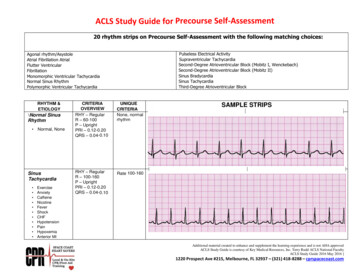
AHA ACLS Study GuideRhythm Strips for practice11Update 01/2021
AHA ACLS Study Guide 1 Update 01/2021 ACLS Course Objectives AHA ACLS book pg. 1 Apply the BLS, Primary and Secondary Assessment sequences for a systematic evaluation of adult patients. Perform prompt, high quality BLS, including prioritizing early chest compressions and integrating early . AHA ACLS book pg. 92-109 Handbook pg. 9-10