Transcription
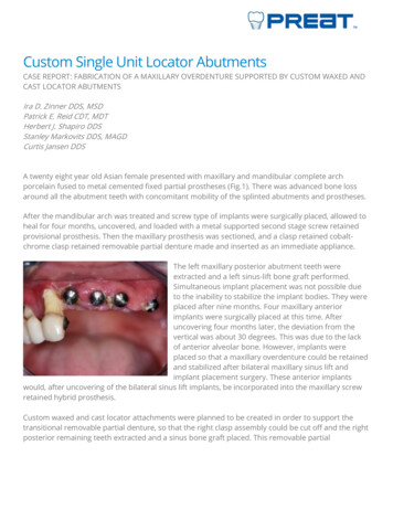
Custom Single Unit Locator AbutmentsCASE REPORT: FABRICATION OF A MAXILLARY OVERDENTURE SUPPORTED BY CUSTOM WAXED ANDCAST LOCATOR ABUTMENTSIra D. Zinner DDS, MSDPatrick E. Reid CDT, MDTHerbert J. Shapiro DDSStanley Markovits DDS, MAGDCurtis Jansen DDSA twenty eight year old Asian female presented with maxillary and mandibular complete archporcelain fused to metal cemented fixed partial prostheses (Fig.1). There was advanced bone lossaround all the abutment teeth with concomitant mobility of the splinted abutments and prostheses.After the mandibular arch was treated and screw type of implants were surgically placed, allowed toheal for four months, uncovered, and loaded with a metal supported second stage screw retainedprovisional prosthesis. Then the maxillary prosthesis was sectioned, and a clasp retained cobaltchrome clasp retained removable partial denture made and inserted as an immediate appliance.The left maxillary posterior abutment teeth wereextracted and a left sinus-lift bone graft performed.Simultaneous implant placement was not possible dueto the inability to stabilize the implant bodies. They wereplaced after nine months. Four maxillary anteriorimplants were surgically placed at this time. Afteruncovering four months later, the deviation from thevertical was about 30 degrees. This was due to the lackof anterior alveolar bone. However, implants wereplaced so that a maxillary overdenture could be retainedand stabilized after bilateral maxillary sinus lift andimplant placement surgery. These anterior implantswould, after uncovering of the bilateral sinus lift implants, be incorporated into the maxillary screwretained hybrid prosthesis.Custom waxed and cast locator attachments were planned to be created in order to support thetransitional removable partial denture, so that the right clasp assembly could be cut off and the rightposterior remaining teeth extracted and a sinus bone graft placed. This removable partial
overdenture was converted to a complete maxillaryoverdenture when the right posterior teeth were extractedand a sinus lift graft placed surgically.Technique for creation of customized angulated maxillary anterior Locator attachments involves thetaking of an elastomeric impression with an open tray at the implant level after healing of the softtissuesFabricate an acrylic resin customized tray with anterior opening for an open-tray impression atimplant level. Screw to place four impression copings with guide pins for an open tray impression.Ensure the seating of the hexes with right angled radiographs.Take an elastomeric impression. Syringe the light bodied material around the four impressioncopings. After set, unscrew the impression copings and remove with the impression.Secure the implant level analogues in place. Spray a separating medium for silicones. Syringe agingival simulation material around the impression copings. Allow the material to set and then boxand pour the cast with die stone. This allows the practitioner to create a soft tissue cast forsimulation of the patient’s gingiva.Verify the hex positions and the positions of the implant head to the impression copings and alterthe cast if necessary.Mount the maxillary and mandibular casts in a centric relation position at the desired VDO.
Castable UCLA types of cylinders were screwed to place on the analogues. Custom waxed angulatedheads are waxed; a stainless steel wire is prepared and placed in each wax up for positioning of theLocator heads, invested and cast in the preselected metal.Use the paralleling mandrel to set the castable threaded inserts (with the threaded male inserted).
Survey and place the Locator female attachments parallel to each other using the parallelingmandrel or black paralleling post.These custom waxed and cast angulated abutments were only necessary on the two central incisorand right lateral incisor implants. The angulation of the left lateral incisor implant did not need acustom abutment. All the abutments were aligned using a parallelometer.After casting, divest but do not place in acid for gold cleaning until the metal wire is removed. Thenthe casting can be placed in acid for cleaning of the metal alloy.Use the Locator Bar tap clean out the cast threads.The threaded Locator female allows retrievability if servicing is required on the attachment.Removal of the investment material from the threaded insert without damage using an ultrasoniccleaner solution, shell blast, bead blast, or use a Fiberglass or Brass Pencil.
Finish and polish the casting in normal manners.
Locator black processing cap males are inserted into the metal housing for chairside pick-up into thedenture. These are snapped onto the Locator females which are already in place in the patient’smouth.Sufficient room is cut out from inside the patient’s removable prosthesis to fit over the Locatorattachment systems.Place a white block-out spacer over the head of each Locator abutment. This spacer will block outthe area surrounding the abutmentEnsure that there is sufficient space between the denture base and the Locator male cap.Cut a lingual escape vent, verify that all undercuts are completely blocked out, and mixautopolymerizing denture resin into a loose mix and place it into the prepared recesses of thedenture base. Then seat the denture into the mouth and hold in place until the resin sets. Removethe denture. The Locator male attachments should be incorporated in the denture base.Carve and polish the maxillary removable denture with the incorporated denture base.Seat metal analogues into the Locator male attachments that have been previously incorporatedinto the denture and pour a mounting cast.Using a face-bow index and verified maxillo-mandibular records the maxillary prosthesis is mountedon a semi-adjustable articulator and the occlusion equilibrated.Adjust the occlusion for bilateral balance, as would be for any other complete removable denture.Insert the overdenture; maintain a labial flange to mask the custom waxed and cast angulatedabutments.
After any posterior surgical procedure, line the denture base with a tissue treatment material as thesurgical area heals.This overdenture will be used until the posterior implants are uncovered.Then a screw retained provisional prosthesis will be made for the maxillary arch.The custom waxed and cast Locator abutments are changed to manufactured angulated abutmentsand incorporated into the screw retained provisional prosthesis.Thus, sequential treatment planning, after a thorough diagnosis, avoids the patient wearing amucosal retained maxillary complete denture. This type of situation requires the restorative dentistto have a clasp retained, cobalt-chrome framework fabricated by the laboratory technician prior toremoval of any teeth, or other surgical procedures, and to explain to the patient what will beperformed.
The posterior cobalt chrome clasp assembly is removed prior to extraction of the maxillary rightposterior teeth. This is done after insertion of the anterior custom waxed and cast angulated Locatorabutments and after patient is acclimated to the prosthesis. After healing of the left posteriorquadrant, four implants are surgically placed using a surgical template for guidance. Four to sixmonths after implant placement, the soft tissues will be opened and the implants uncovered.Healing abutments will be installed by the surgeon, the transitional prosthesis will be relieved andthe right posterior quadrant will have 4 posterior implants placed with a sinus lift procedureFor more information, contact Preat at 1-800-232-7732 or visit preat.com
The left maxillary posterior abutment teeth were extracted and a left sinus-lift bone graft performed. Simultaneous implant placement was not possible due to the inability to stabilize the implant bodies. They were placed after nine months. Four maxillary anterior implants were surgically placed at this time. After