Transcription
Blackwell Science, LtdOxford, UKCMICellular Microbiology 1462-5814 The Authors Journal compilation 2005 Blackwell Publishing Ltd81107119Original ArticleMacrophage vacuoles perforated by Listeria monocytogenesR. Henry et al.Cellular Microbiology (2006) 8(1), 107–119doi:10.1111/j.1462-5822.2005.00604.xFirst published online 1 September 2005Cytolysin-dependent delay of vacuole maturation inmacrophages infected with Listeria monocytogenesRebecca Henry,1,2 Lee Shaughnessy,1Martin J. Loessner,3 Christine Alberti-Segui,4Darren E. Higgins4 and Joel A. Swanson1,21Department of Microbiology and Immunology and2Program in Immunology, University of Michigan MedicalSchool, Ann Arbor, MI 48109-0620, USA.3Institute of Food Science and Nutrition, Swiss FederalInstitute of Technology, CH-8092 Zurich, Switzerland.4Department of Microbiology and Molecular Genetics,Harvard Medical School, Boston, MA 02115-6092, USA.SummaryThe bacterial pathogen Listeria monocytogenes (Lm)evades the antimicrobial mechanisms of macrophagesby escaping from vacuoles to the cytosol, through theaction of the cytolysin listeriolysin O (LLO). Becauseof heterogeneities in the timing and efficiency ofescape, important questions about the contributionsof LLO to Lm vacuole identity and trafficking havebeen inaccessible. Expression of cyan fluorescentprotein (CFP)-labelled endocytic membrane markersin macrophages along with a yellow fluorescent protein (YFP)-labelled indicator of Lm entry to the cytosolidentified compartments lysed by bacteria. Lmescaped from Rab5a-negative, lysosome-associatedmembrane protein-1 (LAMP1)-negative, Rab7-positive, phosphatidylinositol 3-phosphate [PI(3)P]positive vacuoles. Lm vacuoles did not label withYFP-Rab5a unless the bacteria were first opsonizedwith IgG. Wild-type Lm delayed vacuole fusion withLAMP1-positive lysosomes, relative to LLO-deficientLm. Bacteria prevented from expressing LLO untiltheir arrival into LAMP1-positive lysosomes escapedinefficiently. Thus, the LLO-dependent delay of Lmvacuole fusion with lysosomes affords Lm a competitive edge against macrophage defences by providingbacteria more time in organelles they can penetrate.IntroductionMacrophages play a vital role in the innate immuneresponse by internalizing and destroying pathogenicReceived 3 June, 2005; revised 19 July, 2005; accepted 20 July,2005. *For correspondence. E-mail jswan@umich.edu; Tel.( 1) 734 647 6339; Fax ( 1) 734 764 3562. 2005 The AuthorsJournal compilation 2005 Blackwell Publishing Ltdmicrobes by phagocytosis. Sequential interactions of thephagosome with the endocytic pathway lead to changesin the lipid and protein composition of its membrane (Desjardins et al., 1994) and generation of molecules important for host defence, including reactive oxygen andnitrogen intermediates (Shiloh et al., 1999). Eventually thephagosome fuses with lysosomes, intracellular compartments that contain hydrolases which destroy the phagosomal contents.To successfully invade and replicate within macrophages, some intracellular pathogens employ mechanismsto avoid destruction in the harsh environment of lysosomes. The facultative intracellular pathogen Listeriamonocytogenes (Lm) survives within macrophages byproducing a cholesterol-dependent, pore-forming cytolysin, listeriolysin O (LLO), which perforates Lm-containingphagosomes (Lm vacuoles) and allows the bacteria toescape into the nutrient-rich macrophage cytosol (Portnoyet al., 1988; Tweten et al., 2001). After a period of growthin the cytosol, the bacteria recruit and polymerize host cellactin. Rocket-shaped tails of filamentous actin (F-actin)propel the bacteria into neighbouring cells, where theinfectious cycle begins again (Cossart and Lecuit, 1998).Actin nucleation by cytosolic Lm is commonly used as anindicator of Lm escape (Jones and Portnoy, 1994).Listeriolysin O is essential for Lm virulence. Lm lackingLLO (hly Lm) fail to escape vacuoles within macrophagesand are avirulent (Portnoy et al., 1988). LLO acts on theLm vacuole soon after infection. Initial pore formation byLLO occurs within 5 min after internalization of the bacteria (Beauregard et al., 1997), and escape of Lm to themacrophage cytosol occurs within 30 min of internalization (Myers et al., 2003). Therefore, characterization of Lmvacuoles during the first 30 min after infection shouldreveal essential features of Lm survival in macrophages.The extent to which Lm vacuoles interact with theendocytic pathway of macrophages may be important forLm pathogenesis. Modification of vacuole maturation, particularly inhibition of fusion with lysosomes, is a commonstrategy among intracellular pathogens such as Mycobacteria tuberculosis and Salmonella typhimurium (Dereticand Fratti, 1999; Hashim et al., 2000). Vacuoles containing live hly Lm displayed accelerated fusion with earlyendosomes and delayed fusion with lysosomes, compared with vacuoles containing heat-killed Lm (AlvarezDominguez et al., 1997). Prolonged maintenance of the
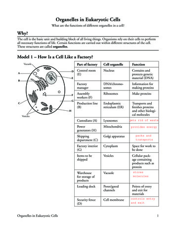
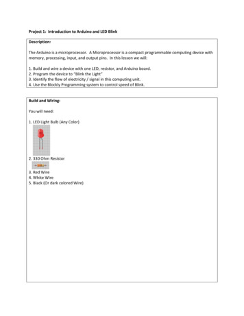
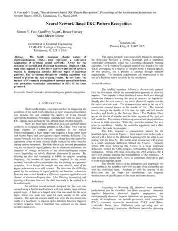
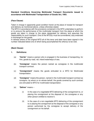
108 R. Henry et al.early endosome marker Rab5a and the late endosomemarker Rab7 on Lm vacuoles may have delayed fusionwith lysosomes (Alvarez-Dominguez et al., 1997; AlvarezDominguez and Stahl, 1999). Alternatively, there is evidence that activation of Rab5a on Lm vacuoles decreasesbacterial escape (Prada-Delgado et al., 2005).In addition to its role in perforating membranes for Lmescape into cytosol, LLO could influence the dynamics ofLm vacuole maturation. This issue is difficult to addressexperimentally. The simplest approach of using fluorescence microscopy of fixed cells to colocalize bacteria withmarkers of endocytic compartments is complicated byseveral factors. First, and most importantly, present methods for distinguishing vacuolar from cytosolic Lm identifycytosolic bacteria by their recruitment of F-actin, a process that occurs long after bacteria escape from the vacuole. By the time cytosolic bacteria can be identified, theyare unlikely to still be associated with the vacuolar compartments from which they escaped. Second, the timingof Lm escape varies between 15 and 30 min after infection, so there is no one time point to fix cells for analysis.Third, only some of the vacuoles are perforated, whileothers retain Lm and may continue to later stages ofvacuole maturation (de Chastellier and Berche, 1994).Because the timing of maturation may vary between thosethat are lysed and those that are not, the two populationsof vacuoles must be distinguished.Issues of heterogeneity in phagosome maturationdynamics have been addressed using live cell imaging.The progressive interactions between phagosomes containing IgG-coated erythrocytes and endocytic compartments were observed in live cells using yellow fluorescentprotein (YFP) chimeras of endocytic markers (Henryet al., 2004). However, applying this technology to studyLm vacuole maturation still requires a method to mark Lmwhen they have gained access to cytosol but not yet shedtheir vacuolar membranes.Here we describe a new indicator of Lm escape thatuses a YFP chimera of the cell wall-binding domain (YFPCBD) of the Lm phage endolysin Ply118 (Loessner et al.,2002). Expression of YFP-CBD within the macrophagecytosol identified cytosolic bacteria shortly after escape.Methods for classification of endocytic compartmentswere used in conjunction with YFP-CBD to distinguishbehaviours of subpopulations of bacteria insidemacrophages.We analysed the progression of Lm vacuoles throughthe endocytic pathway and the escape of Lm to the cytosol. RAW 264.7 macrophages expressed YFP-CBD alongwith one of several cyan fluorescent protein (CFP)labelled chimeras of endocytic pathway markers. Live cellimaging of macrophages infected with wild-type Lm identified recently perforated vacuoles as those containingboth CFP-labelled membrane markers and YFP-CBD-positive bacteria. Employing YFP-CBD as an early indicator of escape not only revealed differences in the maturation of Lm vacuole subpopulations, but also identified thecompartments permissive to Lm escape. Internalized Lmdid not occupy Rab5a-positive compartments, but insteadmoved directly into late endosomal compartments. Vacuoles containing wild-type Lm delayed fusion with lysosome-associated membrane protein-1 (LAMP1)-positivelysosomes, relative to those containing hly Lm. This delayresulted in perforation of Rab7-positive, phosphatidylinositol 3-phosphate [PI(3)P]-positive, LAMP1-negative vacuoles. Although wild-type Lm perforated their vacuolesbefore fusion with LAMP1-positive lysosomes, Lmexpressing LLO under the control of an inducible promoter[inducible LLO (iLLO)] perforated LAMP1-positive compartments, although very inefficiently. Thus, the LLOdependent delay in vacuolar maturation facilitates Lmescape by increasing the duration of Lm residence inpenetrable compartments.ResultsYFP-CBD was an indicator of Lm vacuole escapeThe cell wall-binding domain (CBD) from the Lm phageendolysin Ply118, which binds to a carbohydrate ligand inLm cell walls with high affinity (Loessner et al., 2002), wasdeveloped into a probe for Lm escape to the cytosol.Green fluorescent protein (GFP)-tagged CBD could labelboth wild-type and hly Lm when purified and mixed withbacteria in solution (Fig. 1A and B). The CBD was clonedinto a mammalian expression vector containing YFP. YFPCBD expressed in macrophages was present in the cytosol and the nucleus (Fig. 1C and D). To verify that YFPCBD could be used as an indicator of Lm vacuole escape,macrophages expressing YFP-CBD were infected withwild-type, fluorescent Lm. Phase-contrast and fluorescence images of infected cells were acquired 10 and30 min after infection. YFP-CBD expressed inside infectedmacrophages did not label vacuolar Lm shortly after infection (Fig. 1C). However, by 30 min after infection, bacteriadecorated with YFP-CBD could be detected (Fig. 1D).Bacteria that appeared to be in the macrophage cytosolwere brightly labelled with YFP-CBD, while bacteria withinvacuoles were not. To verify that YFP-CBD was a markerof cytosolic Lm, the actin polymerization by YFP-CBDpositive Lm was measured in cells expressing both YFPCBD and CFP-actin. YFP-CBD-positive Lm labelled withCFP-actin 90 min after infection (Fig. 1E).The timing of YFP-CBD acquisition by cytosolic bacteriawas compared with the timing for F-actin recruitment.While Texas Red (TR)-phalloidin labelled wild-type bacteria 45 min after infection, YFP-CBD-positive bacteria weredetected as early as 15 min after infection (Fig. 2). Mutant 2005 The AuthorsJournal compilation 2005 Blackwell Publishing Ltd, Cellular Microbiology, 8, 107–119
Macrophage vacuoles perforated by Listeria monocytogenes109Fig. 1. YFP-CBD as a marker of Lm delivery into cytosol.A and B. Epifluorescence images of (A) wild-type Lm and (B) hly Lm decorated with HGFP-CBD after incubation with the purified fluorescentprotein.C and D. Phase-contrast (left), SNARF-labelled Lm (middle) and YFP-CBD fluorescence (right) images. (C) Cells viewed at 10 min after infectioncontained bacteria in phase-bright vacuoles (arrows) which were not labelled with YFP-CBD. (D) Cells viewed 60 min after infection showed somebacteria labelled with YFP-CBD (arrows).E. YFP fluorescence image of a RAW 264.7 macrophage showing YFP-CBD-positive cytosolic Lm (left) with corresponding labelling by CFPactin (asterisks). 2005 The AuthorsJournal compilation 2005 Blackwell Publishing Ltd, Cellular Microbiology, 8, 107–119
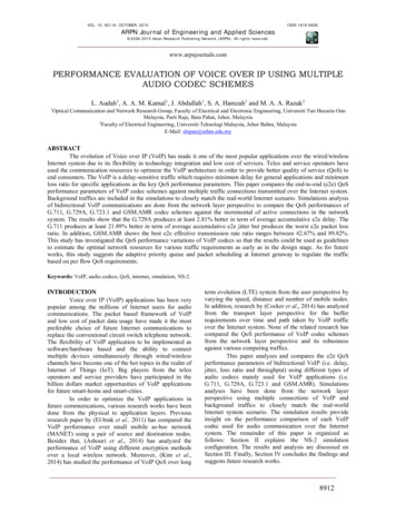
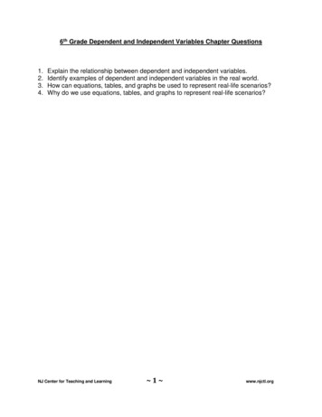
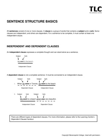
110 R. Henry et al.Fig. 2. YFP-CBD marked cytosolic Lm within 15 min of infection.Macrophages were infected with wild-type or hly Lm for 3 min andwashed, resulting in 1 bacterium per macrophage. Infected cellswere incubated further and fixed at the indicated time points. Bacteriawere identified by DAPI staining; cytosolic bacteria were identified bytheir labelling with either YFP-CBD or TR-phalloidin. At each timepoint 200 infected cells were scored for the presence of YFP-CBDor TR-phalloidin. YFP-CBD labelled wild-type Lm (black circles)between 15 and 30 min, while TR-phalloidin stained wild-type Lm(grey circles) 45–120 min after infection. YFP-CBD did not decoratehly Lm (open circles) at any time. Each data point represents themean standard error of the mean (SEM) of four experiments.hly Lm never labelled with YFP-CBD, consistent with earlier studies showing that these bacteria do not gain accessto macrophage cytosol (Jones and Portnoy, 1994). Thistime-course experiment demonstrated that YFP-CBD provided a more direct and timely assay for Lm vacuoleperforation and escape than actin nucleation.Membrane marker localization to compartmentslysed by LmYFP-CBD was used in conjunction with fluorescent markers of the endocytic pathway to identify the compartmentsperforated by Lm. The endocytic markers included CFPRab5a, CFP-Rab7, LAMP1-CFP and CFP-2xFYVE,which binds to PI(3)P (Gillooly et al., 2000; Stenmarket al., 2002). The overexpression of these CFP chimerasdid not alter Lm vacuole maturation and escape. The rateof wild-type Lm exposure to YFP-CBD was similar in macrophages expressing CFP chimeras or untagged CFP(data not shown). Macrophages were co-transfected withYFP-CBD and one of the CFP-tagged markers ofendocytic compartments. Cells were infected for 3 minwith wild-type Lm, washed free of extracellular bacteria,then phase-contrast, YFP and CFP images of infectedcells were taken at regular intervals (Fig. 3). Within 5 minof infection, intravacuolar bacteria were evident as phasedense rods inside phase-bright vacuoles (Fig. 3A). Thebacteria were present in a phase-bright vacuole between5 and 17 min, after which the vacuole became phasedark. The phase-bright to phase-dark transition was takento indicate vacuole perforation. The endocytic compartment in which the bacteria resided was identified byCFP-Rab7 labelling (Fig. 3B). Coincident with the phasecontrast transition, Lm were decorated with YFP-CBD(Fig. 3C). Bacteria remained associated with the CFPRab7-positive vacuole subsequent to vacuole lysis, asindicated by YFP-CBD (Fig. 3B and C).The presence of Lm in endocytic compartments duringthe acquisition of YFP-CBD labelling allowed identificationof the compartments lysed by Lm. Endosomal markerrecruitment was quantified by measuring the average CFPfluorescence intensity in a small region including the bacterium (Ip) and dividing it by the average CFP fluorescenceintensity of the entire cell (Ic). When the intensity of thephagosome (Ip) equalled the intensity of the cell (Ic) (i.e.Ip/Ic 1.0), the marker was considered not to be enrichedon the phagosome. Control macrophages expressinguntagged CFP consistently showed Ip/Ic values of 1.0Fig. 3. YFP-CBD allowed identification of compartments lysed by Lm. Macrophages were co-transfected with YFP-CBD and CFP-Rab7. Cellswere infected with wild-type Lm, then phase-contrast, YFP and CFP images of infected cells were taken at 2 min intervals. Each column containscomponent images of one time point, indicated in (A) as the time, in minutes, after the start of infection.A. Phase-contrast images show two bacteria in a phase-bright vacuole between 5 and 17 min, after which the vacuole became phase-dark.B. CFP-Rab7 labelling of this vacuole illustrated that the vacuole collapsed after 17 min, but fluorescence persisted. CFP fluorescence appearedto increase over time due to movement of the vacuole within the focal plane.C. Coincident with collapse of the vacuole, the bacteria acquired YFP-CBD. From these images it was apparent that bacteria were present in aRab7-positive compartment during escape to the cytosol. 2005 The AuthorsJournal compilation 2005 Blackwell Publishing Ltd, Cellular Microbiology, 8, 107–119
Macrophage vacuoles perforated by Listeria monocytogenes111(Fig. 4A). Ip/Ic values for Lm vacuoles significantly greaterthan 1.0 signified localization of a CFP chimera to thephagosome. Coincident measurement of YFP-CBDrecruitment to bacteria allowed discrimination of CFPchimera recruitment to vacuoles perforated by Lm (CBDpositive, or CBD ) and intact vacuoles containing CBDnegative (CBD–) Lm.Lm lysed compartments labelled with CFP-Rab7or CFP-2xFYVEWild-type and hly Lm were present in vacuoles labelledwith CFP- or YFP-Rab7 between 5 and 30 min after infection (Fig. 4B and C). The Ip/Ic ratios for vacuoles containinghly Lm were approximately 1.5 from 5 to 30 min afterinfection (Fig. 4B). Intact vacuoles containing wild-typeLm (CBD-negative) displayed Ip/Ic ratios of 1.5–2.2(Fig. 4C). The levels of Rab7 on both intact wild-type andhly Lm vacuoles remained relatively constant. All Lm vacuoles observed were labelled with YFP- or CFP-Rab7 ateach time point. YFP-CBD-positive wild-type Lm weredetectable between 15 and 30 min after infection (Fig. 2).Ip/Ic of YFP-CBD-positive bacteria were 1.5 at 15–20 minafter infection, after which the ratio decreased to 1(Fig. 4D). The high Ip/Ic for CFP-Rab7 when bacteriabegan to acquire YFP-CBD indicated lysis of Rab7positive vacuoles by wild-type Lm. The colocalization ofCFP-Rab7 with YFP-CBD decreased at 25–30 min afterinfection, possibly due to displacement of the bacteriafrom their lysed vacuoles.PI(3)P was recognized by YFP- or CFP-tagged tandemFYVE domains (YFP- or CFP-2xFYVE) (Gillooly et al.,2000; Stenmark et al., 2002). YFP-2xFYVE labelled hlyLm vacuoles at 5–30 min after infection (Fig. 5A). Themarker was also present on intact (CBD-negative) wildtype Lm vacuoles over the same time period (Fig. 5B). AllLm vacuoles were positive for YFP- or CFP-2xFYVE.CFP-2xFYVE localized to CBD-positive bacteria at 15–20 min after infection, then decreased (Fig. 5C). The colocalization of CFP-2xFYVE and YFP-CBD at 15–20 minindicated the presence of PI(3)P on vacuoles perforatedby wild-type bacteria.CFP-Rab5a was absent from Lm vacuolesNeither YFP-Rab5a nor CFP-Rab5a localized to Lm vacuoles from 5 to 30 min. The Ip/Ic ratios in cells expressingYFP- or CFP-Rab5a were near to 1 from 5 to 30 min afterinfection, indicating absence of this marker from Lm vacuoles (Fig. 6A and B). CFP-Rab5a was also not presenton vacuoles containing CBD-positive Lm (Fig. 6C).It was possible that YFP-Rab5a was present on Lmvacuoles transiently and before the earliest time pointmeasured. To test this possibility, phase-contrast and YFPFig. 4. Lm lysed compartments labelled with Rab7. The averagefluorescence intensity of the phagosome (Ip) was divided by theaverage fluorescence intensity of the entire cell (Ic) to producethe cell-normalized phagosome fluorescence (Ip/Ic). Each symbolrepresents the mean values of 45 vacuoles measured inmultiple experiments.A. Ip/Ic ratios for Lm vacuoles in macrophages expressing CFP werenear 1.0 from 5 to 30 min. These values were compared as a negativecontrol to all further experiments (grey dotted line in subsequentpanels).B and C. (B) hly Lm and (C) YFP-CBD-negative, wild-type Lm residedin Rab7-positive vacuoles between 5 and 30 min after infection, asindicated by Ip/Ic values significantly higher than those in cellsexpressing CFP (P 0.01). One hundred per cent of Lm vacuoleswere Rab7-positive.D. YFP-CBD-positive, wild-type Lm displayed Ip/Ic ratios greater than1.5 for CFP-Rab7 from 15 to 20 min, demonstrating that Lm lysedcompartments labelled with CFP-Rab7.The bars indicate SEM. 2005 The AuthorsJournal compilation 2005 Blackwell Publishing Ltd, Cellular Microbiology, 8, 107–119
112 R. Henry et al.of Fc receptors by Lm might localize YFP-Rab5a to Lmvacuoles. The internalization of IgG-opsonized Lm wasobserved in YFP-Rab5a-expressing RAW 264.7 macrophages. YFP-Rab5a did localize transiently to vacuoles containing IgG-opsonized Lm (Fig. 7A and B).Fig. 5. PI(3)P on vacuoles lysed by Lm.A and B. YFP- or CFP-2xFYVE-labelled vacuoles containing (A) hlyLm and (B) YFP-CBD-negative, wild-type Lm at 5–30 min. One hundred per cent of Lm vacuoles were 2xFYVE-positive.C. CFP-2xFYVE and YFP-CBD colocalized 15–20 min after infection,signifying the presence of PI(3)P on vacuoles perforated by wild-typebacteria. At each time point 45 vacuoles were measured.The bars indicate SEM.images were collected during internalization of Lm intoYFP-Rab5a-expressing RAW 264.7 or J774 macrophages. YFP-Rab5a was not present on wild-type Lmvacuoles during internalization or the first few minutessubsequent to vacuole formation in either cell line(Fig. 7A).YFP-Rab5a localized to newly formed phagosomescontaining IgG-opsonized erythrocytes in RAW 264.7macrophages (Henry et al., 2004). Thus, the stimulationFig. 6. Rab5 did not label Lm vacuoles, but localization of Rab5Q79Lto Lm vacuoles did not prevent escape.A. While YFP-Rab5a did not localize to vacuoles containing hly Lm(closed circles), YFP-Rab5Q79L was present from 5 to 20 min (opencircles).B. CFP-Rab5Q79L localized to CBD-negative, wild-type Lm vacuolesfrom 5 to 20 min (open triangles), but CFP-Rab5a did not (closedtriangles).C. CFP-Rab5a was not associated with YFP-CBD-positive Lm at anytime point measured (closed diamonds). CFP-Rab5Q79L associatedwith CBD-positive Lm at 15–20 min (open diamonds), demonstratingthat Lm could lyse vacuoles labelled with Rab5Q79L. At each timepoint 45 vacuoles were measured.The bars indicate SEM. 2005 The AuthorsJournal compilation 2005 Blackwell Publishing Ltd, Cellular Microbiology, 8, 107–119
Macrophage vacuoles perforated by Listeria monocytogenes113uoles by wild-type Lm (Fig. 6C). Moreover, expression ofCFP-Rab5aQ79L did not affect the rate of Lm exposureto YFP-CBD (data not shown). Thus, localization of CFPRab5Q79L to Lm vacuoles did not inhibit escape of bacteria to the cytosol.Wild-type Lm perforated vacuoles before fusion withLAMP1-positive compartmentsVacuoles containing hly bacteria acquired LAMP1-YFPfrom 15 to 25 min after infection, as indicated by Ip/Icvalues near 1.5 (Fig. 8A). The acquisition of LAMP1-CFPby vacuoles containing wild-type Lm was delayed relativeto hly vacuoles. Intact vacuoles containing wild-type LmFig. 7. Rab5a was not present on Lm vacuoles, but did localizetransiently to vacuoles containing IgG-opsonized Lm.A. The internalization of wild-type Lm was observed in RAW 264.7(closed circles) or J774 macrophages (closed triangles). YFP-Rab5awas not associated with wild-type Lm vacuoles during internalizationor the first 5 min subsequent to internalization. YFP-Rab5a associated transiently with vacuoles during entry of IgG-opsonized Lm intoRAW 264.7 macrophages (open circles).B. YFP-Rab5a did not associate with vacuoles containing hly Lm(closed diamonds), but did associate transiently with vacuoles containing IgG-opsonized, hly Lm (open diamonds). The data werealigned by the timing of phase-bright vacuole formation, which wasset at 3 min.At each time point 15 vacuoles were measured. The bars indicateSEM.Rab5Q79L localization to Lm vacuoles did notinhibit escapeThe absence of Rab5a from Lm vacuoles could havefacilitated Lm escape. To determine whether forced localization of Rab5a to vacuoles could inhibit Lm escape, weanalysed Lm vacuoles in macrophages expressing CFPRab5Q79L. This mutant form of CFP-Rab5a is locked inthe GTP-bound state and is recruited to membranes(Roberts et al., 2000). YFP- or CFP-Rab5Q79L waspresent on large endocytic vesicles within macrophages,and localized to vacuoles containing hly and wild-type Lm(Fig. 6A and B). Ip/Ic ratios were significantly greater than1.0 from 5 to 20 min after infection. The decline in Ip/Icvalues at 25 min may be due to fusion of vacuoles withlate endosomes and lysosomes. YFP-CBD-positive bacteria were associated with CFP-Rab5Q79L at 15–20 min,signifying the perforation of CFP-Rab5Q79L-labelled vac-Fig. 8. Wild-type Lm phagosomes showed delayed acquisition ofLAMP1-CFP.A. Vacuoles containing hly bacteria (open circles) acquired LAMP1YFP from 15 to 30 min after infection, as indicated by Ip/Ic values near1.5. Ninety per cent of vacuoles were LAMP1-positive at 15 min, and100% of vacuoles were LAMP1-positive at 30 min. The acquisition ofLAMP1-CFP by vacuoles containing CBD-negative wild-type Lm wasdelayed relative to hly phagosomes. Ip/Ic did not reach a value significantly higher than 1.0 until 25 min (P 0.01). Three per cent ofvacuoles containing CBD-negative wild-type Lm were LAMP1positive at 15 min, and 96% were LAMP1-positive at 30 min.B. Lack of colocalization of LAMP1-CFP with YFP-CBD indicated thatLm escaped to the cytosol before association with LAMP1-CFPlabelled lysosomes. While Ip/Ic ratios for CBD-negative, wild-type Lmwere high from 25 to 30 min, Ip/Ic ratios for CBD-positive wild-type Lmremained low (P 0.01).At each time point 45 vacuoles were measured. The bars indicateSEM. 2005 The AuthorsJournal compilation 2005 Blackwell Publishing Ltd, Cellular Microbiology, 8, 107–119
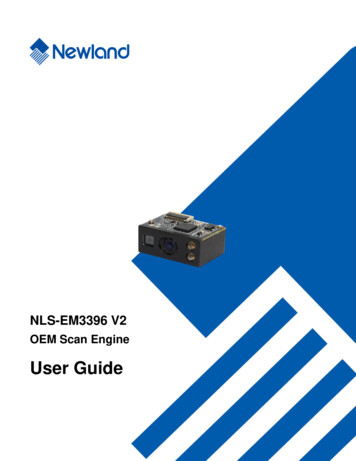
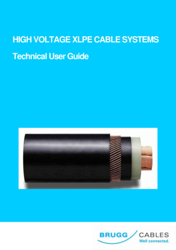
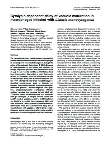
114 R. Henry et al.(CBD-negative) did not show LAMP1-CFP labelling until25 min (Fig. 8A), indicating that wild-type Lm delayedfusion with LAMP1-CFP-positive compartments. Furthermore, LAMP1-CFP did not colocalize with YFP-CBD evenat later time points (Fig. 8B), indicating that Lm perforatedits vacuole before merging with LAMP1-labelled compartments. Thus, even before it created perforations largeenough to admit YFP-CBD, LLO delayed Lm vacuolematuration.Inefficient perforation of LAMP1-positive compartmentsIt was possible that the normal timing of LLO activitycaused perforation of the vacuoles before fusion withLAMP1-positive compartments, but that Lm was nonetheless capable of perforating LAMP1-positive compartments. To examine the efficiency of perforation at laterstages of Lm vacuole maturation, the timing of LLOproduction was manipulated experimentally by placingtranscriptional control of LLO under an isopropyl-βD-thiogalactopyranoside (IPTG)-inducible promoter andincorporating the construct into the chromosome of hlyLm (Dancz et al., 2002). In the absence of IPTG, theiLLO Lm were trapped in vacuoles and could notescape to the cytosol (Fig. 9A and C). Addition ofFig. 9. Perforation of LAMP1-positive compartments by LLO wasinefficient.A. Macrophages expressing YFP-CBD were infected with wild-type oriLLO Lm to produce 1 bacterium per macrophage and were fixed atthe indicated time points. Infected cells were identified by DAPI staining and cytosolic bacteria were identified by YFP-CBD labelling. YFPCBD labelled wild-type Lm (grey circles) by 60 min. Escape of iLLOLm treated with 10 mM IPTG before infection (IPTG 0′, black circles)was reduced relative to wild type. iLLO Lm treated with IPTG at10 min after infection (IPTG 10′, triangles) were present in the cytosolat 60 min. iLLO Lm treated with IPTG at 30 min (IPTG 30′, diamonds)did not become CBD-labelled until 90 min, at which point 0.5% wereYFP-CBD-positive. In the absence of IPTG (open squares, partiallyobscured by IPTG 30′ data), no Lm were CBD-labelled. At each timepoint 100 infected cells were scored for the presence of YFP-CBD.Each data point represents the mean SEM of two experiments.B. Overnight cultures of iGFP Lm or macrophages infected with iGFPLm were fixed at the indicated time points. Intracellular bacteria wereidentified by DAPI staining and GFP-producing bacteria were identified by indirect immunofluorescence. iGFP Lm treated with IPTG inbroth (black circles) stained positive for GFP by 10 min. iGFP Lmtreated with IPTG at 10 min after infection stained positive for GFPfrom 30 to 120 min (black triangles), whereas iGFP Lm treated withIPTG at 30 min stained positive for GFP from 60 to 120 min (blackdiamonds). iGFP Lm did not stain positive for GFP in the absence ofIPTG treatment (open squares). At each time point 100 bacteria werescored for Alexa Fluor 594 staining. Each data point represents themean SEM of two experiments.C. Uninduced iLLO Lm acquired LAMP1-CFP by 20 min after infection, but did not decorate with YFP-CBD at any time (open squares).D. After treatment with IPTG at 10 min after infection, iLLO Lmescaped from LAMP1-CFP-negative compartments at 60–90 min(closed triangles). Many CBD-negative vacuoles acquired LAMP1CFP as early as 20 min (open triangles).E. In cells treated with IPTG at 30 min after infection, iLLO Lmperforated LAMP1-CFP-positive vacuoles at 90–100 min (closeddiamonds). 2005 The AuthorsJournal compilation 2005 Blackwell Publishing Ltd, Cellular Microbiology, 8, 107–119
Macrophage vacuoles perforated by Listeria monocytogenes10 mM IPTG to iLLO Lm immediately before infectionyielded less efficient vacuole perforation than wild-typeLm, but similar kinetics (Fig. 9A). Addition of IPTG tomacrophages at 10 min after infection resulted indelayed perforation, and induction of LLO at 30 minresulted in greatly reduced and delayed perforation.Approximately 0.5% iLLO Lm were CBD-positive at90 min (Fig. 9A).The small number of CBD-positive iLLO Lm observedafter treatment of macrophages with IPTG at 30 min(Fig. 9A) may have resulted from a low percentage ofLLO-producing Lm, due either to limited access of IPTGto Lm vacuoles or to decreased viability of Lm insidethose vacuoles. Attempts to detect LLO in vacuoles byimmunofluorescence were not successful. Instead, theefficiency of protein synthesis induced by IPTG wasinferred using hly Lm that produce GFP [inducible GFP(iGFP) Lm] using the same IPTG-inducible promoter asfound in the iLLO strain. GFP production was detected byimmunofluorescence staining with an anti-GFP antibody,as GFP fluorescence may not be observed until 90 min to4 h after protein synthesis (Zimmer, 2002). iGFP Lmtreated with IPTG in broth stained positive for GFP within10 min after adding IPTG (Fig. 9B). iGFP Lm in macrophages treated with IPTG at 10 min after infection began tostain positive for GFP by 30 min (Fig. 9B). Treatment ofinfected macrophages with IPTG at 30 min led to GFPsynthesis in 15–29% of bacteria (Fig. 9B). The reductionin the number of GFP-positive bacteria at 120 min mayhave been caused by degradation of bacteria within vacuoles. Comparison of the 90 min time points in Fig. 9Aand B shows that prolonged Lm residence in v
The endocytic markers included CFP-Rab5a, CFP-Rab7, LAMP1-CFP and CFP-2xFYVE, which binds to PI(3)P (Gillooly et al., 2000; Stenmark et al., 2002). The overexpression of these CFP chimeras did not alter Lm vacuole maturation and escape. The rate of wild-type Lm exposure to YFP-CBD was similar in mac-rophages expressing CFP chimeras or untagged CFP