Transcription
European Review for Medical and Pharmacological Sciences2022; 26: 278-283Acute embolisms in multiple arterialdistricts following Ad26.COV2-S vaccineV. MURRU1, V. COCCO1, C. MARRAS1, A. SESTU1, F. MARONGIU2,3,L. SABA2,3, M.E. CIANCHETTI2, A. SCUTERI2,3Post-Graduate Medical School of Internal Medicine, University of Cagliari, Cagliari, ItalyUniversity Hospital (AOU) Cagliari, Policlinico Universitario Monserrato, Cagliari, Italy3Dipartimento Scienze Mediche e Sanità Pubblica, Università di Cagliari, Cagliari, Italy12Abstract. – A case of multiple arterial thrombo-sis/embolisms in a 74-year-old Caucasian man withno other cardiovascular risk factors who receivedAd26.COV2-S vaccine 16 days before is reported.The unusual presentation required a longer diagnostic workup. The clinical manifestations and thetherapy-specific response suggest an unusual presentation of Vaccine-induced immune thromboticthrombocytopenia (VITT).Key Words:COVID-19 vaccine, Janssen vaccine, Ad.26.COV2.S,Splenic infarction, Arterial embolism, Thrombosis ofceliac artery, VITT.IntroductionAfter the identifications of the first cases inWuhan, China, in December 2019, coronavirusdisease 2019 (COVID-19) rapidly spread worldwide and took on pandemic proportions. By theend of September 2021, the COVID-19 pandemichas affected more than 229 million people worldwide and 4.71 million have died1.Currently, vaccines represent the most powerful and effective instrument to fight theCOVID-19. In Europe, the European MedicineAgency (EMA) approved four vaccines and fivemore are under rolling review.To date, up to 528 millions of subjects underwent vaccination for COVID-19 in Europe. Theadverse reactions to vaccination should not be intended as an argument against vaccination. Notably, the serious and life-threatening or fatal eventsinvolved arterial and venous thrombosis2.Cerebral venous sinus thrombosis with thrombocytopenia (platelet count 150.000/μL ofblood) have been observed more commonly withCHADOX1 NCOV-19 (AstraZeneca), and Ad26.COV2.S (Janssen – Johnson & Johnson)3.278We report a case of a 74-year-old Caucasianman who developed multiple arterials thrombosesfollowing the Ad26.COV2-S vaccination.Clinical PresentationA 74-year-old Caucasian man was admittedto our Hospital Unit because of abdominal pain,vomit, loss of appetite, constipation, and polyuria,appearing 15 days before hospitalization. He alsoreferred mild hypoesthesia and paresthesia in theright foot and right-side brachialgia. He deniedany fever or cough. The patient gave the consentto access his data during Hospital admission.The medical, surgical, or family history wasnegative for relevant diseases or risk factors (including alcohol or cigarette use), except the presence of treated arterial hypertension (AT-1 blocker). The patient had not previously been exposedto heparin nor reported any previous positiveCOVID-19 molecular test.Thirty days prior to hospital admission, thepatient was vaccinated with the adenoviral vectorvaccine Ad26.COV2-S (Table I). On admission,he had a negative test result to nose-pharyngealswab SARS-CoV-2 by polymerase chain reaction.In ED, the patient was stable (temperature 36.8 C,blood pressure 160/90 mmHg, pulse rate 112beats/minute, oxygen saturation of 96% on roomair). His body mass index (BMI) was 24.2 kg/m2. Cardiovascular system examination noticeda systolic murmur (intensity 2/6) and tachycardia.Crackling sounds were reported at lung auscultation. No alteration in the physical examination ofthe abdomen was observed.An electrocardiogram (ECG) reported sinustachycardia; the PR, QRS, and T-waves were normal with no changes in the ST-segment. The results of blood tests were normal except for a spe-Corresponding Author: Angelo Scuteri, MD, Ph.D; e-mail: angelo.scuteri@unica.it
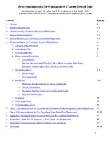
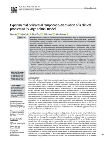
Acute embolisms in multiple arterial districts following Ad26.COV2-S vaccineTable I. Timeline of the patient's story.DateDay ofhospitalizationSequence OutlineJune 14, 2021The patient received the J&J vaccineJune 30, 2021Development of abdominal pain, vomit, loss of appetiteJuly 14, 20210Admittance to the ER for worsening abdominal pain, vomit, loss of appetite,constipation, and polyuriaJuly 16, 20212Acute subclavian artery occlusion - Fogarty arterial embolectomyJuly 19, 20215Subacute superficial femoral artery occlusionJuly 20, 20216Fogarty femoral arterial embolectomyJuly 23, 20219Total body CT with contrastPF4July 26, 202112Haemostasis medical consultationJuly 27, 202113August 12, 202129Discharge of the patientcific mild elevation of amylase, lipase, creatinine,and PCR levels (Table II). Chest x-ray was normal, while direct abdominal x-ray displayed constipation, few air-fluid levels and rectal fecaloma.A subsequent contrast-enhanced abdominalCT showed mesenteric panniculitis and diverticulosis not needing surgical attention for whichhe initiated therapy with Metronidazole 500 mgTable II. Laboratory findings on admission.Laboratory analysisDay 0Haemoglobin (g/dL)Platelet count (x 109/L)Leukocytes (x 109/L)Neutrophil (%)Absolute neutrophil (x 109/L)Lymphocyte (%)Partial thromboplastin time (sec)Prothrombin time (INR)D-dimer (μg/mL)Fibrinogen (mg/dL)Aspartate aminotransferase (U/L)Alanine aminotransferase (U/L)Total Cholesterol (mg/dL)HDL Cholesterol (mg/dL)LDL Cholesterol (mg/dL)Triglycerides (mg/dL)Glucose (mg/dL)HBA1C (%)Creatinine (mg/dL)Amylase (U/L)Lipase (U/L)Pancreatic Amylase (U/L)PCR 421331646.41.48123120105237.2three times daily, Ceftriaxone 2 g once dailyand Enoxaparin 4000 UI once daily. On day 2of hospital stay, the patient presented suddenpain in the right arm, with the disappears of theright radial pulse. Arterial Doppler ultrasounddisplayed acute subclavian artery occlusion inan arterial district without any local lesion oralteration in the whole supra-aortic arterial district. Therefore, the patient had successful Fogarty arterial embolectomy. Enoxaparin 4000UIwas increased twice a day. From day 3 to day5 of hospital stay, the patient presented paraesthesia of the right leg and progressive reductionof the right leg pulse. Arterial Doppler ultrasound showed superficial right femoral arteryocclusion. The patient had another successfullyFogarty arterial embolectomy.Suspecting a cardiac source of embolization(Septal Defects? Paroxysmal Atrial Fibrillation?) thepatient underwent Echocardiography and prolonged(72 hours) Holter ECG, both resulting normal.On day 9 of hospital stay, the persisting anorexia and nausea, leading the patient to significant weight loss and reduced ability to concentratewith psychomotor retardation, induced us to ruleout cancer diagnoses. Thus, a contrast-enhancedtotal body CT was performed. This test showedcoeliac artery occlusion with splenic infarction(Figure 1), and mesenteric panniculitis (not requiring surgical attention).Blood test [homocysteine, lupus anti-coagulant antibodies (LAC), beta-2 glycoprotein antibodies, anti-nuclear antibody (ANA), anti-neutrophil cytoplasmic antibodies (ANCA), IgG4,CA 125, CA 19.9, CEA, PSA total and free, NSE]gastric and colon endoscopy were negative mak279
V. Murru, V. Cocco, C. Marras, A. Sestu, F. Marongiu, L. Saba, M.E. Cianchetti, A. ScuteriABCDFigure 1. Contrast-enhanced CT at admission (A) shows a normal spleen; after 9 days, a large spleen infarction is evident(B) that is attributable to arterial obstruction (indicated by arrow in panel C), confirmed by Maximum Intensity Protection-MIP – reconstruction (D).ing unlikely the presence of autoimmune disease,vasculitis, or primary coagulation disorders.On the same day (day 9), suspecting an unusual VITT (absence of thrombocytopenia andPF4 negative), the patient underwent treatmentwith intravenous immune globulin (0.4 g/Kg/die),Methylprednisolone 80 mg once daily (1 mg/kg/die) and Fondaparinux 7.5 mg once daily.During the last two weeks of hospitalization,the patient had been under close clinical observation, and he started physical/motor rehabilitationto prevent loss of motor function; he also startedthe intravenous steroid tapering. On day 29 thepatient felt better and was discharged at home.After the hospital discharge, it was possibleto perform the anti-PF antibody assay (ASSERACHROM HPIA IgG; absorbance at 405 nm,0.063 e 1.478 for negative or positive case, respectively) on the blood sample drawn on July 28th –prior to any treatment indicated for VITT. Theassay was frankly positive.At an outpatient follow-up visit approximately30 days after hospital discharge, the patient wasin good general condition, feeding normally (3 kg280of weight gain), denying abdominal pain, with awalking autonomy of 50-100 meters. Ultrasoundcontrol showed good patency of the right commonand deep femoral artery. Nevertheless, completeobstruction of the right superficial femoral arteryand the popliteal artery was still evidenced, withthe resumption of blood flow in the anterior tibialartery which appeared to be free.DiscussionAt the beginning of September 2021, the Italian Network of Pharmaco-vigilance reported a total of 1242 adverse reactions out of approximately2 million doses of COVID-19 vaccine Janssenadministered. Of those, 255 were recognized asserious, 953 not serious and 24 were fatal. Amongthese, our case was the first and only report ofsplenic infarction, arterial embolism, and thrombosis of celiac artery.On the 3rd of September 2021, the EudraVigilance, available at www.adrreports.eu, contains1565 thromboembolic events (ADRs) across Eu-
Acute embolisms in multiple arterial districts following Ad26.COV2-S vaccinerope following the use of the Janssen vaccine; despite the report of 7 events of spleen infarction, nocases of celiac artery embolism or thrombosis hasbeen reported.USA’s ADRs are published on the VaccineAdverse Events Reporting System (VAERS),available at vaers.hhs.gov; on the 3rd of September2021 there are 713 thromboembolic events relatedto Janssen vaccine, of which 5 splenic infarctionevents and 3 arterial embolisms are listed.These data confirm once more the safety ofCOVID-19 vaccination. Contemporarily, theysupport the unusual presentation of the adversereaction to COVID-19 vaccines presented by thecase here reported.The COVID-19 pandemic required and is stillrequiring new learning, including the more andmore frequent reports of unusual clinical presentation4. This also concerns the adverse reaction tovaccination: the impact on the thrombosis and hemostasis system includes also hemophilia, as wellas cerebral arterial thrombosis5,6 – though venousand arterial thrombosis recognize different pathways of activation7.The first and major unusual clinical presentation of the adverse reaction to COVID-19 vaccineillustrated by the recent case consist of the arterialand embolic nature of the (multiple) organ damage.Underlying causes of diffuse arterial thrombosis are usually related to embolic phenomenonsecondary to atrial fibrillation, or atheroscleroticplaque rupture, solid organ malignancies, myeloproliferative neoplasms, vasculitis, and coagulation disorder as antiphospholipid antibodysyndrome and hyperhomocysteinemia7,8. Testsfor heritable thrombophilia (Factor V Leiden,Prothrombin G20210A mutation, and deficiencies of Protein C, Protein S, or Antithrombin) arenot indicated8. We gradually exclude all theseetiologies in our patient. Ultrasounds and CT angiography showed healthy arteries without significant atherosclerotic plaques. Echocardiography and Holter ECG excluded atrial fibrillation.Cancer as well was ruled out. Prothromboticscreening for artery thrombosis with normal arterial blood vessels typically relies on the assayof antiphospholipid syndrome, hyperhomocysteinemia, and fibrinogen7-9. All these factors werenot present in our patient.In our case, arterial embolisms in unusualsites (mesenteric panniculitis, splenic infarctionand left renal ischemia, acute subclavian arteryocclusion, and subacute superficial femoral arteryocclusion), was suggestive for VITT10-12.VITT typically manifests in women aged 1849 years, approximately 5-30 days after vaccination with cerebral or venous thromboses; arterialthromboses have also been reported, though theyare rare10-14.The pathophysiology of VITT has been hypothesized to be mediated by immunoglobulin G(IgG) antibodies that recognize PF4 and activateplatelets through their FcγIIa receptors. The useof intravenous immune globulin paralleled itsefficacy in the treatment of autoimmune heparin-induced thrombocytopenia (aHIT – a thrombocytopenic disorder caused by the formationof IgG antibodies against PF4, upon exposure toheparin15) because it inhibits platelet activation10.It is important not to use intravenous immuneglobulin (IVIG) batches obtained from the plasma collected during the SARS-CoV-2 pandemic,to avoid from reducing the vaccine efficacy andinduce vaccination failure in these patients16.At the moment, the pathogenic mechanism ofVITT is unclear. To date, VITT has been describedonly related to adenoviral vector-based DNA virusvaccines, thus implying that the syndrome couldbe related to the vector or other constituents inthe vaccine preparation15. De Bruijn et al17 recently reported the first episode of immune-mediatedthrombotic thrombocytopenic purpura associatedwith an mRNA-based anti- COVID-19 vaccination. Othman et al15 recently reported seven potential mechanisms concerning adenovirus inductionof VITT, from the binding of adenovirus to platelets or anticoagulation factors, to vaccine-inducedCOVID mimicry, to link of adenovirus capsid toPF4, to impurities in vaccine preparation triggering autoantibodies, to possible acute infection withSARS-CoV-2 following vaccine administration.During the recovery, the patient was periodicallytested for COVID-19 infection using molecularPCR test on nasal swab; all tests were negative.At hospital admission, PF4 antibodies weremeasured using the rapid assay HemosIL HITAb(PF4-H) and they were negative ( 1 U/mL). Platton et al18 remarked that these kits have a highsensitivity for HIT, but they have poor sensitivity for VITT in comparison to ELISA, whichwas not available in our laboratory at that time.Subsequently, ELISA assay was positive for anti-PF4 antibodies. Therefore, the patient had 4 outof 5 criteria for VITT. Notably, thrombocytopenia was not observable in our patient, though it isconsidered a typical feature of VITT.Nevertheless, because of strong suspicion ofVITT, the patient received usual therapy based on281
V. Murru, V. Cocco, C. Marras, A. Sestu, F. Marongiu, L. Saba, M.E. Cianchetti, A. Scuteriintravenous immune globulin (0.4 g/Kg/die) for5 days, Methylprednisolone 80 mg once daily (1mg/kg/die) for a week, and Fondaparinux 7.5 mgonce a day19,20.After VITT treatment had finished, the patient had no new thromboembolic events, and hisclinical conditions slowly get better with the disappearance of nausea, improvement of appetite,and weight gain (3 kg after discharge). He hasn’tyet fully regained physical fitness, still presentingclaudication after 50-100 meters of walk.At the moment, there is not enough evidenceto establish a causal relationship between theseevents and the Janssen vaccine.ConclusionsAn increase in vigilance regarding extremelyrare side effects associated with COVID-19 vaccination is needed. VITT is a serious complication of vaccination that is not possible to predictor prevent and that can occur also in the absenceof thrombocytopenia. When patients, among5-30 days post-vaccination, begin to manifestsustained headache, neurologic symptoms/signs,abdominal pain, dyspnea, or limb pain/swelling,platelet count, D-dimer, and PF4 must be measured, and imaging for thrombosis performed14.Reporting unusual adverse events of the vaccinations is crucial to help clinicians better understand the long-term outcomes.AcknowledgmentsV. Murru, V. Cocco, C. Marras, A. Sestu gratefully acknowledge the Autonomous Region of Sardinia (RAS) forthe financial support of their specialist medical training inInternal Medicine.All Authors have given approval of the submitted and finalversions.Author’s ContributionV. Murru drafted the paper and contributed to acquisitionand interpretation of data.V. Cocco revised the paper and contributed to acquisitionand interpretation of data.C. Marras revised the paper and contributed to acquisitionand interpretation of data.A. Sestu revised the paper and contributed to acquisitionand interpretation of data.F. Marongiu revised the paper and contributed to acquisition and interpretation of data.L. Saba revised the paper and contributed to acquisition andinterpretation of data.282M.E. Cianchetti revised the paper and contributed to acquisition and interpretation of data.A. Scuteri revised the paper and contributed to interpretation of data.Conflict of InterestsThe authors declare that they have no conflict of interests.References1) Ministero della Salute. Covid-19 - Situazione nelmondo. Accessed September 27, 2021. dettaglioContenutiNuovoCoronavirus.jsp2) European Medicine Agency. COVID-19 Vaccine Janssen. Accessed September 27, afety-covid-19-vaccines3) Abbattista M, Martinelli I, Peyvandi F. Comparisonof adverse drug reactions among four COVID‐19vaccines in Europe using the EudraVigilance database: thrombosis at unusual sites. J ThrombHaemost 2021; 19: 2554-2558.4) Hunt B. Education of healthcare professionalsand the public about thrombosis and COVID‐19.J Thromb Haemost 2021; 19: 2369-2371.5) Radwi M, Farsi S. A case report of acquired hemophilia following COVID‐19 vaccine. J ThrombHaemost 2021; 19: 1515-1518.6) Blauenfeldt RA, Kristensen SR, Ernstsen SL,Kristensen CCH, Simonsen CZ, Hvas AM.Thrombocytopenia with acute ischemic strokeand bleeding in a patient newly vaccinated withan adenoviral vector-based COVID-19 vaccine. JThromb Haemost 2021; 19: 1771-1775.7) Previtali E, Bucciarelli P, Passamonti SM, Martinelli I. Risk factors for venous and arterial thrombosis. Blood Transfus 2011; 9: 120-138.8) O’Donnell M, Shatzel JJ, Olson SR, DaughetyMM, Nguyen KP, Hum J, DeLoughery TG. Arterialthrombosis in unusual sites: a practical review.Eur J Haematol 2018; 101: 728-736.9) Gavriș CM, Nedelcu LD, Pascu AM. Thrombotic riskin antiphospholipidic syndrome: from hypothesis tocurrent evidence (Review). Exp Ther Med 2021; 21: 1-4.10) Greinacher A, Thiele T, Warkentin TE, WeisserK, Kyrle PA, Eichinger S. Thrombotic Thrombocytopenia after ChAdOx1 nCov-19 Vaccination. NEngl J Med 2021; 384: 2092-2101.11) Schultz NH, Sørvoll IH, Michelsen AE, Munthe LA,Lund-Johansen F, Ahlen MT, Wiedmann M, AamodtAH, Skattør TH, Tjønnfjord GE, Holme PA. Thrombosis and thrombocytopenia after ChAdOx1 nCoV-19Vaccination. N Engl J Med 2021; 384: 2124-2130.12) Scully M, Singh D, Lown R, Poles A, SolomonT, Levi M, Goldblatt D, Kotoucek P, Thomas W,
Acute embolisms in multiple arterial districts following Ad26.COV2-S vaccineLester W. Pathologic antibodies to platelet factor4 after ChAdOx1 nCoV-19 vaccination. N Engl JMed 2021; 384: 2202-2211.13) Favaloro EJ. Laboratory testing for suspectedCOVID-19 vaccine–induced (immune) thromboticthrombocytopenia. Int J Lab Hematol 2021; 43:559-570.14) Iba T, Levy JH, Warkentin TE. Recognizing vaccine-induced immune thrombotic thrombocytopenia. Crit Care Med 2021; 13: 1-7.15) Othman M, Baker AT, Gupalo E, Elsebaie A,Bliss CM, Rondina MT, Lillicrap D, Parker AL.To clot or not to clot? Ad is the question - insights on mechanisms related to vaccine‐induced thrombotic thrombocytopenia. J ThrombHaemost 2021; 19: 2845-2856.16) Karnam A, Lacroix‐Desmazes S, Kaveri S V., Bayry J. Vaccine‐induced immune thrombotic thrombocytopenia: consider IVIG batch in the treatment.J Thromb Haemost 2021; 19: 1838-1839.17) de Bruijn S, Maes MB, De Waele L, Vanhoorelbeke K, Gadisseur A. First report of a de novoiTTP episode associated with an mRNA‐basedanti‐COVID‐19 vaccination. J Thromb Haemost2021; 19: 2014-2018.18) Platton S, Bartlett A, MacCallum P, Makris M,McDonald V, Singh D, Scully M, Pavord S. Evaluation of laboratory assays for anti-platelet factor 4antibodies after ChAdOx1 nCOV-19 vaccination.J Thromb Haemost 2021; 19: 2007-2013.19) Marongiu F, Barcellona D. Trombocitopenia trombotica indotta da vaccino: gli algoritmi per gestirla. Anticoagulazione.it 2021.20) Nazy I, Sachs UJ, Arnold DM, McKenzie SE,Choi P, Althaus K, Ahlen MT, Sharma R, GraceRF, Bakchoul T. Recommendations for the clinical and laboratory diagnosis of VITT againstCOVID‐19: communication from the ISTH SSCsubcommittee on platelet immunology. J ThrombHaemost 2021; 19: 1585-1588.283
July 26, 2021 12 Haemostasis medical consultation July 27, 2021 13 August 12, 2021 29 Discharge of the patient Table II. Laboratory findings on admission. Laboratory analysis Day 0 Haemoglobin (g/dL) 15.5 Platelet count (x 109/L) 244 Leukocytes (x 109/L) 5.170 Neutrophil (%) 58.1 Absolute neutrophil (x 109/L) 2.9 Lymphocyte (%) 28.6