Transcription
FOCUS ARTICLEEuroIntervention 2014;10-online publish-ahead-of-print August 2014 Cardiac arrhythmias in acute coronary syndromes: positionpaper from the joint EHRA, ACCA, and EAPCI task forceBulent Gorenek*† (Chairperson, Turkey), Carina Blomström Lundqvist† (Sweden), Josep Brugada Terradellas†(Spain), A. John Camm† (UK), Gerhard Hindricks† (Germany), Kurt Huber‡ (Austria), Paulus Kirchhof† (UK),Karl-Heinz Kuck† (Germany), Gulmira Kudaiberdieva† (Turkey), Tina Lin† (Germany), Antonio Raviele† (Italy),Massimo Santini† (Italy), Roland Richard Tilz† (Germany), Marco Valgimigli¶ (The Netherlands), Marc A. Vos†(The Netherlands), Christian Vrints‡ (Belgium), and Uwe Zeymer‡ (Germany)Document Reviewers: Gregory Y.H. Lip (Review Coordinator) (UK), Tatjania Potpara (Serbia), Laurent Fauchier (France),Christian Sticherling (Switzerland), Marco Roffi (Switzerland), Petr Widimsky (Czech Republic), Julinda Mehilli (Germany),Maddalena Lettino (Italy), Francois Schiele (France), Peter Sinnaeve (Belgium), Giueseppe Boriani (Italy), Deirdre Lane (UK),and Irene Savelieva (on behalf of EP-Europace, UK)IntroductionIt is known that myocardial ischaemia and infarction leads to severemetabolic and electrophysiological changes that induce silent orsymptomatic life-threatening arrhythmias. Sudden cardiac death ismost often attributed to this pathophysiology, but many patients survive the early stage of an acute coronary syndrome (ACS) reachinga medical facility where the management of ischaemia and infarction must include continuous electrocardiographic (ECG) and hemodynamic monitoring, and a prompt therapeutic response to incidentsustained arrhythmias. During the last decade, the hospital locationsin which arrhythmias are most relevant have changed to include thecardiac catheterization laboratory, since the preferred managementof early acute ACS is generally interventional in nature. However,a large proportion of patients are still managed medically.Both atrial and ventricular arrhythmias may occur in the setting of ACS and sustained ventricular tachyarrhythmias (VAs)may be associated with circulatory collapse and require immediatetreatment. Atrial fibrillation (AF) may also warrant urgent treatment when a fast ventricular rate is associated with hemodynamicdeterioration. The management of other arrhythmias is also basedlargely on symptoms rather than to avert progression to more serious arrhythmias. Prophylactic antiarrhythmic management strategies have largely been discouraged.Although the mainstay of antiarrhythmic therapy used to rely onantiarrhythmic drugs (AADs), particularly sodium channel blockersand amiodarone, their use has now declined, since clinical evidenceto support such treatment has never been convincing. Therapy foracute coronary syndrome and arrhythmia management are nowbased increasingly on invasive approaches. The changes in theclinical approach to arrhythmia management in ACS have been sosubstantial that the European Heart Rhythm Association, the AcuteCardiovascular Care Association and the European Association ofPercutaneous Cardiovascular Interventions established a task forceto define the current position.DOI: 10.4244/EIJY14M08 19* Corresponding author: Prof. Bulent Gorenek MD FACC FESC, Eskisehir Osmangazi University Cardiology Department,Eskisehir, Turkey. Tel: 90 222 2292266; Fax: 90 222 2292266. E-mail: bulent@gorenek.com†EHRA. ‡ ACCA. ¶ EAPCI. Developed in partnership with the European Heart Rhythm Association (EHRA), the Acute Cardiovascular Care Association (ACCA), and the European Association of Percutaneous Cardiovascular Interventions (EAPCI).The article has been co-published with permission in EP-Europace, European Heart Journal - Acute Cardiovascular Care andEuroIntervention. All rights reserved in respect of European Heart Journal - Acute Cardiovascular Care and EuroIntervention. The Authors 2014. For EP-Europace, The Author 2014.1
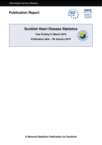
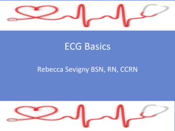
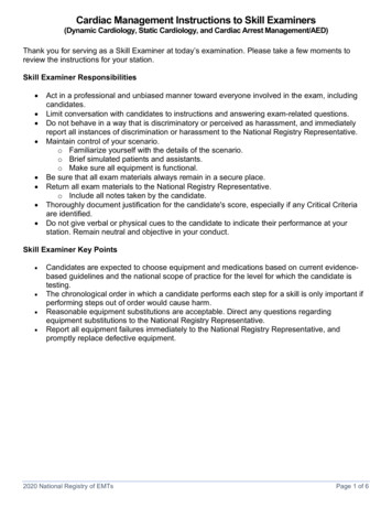
EuroIntervention 2014;10-online publish-ahead-of-print August 2014Mechanisms of ischaemia-related ventriculararrhythmiasGeneration of the ventricular action potential by voltage and substrate dependent ion currents is the basis for the contraction of eachindividual myocardial cell. This ionic balance can be disturbed byinsults, such as ischaemia/reperfusion.Arrhythmogenesis early in the course of an ACS, manifested asoften polymorphic ventricular tachycardia (VT) or ventricular fibrillation (VF) is observed in a minority of patients with acute ischaemiaand is often associated with genetic predisposition.1 Incidence of inhospital mortality due to acute heart failure or VT/VF has declinedmarkedly with the widespread use of reperfusion strategies.Acute myocardial ischaemia leads to adenosine tri-phosphatedeficiency, anaerobic glycolysis causing acidosis, elevation ofextracellular potassium (K ), and lysophosphatidylcholine accumulation. This multifactorial sequence of events results electrophysiologically in (i) ionic imbalance: (a) shorter duration of theaction potential by activation of the substrate related potassium current: IKatp and (b) less reduced resting membrane potential throughinhibition of the inward rectifying potassium current: IK1; (ii) lesscontractile force by events that culminate in the mishandling ofintracellular calcium (Ca2 ); and (iii) a reduced conduction velocitybecause of less functional gap junctions.2Myocardial reperfusion may cause profound electrophysiological alterations, dependent on the prior duration of ischaemia. A VToccurs more frequently with increasing duration of ischaemia, butlater in the course of an ACS, and with extensive myocardial damage, the incidence of VT declines. Important contributors involvedin arrhythmogenesis are the Na /Ca2 exchange pump, the slowlyactivating delayed rectifier K current and phosphorylation of sarcoplasmic reticulum proteins by CAMKII (calcium and calmodulindependent protein kinase II).3 The intracellular Ca2 overload (amongothers caused by reactive oxidative stress) will result in spontaneous Ca2 oscillations (calcium overload paradox) that trigger earlyand delayed after depolarizations induced ectopic beats. In addition,they will contribute to spatial and temporal dispersion of repolarization (variation in the action potential duration) and to re-entrantarrhythmias based on unidirectional conduction block, fractionationof cellular electrograms, and short action potential durations. Forarrhythmogenic mechanisms, the currents flowing from the ischaemic/reperfused to the non-ischaemic zones are most important.The mechanisms responsible for the initiation of these VA may differ based on the underlying disease (Figure 1)4: (intramural) re-entryin ischaemia, whereas triggered activity appears to be the dominantmechanism in reperfusion.5 Also when looking at the responsiblemechanism for VT perpetuation, their role may differ but they contribute both to the severity of the arrhythmia and to sudden cardiac death.Incidence, predictors, and outcomes ofsustained ventricular arrhythmias in patientswith acute coronary syndromeSudden death due to sustained VA is common in patients suffering from an untreated myocardial infarction (MI). In fact, electrical2cardioversion (CV)/defibrillation and management of acute volumeoverload were the principal life-saving measures in the coronarycare units before the advent of reperfusion therapies, beta-blockers, antithrombotic therapy, and statins.6 The electrical changes inacutely ischaemic myocardium, and especially in the border zoneof an evolving MI, initiate and maintain these arrhythmias (seeabove). Prompt and adequate revascularization therapy, usually byinterventional reopening of occluded vessels and stabilization ofthe culprit lesion with a stent, combined with initiation of adequatesecondary prevention therapies (statin, dual antiplatelet therapy,beta-blockers, angiotensin-converting enzyme inhibitors, angiotensin receptor blockers) aimed at preventing subsequent acute coronary events,4 have markedly reduced these life-threatening eventsin modern coronary care units. Patients who present with sustainedVA in the first few days after a MI can hence be characterized by thefollowing features, summarized in Figure 1:(1) Late presenters (i.e. patients in whom reperfusion therapy wasdelayed due to an insufficient chain of care from first onset ofsymptoms to transfer to an acute coronary care centre, or longpatient-related delay, severe acute ischaemia in Figure 1).(2) Patients in whom revascularization was not or only partiallysuccessful due to technical difficulties (severe acute ischaemiain Figure 1).(3) Patients who carry an arrhythmogenic substrate prior to theacute event, either due to a prior infarction or due to a predisposition to electrophysiological instability (‘pre-existing myocardial damage’ in Figure 1).LATE PRESENTERSEven in highly developed healthcare systems, only 70-80%of patients who present with ST-elevation myocardial infarction (STEMI) receive reperfusion therapy.7 Some patients neverreach out for help to the healthcare system, while the diagnosis isdelayed in others. Yet others will present with VF or syncope as theinitial sign of the acute event, necessitating adequate resuscitation.High maximal creatine kinase (CK) values, Q waves on the ECG,markedly reduced left ventricular (LV) function in the sub-acutephase after a MI and a long history of symptoms attributable to theinfarct prior to first treatment are good markers for these patientsonce they have reached the hospital. Patients at high risk in thesetting of an ACS, easily assessed by clinical risk scores such asGRACE or TIMI,8-12 are likely to suffer from larger myocardialdamage, especially when the ECG shows prominent ischaemicchanges, the patient presents with persistent symptoms after initiation of therapy, or CK/troponin release patterns suggest extensivemyocardial damage. These patients are at increased risk of sustained VA.13INCOMPLETE REVASCULARIZATIONRapid revascularization of the culprit lesion is often feasible, butmay sometimes pose technical challenges, e.g. in highly calcified orbifurcated lesions. Furthermore, about 30-40% of STEMI patientsand 70-80% of those with STEMI and cardiogenic shock present
Cardiac arrhythmias in acute coronary syndromesEuroIntervention 2014;10-online publish-ahead-of-print August 2014Figure 1. Scheme of drivers for arrhythmias in acute coronary syndromes.Apre-existing substrate for ventricular arrhythmias, eithersecondary to an old myocardial infarction, due to a cardiomyopathy, or secondary to a genetic predisposition to ventricular arrhythmias,interacts with acute ischaemia, autonomic tone, and acute left ventricular strain to create triggered activity and ventricular arrhythmias.4ARVC: arrhythmogenic right ventricular cardiomyopathy; DCM: dilated cardiomyopathy; HCM: hypertrophic cardiomyopathy;VF: ventricular fibrillation; VT: ventricular tachycardia; WPW: Wolf-Parkinson-White syndrome (Adapted from Heart, Kirchhof P, BreithardtG, Eckardt L. Primary prevention of sudden cardiac death, 92, 1873-8, Copyright 2006, with permission from BMJ Publishing Group Ltd andfrom J Cardiovasc Pathol, 19, Basso C, Rizzo S, Thiene G. The metamorphosis of myocardial infarction following coronary recanalization,22-8, Copyright 2010, with permission from Elsevier).with a relevant stenosis in the non-culprit vessel.14 These patientsremain at risk for recurrent ischaemic events even after the acuterevascularization procedure and seem to be at increased risk forsustained VA.14 A careful review of the coronary anatomy can identify these patients. Emerging data suggest that early completion ofrevascularization may be beneficial, but further data are needed.15ARRHYTHMOGENIC SUBSTRATE PRIOR TO THE INDEX EVENTMost patients survive their first ACS. Owing to progression of arteriosclerosis, a second acute event will often occur despite maximal preventive therapy. Patients who suffer from an acute coronaryevent with pre-existing reduced LV function and myocardial scarsare at risk for sustained VA in the acute and sub-acute phase of3
EuroIntervention 2014;10-online publish-ahead-of-print August 2014a MI. Echocardiographic signs of markedly reduced LV functionor ECG signs of an old MI can identify such patients. Furthermore,patients with increased sympathetic activity, or taken to the extremeof cardiogenic shock, are at increased risk of sustained VA in thesetting of a ‘recurrent’ acute coronary event. Recent evidence supports the assumption that sustained VA in the setting of an ACSis also dependent on an individual genetic predisposition to suchevents. This is most obvious in patients with inherited cardiomyopathies that confer electrical instability (Figure 1),4 but may also bemediated by common genetic variants.1,16Recommendations for evaluation of patients with acute coronarysyndrome at risk for ventricular tachyarrhythmiaPatients with acute coronary syndrome who present with either ofthe following conditions– late from the onset of the symptoms,– incomplete revascularization,– presence of substrate prior to the event,– or those patients with complications,should be considered at increased risk for arrhythmia developmentduring initial evaluation.Antiarrhythmic therapy in patients withsustained ventricular arrhythmias and acutecoronary syndromeThe incidence of sustained VT and VF occurring within 48 h of theonset of an ACS seems to have decreased over the past decades.17-19This is most likely due to the widespread availability of revascularization therapy, limiting the size of infarction and to an increaseduse of beta-blockers.8 However, in a recent retrospective analysisof two randomized trials sustained VA occurred in almost 6% ofpatients in the very early phase of acute MI indicating the significance of VT/VF in this situation.20 The role of AAD therapy for thetreatment of sustained VT/VF in ACS has been strongly debatedand questioned.17 Only limited data exist from clinical trials concerning the use of AADs in ACS. Most of the reports available arelimited by (i) uncontrolled patient selection, (ii) multiple-pragmatictreatment regimens, (iii) small sample size, (iv) variable endpoints,and (v) at least partially by some degree of selection and outcomebias. Controlled randomized trials comparing different AADs inACS are completely lacking.The nature of VT/VF in the setting of ACS is complex, dynamic,and different from the chronic phase of MI (see above).21,22 Variabledegrees of ischaemia and reperfusion may be present which significantly affect both arrhythmia mechanisms and – even moreimportantly – the effects of AADs. Almost all AADs act either ina voltage or rate-dependent manner, and some drugs have bothcharacteristics. Thus, AAD action may be significantly altered inischaemic/reperfused myocardium as compared with non-affectedmyocardium. This may result in significant electrophysiologicalheterogeneity and gradients in conduction and refractoriness.If ischaemia is suspected to be responsible for the arrhythmia, immediate reperfusion is of utmost importance.23 Early use4of beta-blockers in the setting of ACS reduces mortality and theincidence of VA and is therefore recommended.17,24 Similarly, correction of hypomagnesaemia and hypokalaemia is encouragedbecause of the potential contribution of electrolyte disturbances toVA. Statin therapy reduces mortality in patients with coronary heartdisease and is therefore part of the recommended routine medication. Early and intensive statin therapy has been reported to reducethe incidence of premature ventricular complexes (PVCs) and nonsustained VA.25Antiarrhythmic drugs are widely used, but are of modest efficacyand have important side effects. Only sparse data exist to guide AADtherapy for sustained VA related to ACS. However, overwhelmingdata exist that have shown the potential harm of most AAD therapyfor patients with ischaemic heart disease.26 Nevertheless, even withthe advance of non-pharmacological treatment options as catheterablation, temporary or chronic AAD therapy may still be considered for the treatment of refractory VA. Initial attempts to tackleVT/VF in ACS, even when recurrent, should be electrical CV/defibrillation. Antiarrhythmic drug treatment should be consideredonly if episodes of VT/VF are frequent and can no longer be controlled by successive CV/defibrillation. Especially in patients withrecurrent VT/VF triggered by PVCs arising from partially injuredPurkinje fibres, catheter ablation has been shown very effective andshould be considered (see catheter ablation in ACS).27Lidocaine is a class I AAD that may reduce the incidence of VArelated to myocardial ischaemia, although no beneficial effect onearly mortality has been demonstrated.28 Because of potential proarrhythmia, with a trend towards excess mortality, prophylactic treatment with lidocaine has largely been discouraged.17 On the otherhand, prophylactic treatment with lidocaine after cardiac arrest andsuccessful resuscitation has shown significant beneficial effects forboth, recurrence of VA and survival.29 However, others reporteda lower risk of death at 24 h after thrombolysis and prophylacticlidocaine and a neutral effect on overall mortality.30 Recent analysisof GUSTO IIB and III studies has shown reduced early mortality inpatients with VF/VF after acute MI and a neutral effect on 30-daymortality.20 Considering the overall efficacy and side effects, lidocaine should be considered as an AAD for the acute intravenousmanagement of recurrent VT/VF in ACS.Amiodarone is a class III AAD with multiple additional electrophysiological properties. When AAD therapy is necessaryin patients with severe structural heart disease, amiodarone mayhave the most balanced efficacy-to-risk profile. In a pooled database from two similar randomized clinical trials (the EuropeanAmiodarone Myocardial Infarction Trial and the CanadianAmiodarone Myocardial Infarction Trial), that evaluated use ofamiodarone in primary prevention in patients recovering from MI,cardiac death and arrhythmic death or resuscitated cardiac arrestwere significantly lower in patients receiving amiodarone, compared with placebo, if they were also receiving beta-blockers. Thereappeared to be no benefit of amiodarone over placebo in patientsnot receiving beta-blockers.31 However, even amiodarone treatment resulted in excess mortality when given over long periods to
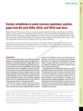
Cardiac arrhythmias in acute coronary syndromesRecommendations for antiarrhythmic therapy in patients withacute coronary syndrome and ventricular tachyarrhythmiaFor patients with acute coronary syndrome (ACS) without ventricular arrhythmias, prophylactic antiarrhythmic drug treatment shouldnot be administered.– If life-threatening ventricular arrhythmias related to ACS occurdespite optimal revascularization, early treatment with betablockers, balancing electrolytes, and sedation to reduce sympathetic drive and/or overdrive stimulation, repetitive electricalcardioversion/defibrillation should be considered first.EuroIntervention 2014;10-online publish-ahead-of-print August 2014patients with advanced heart failure.32 It belongs therefore to thefew AADs that are considered to have no or little effect on longterm prognosis when given to patients with severe structural heartdisease and/or extensive MI. In the context of ACS, amiodaronetherapy – compared with lidocaine – retrospectively has shown anincreased short- and long-term mortality.20 On the other hand, in thesetting of out-of-hospital cardiac arrest, amiodarone was associatedwith better survival to admission rate as compared with lidocaine inpatients with refractory to shock therapy VF.33 Amiodarone shouldbe considered for the suppression (intravenous or oral) and prevention (oral) of recurrent arrhythmias along with beta-blockers.Flecainide, propafenone, and ajmaline exert their antiarrhythmic potential by significant slowing of conduction. In the settingof ACS, these effects may result in aggravation rather than termination of VT/VF. The antiarrhythmic potential of these AADs forthe suppression and termination of VA has been mainly studieduntil the 1990s.34-37 However, in the setting of ACS, these drugsmay cause an aggravation of VT/VF. After the publication of theCAST trial that has shown an increased mortality in patients afterMI treated with encainide, flecainide, or moricizine as comparedwith placebo, further research to class IAAD and VA has largelybeen abandoned.26 Thus, these drugs should not be used in ACS.Dofetilide and azimilide are class III AADs that prolong cardiac repolarization and refractory period with proven efficacy onthe suppression of VA.38-40 The treatment with azimilide did notaffect the mortality of patients with a recent MI and an LV ejectionfraction of 15% to 35%.41 Only few data exist on the use of dronedarone for the treatment of VA; however, an increased mortalityhas been reported in patients with AF and heart failure treatedwith dronedarone.42 All class III AADs increase the QT intervalwith a risk for torsade de pointes tachycardias. However, none ofthese drugs had been specifically investigated for the treatmentof VA related to ACS and can thus not be recommended for thisindication.Ranolazine is a piperazine derivative with a chemical structuresimilar to that of lidocaine. The MERLIN-TIMI 36 study randomized 6560 patients with ACS to ranolazine or placebo.43 Althoughthere was no significant difference in the combined primary endpoint of cardiovascular death, MI, or recurrent ischaemia, the incidence of non-sustained VT ( 30 s) was significantly reduced byranolazine compared with placebo.43,44 The role of ranolazine as anAAD is nevertheless still investigational.45– If antiarrhythmic drug therapy is necessary on top of thesemeasures for the acute treatment of recurrent ventriculararrhythmias related to ACS after failure or non-availabilityof other treatment capabilities, administration of intravenousamiodarone is reasonable, followed by intravenous lidocaine,if necessary (Figure 2).– When experience is available, early catheter ablation shouldbe considered when other treatments fail (see below).– Ventricular tachyarrhythmia in the first minutes after successful reperfusion therapy can be transient without need fortreatment, known as reperfusion arrhythmias.– If frequent premature ventricular complexes and non-sustained ventricular tachycardia continue despite successfulreperfusion therapy under sufficient beta-blocker therapy,they should only be treated if hemodynamically important.This treatment should follow the same principles as the treatment of sustained VA.Recurrent VT/VF and Electrical Storm in ACSCardioversion/defibrillationOverdrive pacingAttempt complete revascularizationTreat ischaemiaCorrect electrolyte imbalanceβ-blocker therapyDeep sedationRecurrent VT/VFElectrical StormAmiodaroneLidocaineConsider catheter ablationAmiodaroneConsider ICD reprogrammingConsider catheter ablationConsider LVAD implantationFigure 2 Treatment recommendations for recurrent VT/VF andelectrical storm in ACS. ACS: acute coronary syndrome; ICD:implantable cardioverter-defibrillator; LVAD: left ventricular assistdevice; VF: ventricular fibrillation; VT: ventricular tachycardiaHow to manage electrical storm andinappropriate implantable cardioverterdefibrillator shocks in patients with acutecoronary syndromeIn patients with ACS, electrical storm and inappropriate implantable cardioverter-defibrillator (ICD) shocks are associated with poorprognosis.46-49Electrical storm is defined as the three or more episodes of VTor VF in any 24-hour period.46,50 It results from interplay betweenpre-existing vulnerable substrate and acute triggers. It is a rare but5
EuroIntervention 2014;10-online publish-ahead-of-print August 2014very serious event and is associated with a poor prognosis.51 Mostinvestigations failed to reveal any clear cause for the developmentof electrical storm. Acute ischaemia is more likely to induce VF orpolymorphic VT than monomorphic VT.50The majority of inappropriate shocks were related to supraventricular tachycardia.48,49 Both appropriate and inappropriate implantable ICD shocks were significant predictors of death, whereas nochange was noted in mortality among those with anti-tachycardiapacing (ATP)-treated arrhythmias.52ACUTE TREATMENT AND METHODS TO REDUCE INCIDENCEOF IMPLANTABLE CARDIOVERTERDEFIBRILLATOR SHOCKElectrical storm constitutes a critical situation both on managementof hemodynamically unstable arrhythmias and because it is associated with significant elevated sympathetic tone, which facilitatefurther arrhythmias.The patient has to be hospitalized and monitored in an intensivecare unit. The most effective treatment is to reverse the ischaemiawith emergency coronary revascularization or with anti-ischaemic,antithrombotic (antiplatelet, anticoagulant) agents. The intravenousadministration of magnesium and potassium may be undertaken inpatients with QT prolongation or hypokalemia. The suppression ofmalignant arrhythmias is an accepted indication for placement ofa percutaneous ventricular assist device53 (see below).The key intervention in electrical storm is blocking the sympathetic system through the intravenous administration of beta-blockers, especially propranolol;54 combined with sedatives, such asbenzodiazepine. Intravenous analgesics and sedatives should alsobe given. Amiodarone has moderate negative inotropic effects, isrelatively safe in patients who have heart failure and is effective asadjunctive therapy to prevent recurrent ICD shocks.55 Trials havenot confirmed lidocaine is superior to other antiarrhythmics.56 If anICD fails to convert a life-threatening rhythm, external defibrillation pads should be ready for use.Implantable cardioverter-defibrillator programming is probably a key issue to prevent electrical storm.57 Although all devicesare pre-programmed, it is important to review the device settingsbecause these settings may be inappropriate for the individualpatient.50 Patients should receive an ICD with ATP programmingeven if the clinical presentation was VF, or VT was not inducedat the electrophysiological study (EPS).58 Programming ICD todeliver ATP for VT faster than 200/min is safe, reduces the needfor shocks and improves mortality compared with conventionaltherapy.59 The number of VT cycles needed for detection can beincreased from nominal values to allow spontaneous termination, and sustained rate duration should be prolonged or disabled. Redetection can carefully be prolonged to reduce the risk ofinappropriate detection of non-sustained VT in many patients.50,60In some cases, it may be advisable to turn off the ICD in orderto avoid multiple ineffective shocks provided that the patient isstrictly monitored in an intensive care unit and that CV/defibrillation might be provided in a timely fashion in case of life-threatening arrhythmias.6On the basis of the observation that VT tends to be abrupt inonset and somewhat regular (high stability), sinus tachycardia tendsto accelerate gradually, and AF is irregularly irregular (low stability), it was found that programming sudden onset and stability criteria helped to discriminate supraventricular arrhythmias and reducedthe risk of inappropriate shock.61 In addition, just as the surfaceQRS complex during VT generally differs from that during sinusrhythm, discrimination algorithms can be used.Radiofrequency ablation of PVC originating from the Purkinjefibre network holds great promise in the treatment of electricalstorm. The typical indication for ablation is VT refractory to medical therapy in patients who also receive multiple ICD shocks.52 Inpatients with electrical storm, catheter ablation should be considered as a treatment option at an early stage.27,62Recommendations for management of electrical storm and inappropriate shocks in patients with acute coronary syndrome– Initial management of patients with electrical storm requiresidentifying and correcting underlying ischaemia.– Amiodarone, beta-blockers and electrolyte correction asneeded form the cornerstone of antiarrhythmic therapy.– Patients who have implantable cardioverter-defibrillator mayrequire device reprogramming.– A focal rescue ablation approach targeting the trigger for ventricular fibrillation early after acute myocardial infarctionseems promising (Figure 2).Catheter ablation of sustained ventriculararrhythmias in acute coronary syndromeIncessant sustained VA that occurs in ACS is a life-threatening condition, carrying a high morbidity and mortality rate. It can be difficult to control with AADs, as class I AADs and amiodarone efficacyin reduction of mortality is controversial and time is required foramiodarone to reach therapeutic levels to provide effective rhythmcontrol. Catheter ablation of VA during the acute phase of an ACSis rarely performed, and most ablations for incessant VA occur postMI. However, in patients with intractable, drug-refractory VA, catheter ablation after treating the underlying ischaemia can play animportant role and these patients should be referred to a specializedablation centre.The two main mechanisms implicated in the induction of sustained VA in ACS are macro-re-entry from heterogeneous substrate,and after-depolarizations and triggered activity from impaired butischaemia-resistant Purkinje fibres within areas of myocardialischaemia leading to PVC,27 and both can be targeted for ablation(Figure 3).Radiofrequency ablation using an irrigated-tip ablation catheter, in conjunction with a 3D-electroanatomical mapping systemis most commonly undertaken. This is done via either an antegrade transseptal, retrograde aortic or a combined approach. Inthe majority of cases due the sub-endocardial location of thePurkinje fibres and the ischaemic myocardial substrate, endocardial mapping, and ablation suffices. Mapping of PVCs requires
Cardiac arrhythmias in acute coronary syndromest
symptoms to transfer to an acute coronary care centre, or long patient-related delay, severe acute ischaemia in . Figure 1). (2) Patients in whom revascularization was not or only partially . successful due to technical difficulties (severe acute ischaemia in . Figure 1). (3) Patients who carry an arrhythmogenic substrate prior to the