Transcription
Original Researchpublished: 30 October 2017doi: 10.3389/fimmu.2017.01419HYoshihito Minoda1†, Isaac Virshup 2†, Ingrid Leal Rojas1, Oscar Haigh1, Yide Wong 3,John J. Miles 3, Christine A. Wells 2,4 and Kristen J. Radford 1*Cancer Immunotherapies Laboratory, Mater Research Institute, University of Queensland, Translational Research Institute,Brisbane, QLD, Australia, 2 The Centre for Stem Cell Systems, Anatomy and Neuroscience, Faculty of Medicine, Dentistryand Health Sciences, The University of Melbourne, Melbourne, VIC, Australia, 3 Centre for Biodiscovery and MolecularDevelopment of Therapeutics, AITHM, James Cook University, Cairns, QLD, Australia, 4 Walter and Eliza Hall Institute ofMedical Research, Melbourne, VIC, Australia1Edited by:Manfred B. Lutz,University of Würzburg, GermanyReviewed by:Diana Dudziak,University Hospital of Erlangen,GermanyElodie Segura,Institut Curie, France*Correspondence:Kristen J. Radfordkristen.radford@mater.uq.edu.au†These authors have contributedequally to this work.Specialty section:This article was submitted toAntigen Presenting Cell Biology,a section of the journalFrontiers in ImmunologyReceived: 12 July 2017Accepted: 12 October 2017Published: 30 October 2017Citation:Minoda Y, Virshup I, Leal Rojas I,Haigh O, Wong Y, Miles JJ, Wells CAand Radford KJ (2017) HumanCD141 Dendritic Cell andCD1c Dendritic Cell UndergoConcordant Early GeneticProgramming after Activationin Humanized Mice In Vivo.Front. Immunol. 8:1419.doi: 10.3389/fimmu.2017.01419Human immune cell subsets develop in immunodeficient mice following reconstitutionwith human CD34 hematopoietic stem cells. These “humanized” mice are useful models to study human immunology and human-tropic infections, autoimmunity, and cancer.However, some human immune cell subsets are unable to fully develop or acquire fullfunctional capacity due to a lack of cross-reactivity of many growth factors and cytokinesbetween species. Conventional dendritic cells (cDCs) in mice are categorized into cDC1,which mediate T helper (Th)1 and CD8 T cell responses, and cDC2, which mediateTh2 and Th17 responses. The likely human equivalents are CD141 DC and CD1c DC subsets for mouse cDC1 and cDC2, respectively, but the extent of any interspeciesdifferences is poorly characterized. Here, we exploit the fact that human CD141 DCand CD1c DC develop in humanized mice, to further explore their equivalency in vivo.Global transcriptome analysis of CD141 DC and CD1c DC isolated from humanizedmice demonstrated that they closely resemble those in human blood. Activation of DCsubsets in vivo, with the TLR3 ligand poly I:C, and the TLR7/8 ligand R848 revealedthat a core panel of genes consistent with DC maturation status were upregulated byboth subsets. R848 specifically upregulated genes associated with Th17 responses byCD1c DC, while poly I:C upregulated IFN-λ genes specifically by CD141 DC. MYCLexpression, known to be essential for CD8 T cell priming by mouse DC, was specificallyinduced in CD141 DC after activation. Concomitantly, CD141 DC were superior toCD1c DC in their ability to prime naïve antigen-specific CD8 T cells. Thus, CD141 DC and CD1c DC share a similar activation profiles in vivo but also have induce uniquesignatures that support specialized roles in CD8 T cell priming and Th17 responses,respectively. In combination, these data demonstrate that humanized mice provide anattractive and tractable model to study human DC in vitro and in vivo.Keywords: humanized mice, dendritic cells, CD141 DC, CD1c DC, poly I:C, R848, microarrayFrontiers in Immunology www.frontiersin.org1October 2017 Volume 8 Article 1419
Minoda et al.Transcriptome of In Vivo Activated Human DCINTRODUCTIONcan cross-present some forms of Ag to CD8 T cells (21); in theabsence of inducing T regulatory cells (22). Indeed CD1c DCshave also been trialed as vaccine candidates (23). Thus, there iscurrently no clear consensus as to whether targeting specific ormultiple human DC subsets will be most beneficial for targetedimmunotherapy (22).Effective preclinical models to study human CD141 DC andCD1c DC in vivo are needed in order to further understand fundamental human DC biology and evaluate new immunothera peutics. Transfer of human CD34 HSC into immunodeficientmice lacking T, B, and NK cells leads to stable long-term engraftment of human HSC and differentiation of human immune cellsubsets. These “humanized”(hu) mice are emerging as a powerfultool to study the human immune system and are being increasinglyused to model human-tropic infectious diseases, hematopoiesis,autoimmunity, and cancer and to evaluate new drugs, vaccines,and immunotherapeutics (24–26).One of the current limitations of hu mouse models is thedefective development and/or function of some human leukocyte compartments, arising from a lack of cross-reactivitybetween mouse and human cytokines and growth factors(24–27). This is most notable within the monocyte/macrophagelineages, which require the addition of human cytokines topromote development and acquire functional capacity. MouseRag2 / Il2rg / strains with human cytokine genes “knocked in”are under development, these strains accommodate enhancedmonocyte/macrophage and NK cell lineage development (26).In contrast, we and others have shown that human CD141 and CD1c DC subsets develop in the BM, spleen, and lungsfollowing human CD34 reconstitution in a number of immunodeficient mouse strains, making this an attractive modelto study human cDC function in vivo (28–30). Although theCD141 DC and CD1c DC that develop in these mice exhibitmany of the phenotypic and functional characteristics of theirhuman blood counterparts, the extent to which they recapitulatehuman DC functionally has not been fully defined. In this study,we examined the global transcriptome of the CD141 DC andCD1c DC that develop and become activated in vivo in hu miceto establish the extent of their similarity with their human bloodcounterparts. We then used this model to identify early changesin gene expression associated with activation of human CD141 DC and CD1c DC in vivo. These data validate hu mice as apowerful model to study human DC and identify common andunique pathways associated with in vivo activation.Dendritic cells (DCs) are specialized antigen (Ag)-presentingcells that initiate and direct immune responses (1, 2). DC are aheterogenous cell population comprised of distinct subsets thatharbor specialized capacity to drive specific immune responses.DC develop from CD34 hematopoietic stem cells (HSC) inthe bone marrow (BM) that develop into the common myeloidprogenitor, giving rise to the more restricted macrophage/DCprogenitor (MDP). The MDP can develop into monocytes, orthe common DC progenitor (CDP). The CDP give rise to plasmacytoid (p) DC and two “classical” or “conventional” (c) DCsubsets now referred to as “cDC1” and “cDC2.” While surfacemarker expression has been the convention for characterizationof conventional DC subsets, gene expression studies are redefining functional characteristics within these subsets (1, 3–5). pDCsare major producers of type-I IFN and are considered importantfor anti-viral immunity, the cDC1 and 2 subsets are critical forshaping adaptive immunity to intracellular and extracellularpathogens, respectively.In mice, the cDC1 subset comprises the lymphoid residentCD8 DC and the tissue resident CD103 DC. These DCsrequire FLT3L and transcription factors IRF8, ID2, and Batf3for their development. cDC1 are required for priming protective CD8 T cell responses against cancer and for the efficacyof cancer immunotherapies (6). cDC1 produce high levels ofIL-12p70, induce T helper (Th)-1 responses, and cross-presentexogenous Ag for priming of CD8 cytolytic T cell responses(7–9). Hallmarks of these functions include the expression ofchemokine receptor, XCR1, and adhesion molecule nectin-likeprotein 2 (Necl2/CADM1), both of which play important roles inCD8 T cell priming (10–12). The presence of C-type lectin-likereceptor, Clec9A, is essential for recognition and cross-primingof necrotic cell Ag, as is expression of TLR3, which enhancescross-priming and induces large amounts of IFNλ followingligation with agonists such as poly I:C (11, 13–15); and reviewed inRef. (16). The human equivalent of cDC1 are defined as CD141 (BDCA3) DC and share many similarities with mouse cDC1,including expression of XCR1, CADM1, Clec9A, and TLR3,Type III IFN production in response to TLR3 ligation, and crosspresentation of Ag from necrotic cells (2). There is therefore astrong rationale to develop similar strategies to target the humancDC1 equivalent for new cancer immunotherapies (16).The cDC2 subset is also referred to as CD11b DC in themouse lymphoid and non-lymphoid tissues (1, 4). These DCsare FLT3L and IRF4 dependent and share significant overlap inphenotype with cells of the monocyte lineage (17). Monocytederived DCs contribute to numerous DC niches throughout thebody under inflamed conditions [reviewed in Ref. (18)], andare phenotypically distinct from cDC1 and cDC2 by a lack ofexpression of Flt3, and the zinc finger transcription factor zbtb46(19). cDC2 play a key role in driving adaptive immune responsesto extracellular pathogens, owing to their capacity to promoteTh2 and Th17 responses. The human equivalents of mousecDC2 are defined as CD1c (BDCA-1) DC (2), and while thereis evidence to suggest that they promote Th17 responses (20),they also produce high levels of IL-12, induce Th1 responses andFrontiers in Immunology www.frontiersin.orgMATERIALS AND METHODSGeneration of Hu Mice and Isolation of DCCord blood was obtained with written informed consent fromthe Queensland Cord Blood Bank with approval from the MaterAdult Hospital Human Ethics Committee. CD34 hematopoieticprogenitor cells were isolated by density gradient enrichment followed by a positive selection using a CD34 isolation kit (MiltenyiBiotec) as previously described (30). NSG-A2 mice (stock no.014570) were purchased from Jackson Laboratories. 2–5-day-oldNSG-A2 pups received 10 Gy total body irradiation 4 h prior2October 2017 Volume 8 Article 1419
Minoda et al.Transcriptome of In Vivo Activated Human DCto intrahepatic injection of human CD34 cells. Engraftment ofhuman CD45 cells was confirmed 10–12 weeks later, after whichhu mice received 2 s.c. doses of human recombinant huFLT3-L(BioXcell) 4 days apart prior to experimentation. Engrafted micewere injected retro-orbitally with 50 µg poly IC (Invivogen) or20 µg R848 (Invivogen) alone or in combination and mice wereeuthanized 2 h later. This study was carried out in accordancewith the recommendations of the Australian code for the care anduse of animals for scientific purposes (8th Edition). The protocolwas approved by the University of Queensland Animal EthicsCommittee.differentially expressed by CD141 DC and CD1c DC wereidentified using ANOVA analysis with p value cut-off thresholdof Bonferroni correction p 0.01 and log(2) 3-fold difference.The data have been deposited in the GEO database, accession# GSE99666, and on Stemformatics http://www.stemformatics.org/datasets/search?ds id 6612. Publically available data setsand gene signatures for human DC subsets GSE35457 (17) wereprocessed similarly for comparison.Cytokine Chemokine and ActivationMarker AssaysDendritic cell subsets purified from hu mice were exposed toTLR adjuvants for 18 h in vitro, or cultured in media alone for18 h. Costimulatory molecule expression on the cell surface wasquantified by flow cytometry. Culture supernatants and serumsamples from the mice were analyzed for human cytokines usinga LEGENDplex flow cytometry bead array kit (Biolegend).Human IFN-λ was measured by ELISA (R&D Systems).Flow CytometrySingle cell suspensions of BM, liver, lung, spleen, and peripheralblood from engrafted mice were blocked with rat and mouseserum then labeled with Live Dead Aqua (Life Technologies),anti-mouse CD45 PerCP Cy5.5, anti-human CD45 APC Cy7,HLA DR PE Cy7, CD19/20 Pacific blue and either CD141 APC,CD123 PE (all from BioLegend), and CD1c FITC (Abcam) toidentify DC, or CD3 Pacific blue, CD8 PE Cy7, CD14 APC (allfrom BioLegend), and CD4 FITC (BD Biosciences) for otherleukocytes (Figure S1 in Supplementary Material). Absolutecell counts were determined by the addition of 5,000 Trucountbeads (BD Biosciences) per tube. Data were acquired on a Cyanflow cytometer (Beckman Coulter) and analyzed using Flow Josoftware (Tree star, version 8).CD8 T Cell Priming AssaysHLA-A2 DC subsets isolated from hu mice were pulsed withthe HLA-A2-restricted MART-1 peptide, ELAGIGILTV (1 µM)in the presence of poly IC and R848 for 2 h at 37 C. Naïve CD8 T cells from autologous cord blood mononuclear cells werepurified by negative selection using CD8 T cell isolation kit(Miltenyi Biotec). Peptide-pulsed DCs were cultured with naïveCD8 T cells in the presence of IL-2 (Hoffman-LaRoche) andT cell growth factor for 21 days. MART-1-specific CD8 T cellswere quantitated by flow cytometry using a MART-1/HLA-A2specific pentamer-APC (ProImmune), Live Dead aqua, andanti-human CD19-Pacific blue, CD14-Pacific blue, CD3-PE, andCD8-FITC antibodies (BioLegend). Samples were acquired ona LSR Fortessa flow cytometer (BD Biosciences) and analyzedusing Flow Jo software (Tree star, version 8). For polyfunctionalT cell assays, the CD8 T cell cultures were restimulated for 6 h inthe presence of absence of MART-1 peptide (1 µM) with brefeldinA, GolgiStop, and anti-CD107a-FITC (BD Biosciences). Cellswere then washed and labeled with anti-CD4 BV711, anti-CD8APC Cy7, and Live dead followed by permeabilization (Cytofix/Cytoperm; BD Biosciences) and stained with anti-TNFα APC,anti-IFNγ PE, and anti-IL-2 Alexa700. Samples were acquiredon a LSR Fortessa cytometer (BD Biosciences) and analyzedusing Flow Jo software (Tree star, version 8). Boolean gating wasperformed to measure the frequency of each response based onall possible functional combinations of IFNγ, TNFα, IL-2, andCD107a. The data were analyzed and graphed using SimplifiedPresentation of Incredibly Complex Evaluations (SPICE) software, version 5.3.DC Isolation from Hu MiceHuman DCs were enriched from single cell BM suspensionsby first labeling with Ab specific for human CD3, CD14, CD19,CD20 (all from Beckman Coulter), CD34 (BD BioSciences), andmouse CD45 (BD BioSciences) and Ter119 (BioLegend) followedby depletion of bound cells using sheep anti-rat IgG Dynabeads(Invitrogen) as previously published (30). Cells were then labeledwith Live Dead aqua, anti-mouse CD45 PerCP Cy5.5, antihuman CD45 APC Cy7, CD3/CD14/CD19/CD20 Pacific blue,HLA DR PE Cy7, CD123 PE or PerCP Cy5.5, CD1c FITC, andCD141 APC and sorted using a Moflo Astrios (Beckman Coulter)(Figure S2 in Supplementary Material).Gene Expression AnalysisTotal RNA from purified DC subsets was prepared by resuspen ding between 1 105 and 1 106 cells in TRIzol Reagent (LifeTechnologies) followed by chloroform extraction and isopropanol precipitation. RNA quantity and integrity was measuredusing the Bioanalyzer RNA Nano chip (Agilent Technologies),with all RNA integrity numbers ranging between 8.4 and 9.7.cDNA was generated from 450 ng RNA and converted to cRNAusing an Illumina TotalPrep RNA Amplification Kit (Ambion).A 14 h in vitro transcription was performed. Amplified cRNA(750 ng) for each sample was hybridized onto HumanHT-12 v4Expression BeadChip Kit (Illumina) according to the manufacturer’s protocol and scanned on an Illumina BeadArray Reader(Illumina). Quality control, normalization, and log 2 transformation of the raw expression data were performed using the LumiBioconductor package (31) and integrated into the Stemformaticsplatform www.stemformatics.org (32) for visualization. TranscriptsFrontiers in Immunology www.frontiersin.orgRESULTSHu Mouse CD141 DC and CD1c DCAre Closely Related to Human BloodEquivalentsRobust engraftment of human CD45 cells was observed10–14 weeks after reconstitution of 2–5-day-old NSG-A2 pups3October 2017 Volume 8 Article 1419
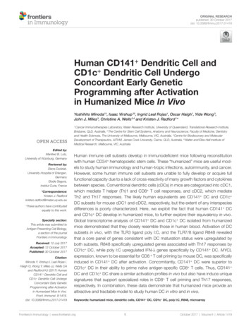
Minoda et al.Transcriptome of In Vivo Activated Human DCwith human cord blood-derived CD34 HSC (Figure 1A;Figure S1 in Supplementary Material). Human CD45 cells weremost prevalent in the spleen and liver, where they comprised57.34% 17.97 (mean SD, n 9) and 67% 21.45 (mean SD,n 5) of mononuclear cells, respectively. Human CD45 cellscomprised 39.88% 15.66 (mean SD, n 5) of mononuclearcells in the BM and could also be detected in lower proportions in the lungs and blood (Figure 1B). Human CD141 andCD1c DC subsets were found in all organs examined, with thelargest numbers residing in the BM compartment (Figure 1C).Human B cells, CD14 monocytes, plasmacytoid DC, and CD4 and CD8 T cells also developed in this model (Figure S1 inSupplementary Material). Thus, NSG-A2 mice support robust,multi-lineage development of human leukocytes, includingCD141 and CD1c DC.We compared transcriptomes of BM-derived CD141 andCD1c DC from hu mice with equivalents from human blood(17). We identified 316 genes that were significantly differentially expressed (log-3 fold change, adjusted p 0.01) betweenCD141 and CD1c DC regardless of tissue source (Table S1 inSupplementary Material). Consistent with known DC functions,gene set enrichment analysis of these subsets ranked C-typelectins, endosomal TLR pathways, and the AIM2 inflammasomeas most discriminating (Table S2 in Supplementary Material).Figure 1 Development of CD141 and CD1c dendritic cell (DC) in hu mice. (A) Gating strategy used to define DC subsets in the bone marrow (BM) of humice. After gating on live singlet cells, DCs were defined as hu CD45 HLA-DR , CD19/20 , and CD1c or CD141 . (B) Frequency of human leukocytes, CD141 DC and CD1c DC engrafted in the organs of NSG-A2 mice expressed as % mononuclear cells (huCD45) or as % hu CD45 cells (CD141 and CD1c DC).(C) Frequency of human leukocytes, CD141 DC and CD1c DC expressed as absolute cell numbers per organ. BM data are quantitated per tibia and femur,and blood per ml.Frontiers in Immunology www.frontiersin.org4October 2017 Volume 8 Article 1419
Minoda et al.Transcriptome of In Vivo Activated Human DCof CD36 and CD163 along with inflammatory genes includingCD14, S100A9, S100A8 (5). Hu mouse CD1c DC expressedmuch higher levels of the CD1c A non-inflammatory markersthan hu mouse CD141 DC, but also expressed the inflammatory markers CD163, CD14, S100A9, and S100A8, which werefurther upregulated after activation (Figure S5 in SupplementaryMaterial). These data indicate that the hu mouse CD1c DCisolated here likely contain both inflammatory and non-inflammatory CD1c DC subsets. Because CD1c DC have knownoverlapping features with monocytes (20), we used differentialexpression analysis to identify genes that CD1c DC and mono cytes co-express. More than 820 differentially expressed geneswere identified by comparing hu mouse CD1c DC, bloodCD1c DC, and blood monocyte subsets with CD141 DC andpDC (log-3 fold change, adjusted p 0.01, and Figure S6 inSupplementary Material). Genes shared between monocytesand CD1c DC included CSF1R, SIRPA, IL1B, NLRP3, CASP1,FCGR2A, and FCGR2B.Hierarchical clustering analysis of the differentially expressedgenes revealed clustering of hu mouse CD141 DC with humanblood CD141 DC, and hu mouse CD1c DC with human bloodCD1c DC (Figure 2A). In an unsupervised clustering approach,the first component of a principal component analysis was concordant with batch, tissue, and species source (hu mice versushuman blood). However, clustering of primary human CD141 DC with hu mouse CD141 DC, and CD1c DC across the twodatasets with human peripheral blood monocytes was evidentbetween the first and second principal components (Figure S3 inSupplementary Material).Amongst the 156 genes preferentially expressed by humouse CD141 DC and blood CD141 DC were genes previously associated with this subset including CLEC9A, CADM1,ID2, BATF3, RAB7B, AIM2, BTLA, SEPT3, CLNK, and GSAM(Figure 2B; Table S1 in Supplementary Material). IRF8, TLR3,and XCR1 were also more highly expressed by CD141 DC(Figure 2B; Figure S4 in Supplementary Material). Genesknown to be expressed by the CD1c DC lineage, includingIRF4, SIRPA, CLEC10A, FCGR2B, FCER1A, TLR5, and TLR7were amongst the 160 top ranked differentially expressed genesin hu mouse and human blood CD1c DC (Figure 2B; Table S1 inSupplementary Material). CD1c blood DCs have recently beensubdivided into two subsets defined as “non-inflammatory”or “CD1c A”, defined by expression of FCER1A, CLEC10A, andFCGR2B; and “inflammatory” or “CD1c B” defined by expressionR848 and Poly I:C Induce Common GeneExpression Profiles in CD141 DC andCD1c DC In VivoTLR expression and maturation by human DC are well characterized in vitro but their responses in vivo are not. The TLR3ligand, poly I:C, and the TLR7/8 ligand, R848, have been usedFigure 2 Hu mouse CD141 dendritic cell (DC) and CD1c DC subsets cluster with their human blood counterparts. Hierarchical Cluster (Pearson correlation) ofgene expression data showing (A) differentially expressed genes with a log-3 fold change (lfc) over one with adjusted p value 0.01. (B) Expression of genes knownto be associated with CD141 DC and CD1c DC. Sample groups are colored at the bottom of the sample tree by ClustDiff (CD1c green; CD141 blue); hostspecies (humanized mouse green; human cells blue) and treatment (R848 alone blue; Poly I:C green; Poly I:C R848 combination pink; untreated orange). Thecolor of expression scores is scaled by Z-score, per row.Frontiers in Immunology www.frontiersin.org5October 2017 Volume 8 Article 1419
Minoda et al.Transcriptome of In Vivo Activated Human DC(29% of pathway genes represented, p 9.05E-04), canonicalNF-κB pathway (17% of pathway genes represented, p 0.027),RIG-I/MDA5, and TLR signaling (Figures 3A,B; Table S3 inSupplementary Material). Twenty five of these genes belong toa core set of NK-κB or IFN-stimulated genes (ISG) associatedwith DC maturation in both mouse and human irrespective ofstimuli (Table S4 in Supplementary Material) (36). Most of thegenes differentially expressed after activation were upregulated tosimilar degree by CD141 DC and CD1c DC in response to allas adjuvants to activate human DC alone or in combination(21, 28, 33, 34). Because cytokines can be detected in mouseserum within 1 h of adjuvant injection (35), we chose a 2 h timepoint to investigate the early changes in gene expression associated with activation of BM derived CD141 DC and CD1c DC in vivo. A one-way AVONA analysis (adjusted p 0.05)revealed differential expression of 408 genes as a result of activation. These genes were involved in immune signaling pathwaysincluding Tnfr2 signaling, IFNα/β signaling, Tnfr1 signalingFigure 3 Changes in gene expression by hu mouse dendritic cells (DC) as a result stimulation with poly I:C and or R848 in vivo. Expression of CD1c DC andCD141 DC genes following 2 h activation with poly I:C and/or R848 in vivo in hu mice. Boxes represent 25th–75th percentiles minimum and maximum valueswith line at the median from DC sorted from 2 to 3 individual mice. Expression of selected differentially expressed genes (A) upregulated by all TLR stimuli and(B) more highly expressed by R848 activated DC. Y-axis (normalized, log (2) expression).Frontiers in Immunology www.frontiersin.org6October 2017 Volume 8 Article 1419
Minoda et al.Transcriptome of In Vivo Activated Human DCCD1c DC Upregulate Th17 PromotingCytokines after Activation In Vivowith R848stimulatory conditions. These included TNFAIP3, TRAF1, IFIT1,ITIF2, ISG15, and OASL (Figure 3A). A small subset of geneswere significantly upregulated by both CD141 DC and CD1c DC in response to R848 alone or combined with poly I:C but wereonly marginally upregulated by poly I:C alone. These includedimmunoregulatory molecules LTA, NFIL3, CD274, and IGFB4(Figure 3B). Conversely, no genes were found to be significantlyupregulated on both subsets activated with poly I:C that were notalso upregulated by R848.Poly I:C and R848 alone or combined induced expression ofcostimulatory molecule genes CD40, CD80, and CD83 to similarlevels by both CD141 DC and CD1c DC (Figure 4A). Geneexpression correlated with increased protein expression of CD40,CD80, and CD83 on the surface of purified CD141 DC andCD1c DC after in vitro activation (Figure 4B). However, cellsurface expression of CD80 was significantly higher on activatedCD141 DC compared to activated CD1c DC, and while CD83expression was also higher on activated CD141 DC, this did notreach statistical significance. Similarly, chemokine genes CXCL9and CXCL10 were upregulated by both DC subsets followingactivation but only CXCL10 reached statistical significance(Figure 5A). Upregulation of CXCL9 and CXCL10 correlatedwith detectable levels of these chemokines in the serum of humice 2 h after activation (Figure 5B). In vitro, purified CD141 DC were the main producers of CXCL9 while both subsetsproduced high levels of CXCL10 (Figure 5C). Collectively, thesedata highlight both common and distinct pathways of CD141 DC and CD1c DC that are activated by R848, poly I:C, or thecombination.The mouse CD11b equivalents of CD1c DC promote Th17responses (20). Consistent with this, we found genes encoding theTh17-promoting cytokines IL1B and IL6 were upregulated by bothBM derived DC subsets after activation, but levels expressed byactivated CD1c DC were fourfold and eightfold higher than similarlyactivated CD141 DC, respectively (Figure 6A). Importantly,expression of IL23A was 19-fold higher in CD1c DC activatedwith R848 alone or combined with poly I:C compared to similarlyactivated CD141 DC. IL-1β, IL-6, and IL-23 were detectable inthe serum of hu mice and although purified hu mouse CD1c DCproduced higher amounts of these cytokines in vitro comparedto CD141 DC, the levels did not reach statistical significance(Figures 6B,C). TNF and IL-12A were similarly upregulatedby in vivo activated CD141 DC and CD1c DC with cytokinesdetectable in the serum. Higher levels of TNF and IL-12p70 wereproduced by in vitro cultured CD1c DC compared with CD141 DC, although this did not reach statistical significance (Figure 6C).CD141 DC Upregulate Type III IFN afterActivation In Vivo with Poly I:CCD141 DC have previously been shown to produce IFNα afterpoly I:C activation in vivo (28). Here, we found genes for IFNαsubtypes IFNA2, IFNA7, and IFNA21 were upregulated to a similardegree by both subsets, most notably in response to R848 alone orcombined with poly I:C, while genes encoding most other IFNαFigure 4 Expression of costimulatory molecules after activation of hu mouse CD141 dendritic cells (DC) and CD1c DC. (A) Normalized log 2 expression ofCD1c DC and CD141 DC genes following 2 h activation with poly I:C and/or R848 in vivo in hu mice. Boxes represent 25th–75th percentiles minimum andmaximum values with line at the median from DC sorted from 2 to 3 individual mice. (B) Cell surface protein expression of costimulatory molecules by purified DCsubsets (0 h) and after 18 h culture in vitro in medium alone or with poly I:C R848. Shown is the mean SEM fold upregulation of the mean fluorescence intensityof each costimulatory molecule relative to an isotype control.Frontiers in Immunology www.frontiersin.org7October 2017 Volume 8 Article 1419
Minoda et al.Transcriptome of In Vivo Activated Human DCFigure 5 Expression of chemokines CXCL9 and CXCL10 by hu mouse CD141 dendritic cells (DC) and CD1c DC. (A) Normalized log 2 expression of CD1c DC and CD141 DC genes following 2 h activation with poly I:C and/or R848 in vivo in hu mice. Boxes represent 25th–75th percentiles minimum and maximumvalues with line at the median from DC sorted from 2 to 3 individual mice (B) Chemokine protein levels in the serum of hu mice 2 h after activation. (C) Production ofchemokine protein by purified hu mouse DC 18 h after activation in vitro, versus control [cultured in medium alone in vitro (cont)].compared to similarly activated CD1c DC (Figure 7A). Thiscorrelated with detection of IFNλ in the serum and in the supernatants of purified CD141 DC activated in vitro (Figure 7C).These data confirm that CD141 DC are major producers of IFNλin vivo.subtypes were below detectable levels in both BM-derived DCsubsets (Figure 7A). Although large amounts of IFNα were detec table in the serum of mice treated with R848 and R848 poly I:C,no IFNα was detected in the serum of mice treated with polyI:C alone (Figure 7B). Moreover, IFNα was not detectable in thesupernatants of purified CD141 or CD1c DC activated in vitro(data not shown). IFNB1 was upregulated by both subsets inresponse to all stimuli. CD141 DC are known to produce largeamounts of IFNλ in response to poly I:C (14) and consistent withthis, IFNλ genes IL28A, IL28B, and IL29 were expressed 13-,17-, and 8-fold higher by poly I:C R848-activated CD141 DCFrontiers in Immunology www.frontiersin.orgCD141 DC Upregulate MYCL afterActivation and Prime Naïve CD8 T CellsMYCL is required for optimal CD8 T cell priming by mouse DC(37) and was expressed at 10-fold higher levels by CD141 DC8October 2017 Volume 8 Article 1419
Minoda et al.Transcriptome of In Vivo Activated Human DCFigure 6 Expression of inflammatory cytokines by hu mouse CD141 dendritic cells (DC) and CD1c DC. (A) Gene expression levels 2 h after activation of DCsubsets in vivo. Data are presented as 25th–75th percentiles minimum and maximum values with line at the median of normalized log 2 gene expression by DCsubsets sorted from 2 to 3 individual mice. (B) Cytokine levels in the serum of hu mice 2 h after activation. (C) Production of cytokines by purified hu mouse DC 18 hafter activation in vitro, versus control [cultured in me
beads (BD Biosciences) per tube. Data were acquired on a Cyan flow cytometer (Beckman Coulter) and analyzed using Flow Jo software (Tree star, version 8). Dc isolation from hu Mice Human DCs were enriched from single cell BM suspensions by first labeling with Ab specific for human CD3, CD14, CD19,